. 2008 Apr 2;3(4):e1886.
doi: 10.1371/journal.pone.0001886.

Paracrine factors of mesenchymal stem cells recruit macrophages and endothelial lineage cells and enhance wound healing

Affiliations

Paracrine factors of mesenchymal stem cells recruit macrophages and endothelial lineage cells and enhance wound healing

Liwen Chen et al. PLoS One. .

Erratum in

Abstract

Bone marrow derived mesenchymal stem cells (BM-MSCs) have been shown to enhance wound healing; however, the mechanisms involved are barely understood. In this study, we examined paracrine factors released by BM-MSCs and their effects on the cells participating in wound healing compared to those released by dermal fibroblasts. Analyses of BM-MSCs with Real-Time PCR and of BM-MSC-conditioned medium by antibody-based protein array and ELISA indicated that BM-MSCs secreted distinctively different cytokines and chemokines, such as greater amounts of VEGF-alpha, IGF-1, EGF, keratinocyte growth factor, angiopoietin-1, stromal derived factor-1, macrophage inflammatory protein-1alpha and beta and erythropoietin, compared to dermal fibroblasts. These molecules are known to be important in normal wound healing. BM-MSC-conditioned medium significantly enhanced migration of macrophages, keratinocytes and endothelial cells and proliferation of keratinocytes and endothelial cells compared to fibroblast-conditioned medium. Moreover, in a mouse model of excisional wound healing, where concentrated BM-MSC-conditioned medium was applied, accelerated wound healing occurred compared to administration of pre-conditioned or fibroblast-conditioned medium. Analysis of cell suspensions derived from the wound by FACS showed that wounds treated with BM-MSC-conditioned medium had increased proportions of CD4/80-positive macrophages and Flk-1-, CD34- or c-kit-positive endothelial (progenitor) cells compared to wounds treated with pre-conditioned medium or fibroblast-conditioned medium. Consistent with the above findings, immunohistochemical analysis of wound sections showed that wounds treated with BM-MSC-conditioned medium had increased abundance of macrophages. Our results suggest that factors released by BM-MSCs recruit macrophages and endothelial lineage cells into the wound thus enhancing wound healing.


Conflict of interest statement

Competing Interests: The authors have declared that no competing interests exist.

Figures

Figure 1
Figure 1. mRNA levels of cytokines and extracellular matrix molecules in BM-MSCs and fibroblasts.
Total RNA extracted from BM-MSCs (MSC) or dermal fibroblasts (FB) treated under hypoxic conditions was analyzed by Real-Time PCR for mRNA expression of genes as indicated in the figure. Fold changes vs dermal fibroblasts are shown. Data are mean±SD; n = 3; *P<0.05 vs FB. KGF, keratinocyte growth factor; HB-EGF, heparin-binding EGF-like growth factor; TGFb1, Transforming growth factor-β1; Ang, angiopoietin; SDF1, stromal cell-derived factor-1; MIP, macrophage inflammatory protein; SCF, stem cell factor; EPO, erythropoietin; TPO, thrombopoietin; G-CSF, granulocyte colony stimulating factor; MCP1, monocyte chemotactic protein-1; MIG, monokine induced by gama interferon.
Figure 2
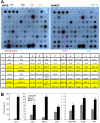
Figure 2. Protein levels of cytokines in BM-MSC-conditioned medium.
(A) Antibody-based protein array analysis human dermal fibroblast (FB)- or BM-MSC-conditioned medium under hypoxic conditions. Similar results were obtained from three independent experiments and results from one of them are shown. The abbreviations are donated in Table 2. (B) ELISA measurement of cytokines in murine fibroblast- or BM-MSC- conditioned medium under hypoxic conditions. Data are expressed as means±SD (n = 3, *P<0.01). PDGF-BB, platelet-derived growth factor-BB. Other abbreviations can be found in the legend for Figure 1.
Figure 3
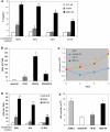
Figure 3. Effects of BM-MSC-conditioned medium on cell migration and proliferation.
(A) migration of CD14+ monocytes. CD14+ monocytes were isolated as described in “Methods” and equal numbers of the cells were loaded to the top chambers. Control (CTL) vehicle medium, fibroblast (FB-M)- or BM-MSC (MSC-M)-conditioned medium at different concentrations were added to the bottom chambers. Cells migrated into the bottom chambers were counted. Triple wells were used. Data shown represent mean±SD of 3 independent experiments (P<0.01). (B) Keratinocyte migration. Equal numbers of murine dermal keratinocytes were added to the top chambers. Media in the bottom chambers were as indicated. Cells migrated to the down-side of the filter were stained, photographed (6 fields per well) and counted. Triple wells were used for each treatment and data shown represent mean±SD of 3 independent experiments (P<0.001). (C) Keratinocyte proliferation. 0.5×10⁵ murine dermal keratinocytes per well were incubated with vehicle-M, FB-M, MSC-M or keratinocyte SFM supplemented with EGF (5 ng/ml, EGF) for different times and cell numbers were counted. Triple wells were used for each treatment. Values shown represent mean±SD of 4 independent experiments (* P<0.01). (D) HUVEC migration. The bottom chambers contained vehicle-M, FB-M or MSC-M at various dilutions. Cells migrated to the down-side of the filter were stained, photographed (6 fields per well) and counted. Triple wells were used for each treatment and data shown represent mean±SD of three independent experiments (*P<0.01 vs vehicle-M; #P<0.01 vs FB-M) (E) HUVEC proliferation. Equal numbers of HUVECs were grown in vehicle-, FB- or MSC-conditioned basal endothelial growth medium (EGM-2) supplemented with 2% FBS or complete EGM-2 and incubated for 3 days. Cell numbers were counted. Experiments were performed in triplicate wells (n = 3, * P<0.001).
Figure 4
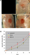
Figure 4. Effect of BM-MSC-conditioned medium on wound closure.
(A) Representative images of wounds before treatment or 7 days after treatment with vehicle control medium (vehicle-M), concentrated fibroblast (FB-M)- or BM-MSC-conditioned medium (MSC-M). (B) Measurement of wound sizes at different times (n = 6 to 13, *P<0.05, **P<0.001). A control panel has been redacted from this figure; see the published Correction notice for more information.
Figure 5
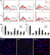
Figure 5. Analysis of cells in wounds.
(A) FACS analysis of cells derived from each wound indicated that wounds treated with concentrated BM-MSC-conditioned medium (MSC-M) at 7 or 14 days had increased percentages of CD4/80 positive monocytes/macrophages compared to wounds treated with vehicle control medium (vehicle-M) or concentrated fibroblast-conditioned medium (FB-M). (B&C) Percentages of cells in wound after FACS analysis (n = 5∼6, *P<0.05). (D) Representative images of confocal microscopy of day 7 wounds treated with vehicle medium, concentrated fibroblast- or BM-MSC-conditioned medium after immunostaining for CD68 (red). Nuclei were stained blue with Hoechst. scale bar, 20 µm.

Similar articles

Cited by

References

    1. Martin P. Wound healing–aiming for perfect skin regeneration. Science. 1997;276:75–81. - PubMed
    1. Singer AJ, Clark RA. Cutaneous wound healing. N.Engl.J Med. 1999;341:738–746. - PubMed
    1. Falanga V. Wound healing and its impairment in the diabetic foot. The Lancet. 2005;366:1736–1743. - PubMed
    1. Boulton AJ, Vileikyte L, Ragnarson-Tennvall G, Apelqvist J. The global burden of diabetic foot disease. Lancet. 2005;366:1719–1724. - PubMed
    1. Dominici M, Le BK, Mueller I, Slaper-Cortenbach I, Marini F, et al. Minimal criteria for defining multipotent mesenchymal stromal cells. The International Society for Cellular Therapy position statement. Cytotherapy. 2006;8:315–317. - PubMed

Publication types

Substances