. 2008 Apr 2;3(4):e1887.
doi: 10.1371/journal.pone.0001887.

Different effect of proteasome inhibition on vesicular stomatitis virus and poliovirus replication

Affiliations

Different effect of proteasome inhibition on vesicular stomatitis virus and poliovirus replication

Nickolay Neznanov et al. PLoS One. .

Abstract

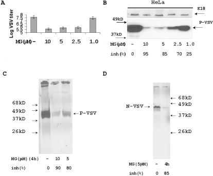
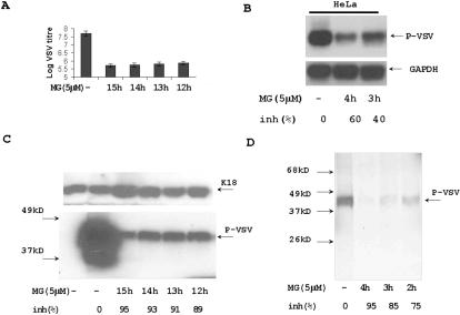
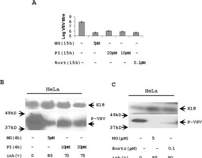
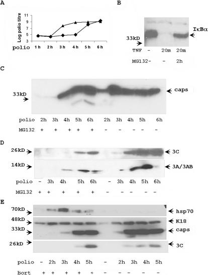
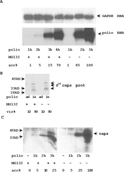
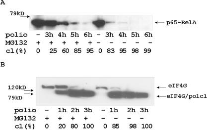
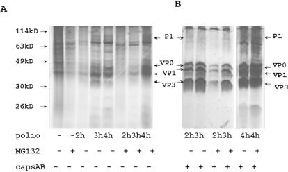
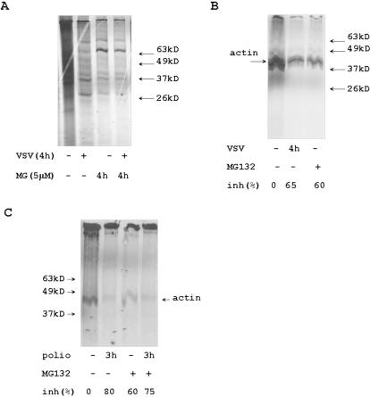
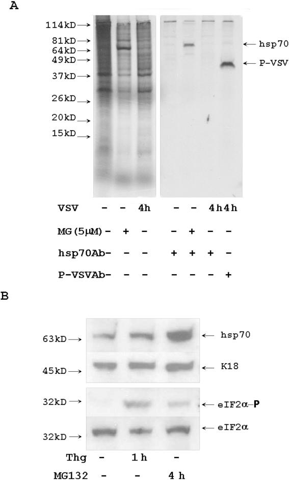
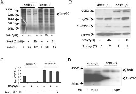
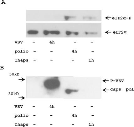
Proteasome activity is an important part of viral replication. In this study, we examined the effect of proteasome inhibitors on the replication of vesicular stomatitis virus (VSV) and poliovirus. We found that the proteasome inhibitors significantly suppressed VSV protein synthesis, virus accumulation, and protected infected cells from toxic effect of VSV replication. In contrast, poliovirus replication was delayed, but not diminished in the presence of the proteasome inhibitors MG132 and Bortezomib. We also found that inhibition of proteasomes stimulated stress-related processes, such as accumulation of chaperone hsp70, phosphorylation of eIF2alpha, and overall inhibition of translation. VSV replication was sensitive to this stress with significant decline in replication process. Poliovirus growth was less sensitive with only delay in replication. Inhibition of proteasome activity suppressed cellular and VSV protein synthesis, but did not reduce poliovirus protein synthesis. Protein kinase GCN2 supported the ability of proteasome inhibitors to attenuate general translation and to suppress VSV replication. We propose that different mechanisms of translational initiation by VSV and poliovirus determine their sensitivity to stress induced by the inhibition of proteasomes. To our knowledge, this is the first study that connects the effect of stress induced by proteasome inhibition with the efficiency of viral infection.


Conflict of interest statement

Competing Interests: The authors have declared that no competing interests exist.

Figures

Figure 1
Figure 1. Proteasome inhibitor MG132 suppresses VSV replication and protein synthesis.
(A) Titration of VSV in MG132 treated cells. HeLa cells were infected with VSV (MOI = 1) for one hour with additional washing and incubated overnight. Medium from control VSV infected cells and VSV infected cells treated with 10 µM, 5 µM, 2.5 µM, and 1 µM of MG132 were used for plaque assay to detect virus replication. Results represent average data of two experiments. (B) Western blotting with anti-P-protein antibodies. HeLa cells were infected with VSV (MOI = 5) for 1 hour. After changing the medium, MG132 was added in the indicated concentrations and the cells were incubated for additional 4 h. Total protein extracts (5 µg) were analyzed with anti-P-protein Abs. Keratin 18 (K18) was a protein loading control. Intensity of each band was estimated with ImageJ software to calculate percentage of viral protein synthesis inhibition. (C) Immunoprecipitation of S35 labeled P-protein. HeLa cells were infected with VSV (MOI = 5) for 4 h. Proteins were labeled with S35 methionine/cysteine for last 30 min of infection and VSV P-protein was precipitated with specific antibodies from cytoplasmic protein extracts and analyzed by electrophoresis and autoradiography. MG132 was added for 4 h in indicated concentrations. (D) Immunoprecipitation of S35 labeled N-protein. The protein extracts described in panel C were precipitated with antibodies specific to N-protein. Proteins were analyzed by electrophoresis and autoradiography.
Figure 2
Figure 2. The effect of MG132 on VSV replication at different time of infection.
(A) Titration of VSV virus from medium of overnight infected HeLa cells. HeLa cells were infected with VSV MOI = 1. The incubation of the cells with virus lasted one hour with additional washing. 5 µM of MG132 were added to cells at time of infection (15 h), 1 h (14 h), 2 h (13 h), and 3 h (12 h) after VSV infection. Results represent average data of two experiments. (B) VSV mRNA synthesis in MG132 treated cells. Northern blot analysis of 10 µg of total RNA from VSV (MOI = 5) infected for 4 h cells treated with MG132 at a time of infection (4 h), or 1 h after infection (3 h). Hybridization with P32 labeled P-protein cDNA probe. RNA loading was standardized by hybridization with GAPDH-gene probe. The hybridization signal of each band was estimated by ImageJ software to calculate percentage of RNA synthesis inhibition. (C) Immunoblotting with anti P-protein Abs. HeLa cells were infected with VSV (MOI = 1) and treated with 5 µM of MG132 as indicated in panel A. Total protein extracts (5 µg) from these cells were purified and tested by Western blotting with anti-P-protein Abs. Keratin 18 was a protein loading control. (D) Immunoprecipitation of S35-methionine labeled P-protein from VSV infected cells. HeLa cells were infected with VSV (MOI = 5) and treated with 5 µM of MG132 at time of infection (4 h), 1 h after infection (3 h), or 2 h after infection (2 h). After 4 h of infection the cells were incubated with S35-methionine/cysteine for 30 min. Cytoplasmic protein extracts were purified and VSV P-protein was precipitated with anti-P-protein Abs. The efficiency of P-protein synthesis was estimated by electrophoresis and autoradiography.
Figure 3
Figure 3. Different proteasome inhibitors affect VSV replication.
(A) Proteasome inhibitor 1 and Bortezomib decreased VSV replication. Titration of VSV from the medium of overnight infected HeLa cells. VSV infection (MOI = 1) for one hour was substituted by the regular medium with indicated concentration of proteasome inhibitors. VSV was titrated by plaque assay after overnight growth. (B) Analysis of P-protein synthesis in the cells treated with proteasome inhibitor 1. HeLa cells were infected with VSV (MOI = 5) for 4 h and treated with proteasome inhibitor 1 (PI) or MG132 (MG) at a time of VSV infection. The total protein extracts (5 µg) from these cells were analyzed by Western blotting with anti-P-protein Abs. The concentrations of proteasome inhibitors varied from 5 to 20 µM. Keratin 18 (K18) was a protein loading control. (C) Bortezomib suppressed VSV replication. HeLa cells were infected with VSV, treated with Bortezomib (100 nM) and MG132 (5 µM), and analyzed as described in panel B. K18 was a protein loading control.
Figure 4
Figure 4. Proteasome inhibitors delay the replication of poliovirus.
(A) HeLa cells (triangles) and HeLa cells pre-treated for 2 h with 5 µM proteasome inhibitor MG132 (squares) were infected with poliovirus strain Mahoney (MOI = 5) for 1 h. After replacement of medium, the accumulation of virus in medium was estimated by titration. (B) MG132 inhibits TNF-specific degradation of IκBα. Control HeLa cells and HeLa cells pretreated with 5 µM MG132 for 2 h were incubated with 1 ng/ml of human TNF for 20 min. 10 µg of total protein extracts were analyzed with anti-IκBα Abs. (C) HeLa cells and HeLa cells pre-treated with MG132 were infected with poliovirus (MOI = 5) for 1 h. After medium replacement, protein extracts were collected at different times of infection. The accumulation of poliovirus capsid proteins was tested in Western blotting experiments from 10 µg of protein extracts. (D) The protein extracts described in section B were tested with anti-proteins 3C and 3A Abs. The accumulation of poliovirus proteins 3C, 3A and 3AB were detected in 10 µg of protein extracts. (E) Bortezomib treatment attenuated poliovirus replication. HeLa cells were pretreated with Bortezomib for 2 h, then infected and analyzed as described in panels C and D. K18 was a loading control. Hsp70 is a control of Bortezomib activity.
Figure 5
Figure 5. (A) The accumulation of poliovirus RNA was delayed but not abolished in MG132 treated poliovirus-infected cells.
Northern blot hybridization of 5 µg of total RNA from poliovirus infected cells with poliovirus protein 3C hybridization probe. Hybridization with GAPDH gene was a RNA loading control. (B) The inhibition of proteasome activity does not affect the entrance of poliovirus into the cells. MG treated and control HeLa cells were pre-incubated with S35-labeled poliovirus (MOI = 100) for 1 h at 4°C. To estimate adsorption background, cells (ad) were washed with cold PBS. Virus internalization (in) was estimated by accumulation of S35-labeled poliovirus capsid proteins during additional 1 h incubation at 37°C. S35-labeled proteins were analyzed by electrophoresis and autoradiography. (C) Poliovirus capsid proteins accumulate slower in MG132 pretreated cells. The extracts from poliovirus-infected cells were analyzed with anti-poliovirus capsid Abs. Control or MG132 2 h pretreated cells were incubated with poliovirus (MOI = 5) for 1 h. Virus containing medium was washed out and cells were incubated for indicated time. 10 µg of protein from infected cells were analyzed by Western blotting with anti-poliovirus capsid Abs.
Figure 6
Figure 6. Proteolytic cleavage of p65-RelA and eIF4G occurred later during poliovirus infection of the cells with inhibited proteasome activity.
HeLa cells and MG 132 2 h pretreated HeLa cells were infected with poliovirus (MOI = 5) for 1 h. After change of medium, total protein extracts were collected every hour and tested with anti-p65-RelA C-terminus specific Abs (A) or with anti eIF4G N-terminus specific Abs (B). 10 µg of protein were tested in Western blotting experiments.
Figure 7
Figure 7. Poliovirus protein synthesis was delayed in MG132-pretreated cells.
Control HeLa cells and HeLa cells pre-treated with MG132 for 2 h were infected with poliovirus (MOI = 5) for 2, 3 and 4 h. All cells were incubated in methionine/cysteine free medium supplemented with S35-methionine/cysteine for last 30 min before harvesting. To study general translation, 10 µg of cytoplasmic protein extracts were separated by electrophoresis and analyzed by autoradiography (A). To study poliovirus capsid protein accumulation, capsid proteins were precipitated by specific Abs from 100 µg of cytoplasmic protein extracts and analyzed by electrophoresis and autoradiography (B).
Figure 8
Figure 8. The effect of MG132 and virus infection on cellular protein synthesis.
(A) Protein extracts were purified from control HeLa cells, cells infected with VSV for 4 h, cells treated with 5 µM of MG132 for 4 h, and cells infected with VSV and treated with MG132 for 4 h. All cells were incubated with S35 methionine/cysteine for last 30 min before the protein extracts purification. Cytoplasmic protein extracts were analyzed by electrophoresis and autoradiography. (B) Cytoplasmic protein extracts from control, VSV infected, and MG treated cells were precipitated with anti-actin Abs, and the complexes were purified on protein A agarose. S35 labeled actin was analyzed by electrophoresis and autoradiography. (C) Cytoplasmic S35-labeled protein extracts from MG-treated and poliovirus-infected cells were precipitated with anti-actin Abs and analyzed as described in panel B. All protein bands' intensity was detected by ImageJ software to calculate percentage of protein synthesis inhibition.
Figure 9
Figure 9. Treatment with MG132 activates stress.
(A) Inhibition of proteasome activated hsp70 synthesis. Control HeLa cells, cells treated for 4 h with MG132, and 4 h VSV-infected cells were incubated for last 30 min with S35 methionine/cysteine. Cytoplasmic proteins were precipitated with anti-hsp70 and anti-P-VSV Abs. Precipitated proteins were analyzed by electrophoresis and autoradiography. (B) MG132 stimulated eIF2α phosphorylation. HeLa cells were treated with 1 µM of thapsigargin for 1 h and with 5 µM of MG132 for 4 h. 10 µg of protein extracts were analyzed with Abs specific for eIF2α and eIF2α- phosphate (eIF2α-P). Hsp70 is a marker of MG132 activated stress. Keratin 18 (K18) is a loading control.
Figure 10
Figure 10. Inhibition of VSV replication in MG132 treated fibroblasts depends on GCN2.
(A) Attenuation of translation in MG132- (MG), and Bortezomib (Bort) -treated cells is GCN2-dependent. Control wt GCN2+/+ MEF and GCN2−/− MEF, or cells treated with proteasome inhibitors for 4 h were incubated with S35-methionine/cysteine for 30 min. Protein synthesis was estimated by electrophoresis and autoradiography. (B) Western immunoblotting analysis of GCN2-dependent phosphorylation of eIF2α in response to MG132. 10 µg of protein extracts from control and MG132 treated cells were analyzed with indicated antibodies. Efficiency's fold of eIF2α phosphorylation (Phosp(x)) was estimated with ImageJ software. (C, D) Replication of VSV was not affected by proteasome inhibitors in GCN2−/− MEF. Proteasome inhibitors were added 1 h after infection with VSV (MOI = 1) and cells were incubated over night. Replication of VSV was estimated by titration in two experiments (C), or by Western immunoblotting with anti P-VSV protein Abs (D). Tubulin (tub) is a protein loading control.
Figure 11
Figure 11. Different activation of eIF2α phosphorylation by VSV and poliovirus infections.
HeLa cells were infected with VSV for 4 h, infected with poliovirus for 4 h, or treated with 1 µM of thapsigargin for 1 hour. Cytoplasmic protein extracts from these and control cells were analyzed with Abs against eIF2α and phosphorylated form of eIF2α (panel A). Same membrane was analyzed with Abs against VSV P- protein and poliovirus capsid proteins (panel B).

Similar articles

Cited by

References

    1. Weissman AM. Themes and variations on ubiquitination. Nat Rev Mol Cell Biol. 2001;2:169–178. - PubMed
    1. Kloetzel PM. Antigen processing by the proteasome. Nat Rev Mol Cell Biol. 2001;2:179–187. - PubMed
    1. Roos-Mattjus P, Sistonen L. The ubiquitin-proteasome pathway. Ann Med. 2004;36:285–295. - PubMed
    1. Krebs DL, Hilton DJ. SOCS proteins: negative regulators of cytokine signaling. Stem Cells. 2001;19:378–387. - PubMed
    1. Caraglia M, Marra M, Pelaia G, Maselli R, Caputi M, et al. Alpha-interferon and its effects on signal transduction pathways. J Cell Physiol. 2005;202:323–335. - PubMed

Publication types