. 2008 Apr 2;3(4):e1926.
doi: 10.1371/journal.pone.0001926.

Alignment of non-covalent interactions at protein-protein interfaces

Affiliations

Alignment of non-covalent interactions at protein-protein interfaces

Hongbo Zhu et al. PLoS One. .

Abstract

Background: The study and comparison of protein-protein interfaces is essential for the understanding of the mechanisms of interaction between proteins. While there are many methods for comparing protein structures and protein binding sites, so far no methods have been reported for comparing the geometry of non-covalent interactions occurring at protein-protein interfaces.

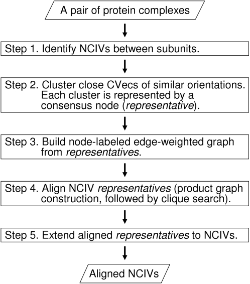
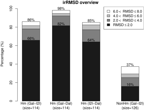
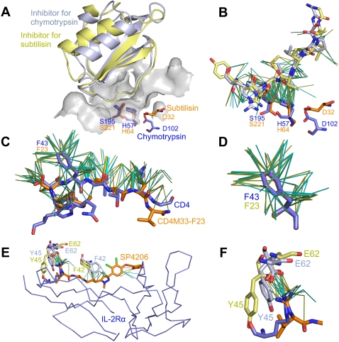
Methodology/principal findings: Here we present a method for aligning non-covalent interactions between different protein-protein interfaces. The method aligns the vector representations of van der Waals interactions and hydrogen bonds based on their geometry. The method has been applied to a dataset which comprises a variety of protein-protein interfaces. The alignments are consistent to a large extent with the results obtained using two other complementary approaches. In addition, we apply the method to three examples of protein mimicry. The method successfully aligns respective interfaces and allows for recognizing conserved interface regions.

Conclusions/significance: The Galinter method has been validated in the comparison of interfaces in which homologous subunits are involved, including cases of mimicry. The method is also applicable to comparing interfaces involving non-peptidic compounds. Galinter assists users in identifying local interface regions with similar patterns of non-covalent interactions. This is particularly relevant to the investigation of the molecular basis of interaction mimicry.


Conflict of interest statement

Competing Interests: The authors have declared that no competing interests exist.

Figures

Figure 1
Figure 1. Flow chart of Galinter.
(NCIV: non-covalent interaction vector; CVec: contact vector)
Figure 2
Figure 2. Overview of irRMSD values for pairwise comparison of protein-protein interfaces.
Most interfaces for non-homologous complexes cannot be compared using backbone alignment method. Thus for the alignments of non-homologous complex interfaces, only an overview of irRMSD values for the comparison between Galinter and I2I-SiteEngine are shown. (Hm: S/D-homologous; NonHm: non-homologous; Gal: Galinter; I2I: I2I-SiteEngine; Dal: DaliLite)
Figure 3
Figure 3. Analysis of mimicry cases.
Every example is shown with two representations in the same orientation. In all representations, the homologous side is in light blue and light yellow at the top, the mimic side is shown in dark blue and orange at the bottom. NCIVs at interfaces are shown as thin lines. A) Superposed inhibitors and catalytic triads for chymotrypsin (1acb) and subtilisin (1lw6) according to the Galinter alignment. The inhibitor for Chymotrypsin is shown in light blue and the inhibitor for subtilisin is shown in light yellow. The catalytic triads of chymotrypsin and subtilisin are shown as sticks in dark blue and orange, respectively. The chymotrypsin binding site is shown as a gray surface. B) Superposed NCIVs for chymotrypsin/inhibitor interface (1acbEI) and subtilisin/inhibitor interface (1lw6EI) according to the Galinter alignment. Only matched NCIVs are shown. Chymotrypsin/inhibitor NCIVs are shown in cyan, and subtilisin/inhibitor NCIVs are shown in yellow. C) Superposed NCIVs for CD4/gp120 interface (1rzjCG) and CD4M33-F23/gp120 interface (1yymMG) according to the Galinter alignment. CD4 is shown in dark blue and CD4M33-F23 is in orange. Only matched NCIVs are shown. CD4/gp120 NCIVs are shown in cyan, and CD4M33-F23/gp120 NCIVs are in yellow. Hydrogen bonds are shown as thick lines. D) An enlarged view of the matched NCIVs involving the hot spot phenylalanines. E) Superposed NCIVs according to the Galinter alignment of IL-2Rα/IL-2 interface (1z92BA) in dark and light blue, and of SP4206/IL-2 interface (1py2_A) in orange and light yellow. Only matched NCIVs are shown. IL-2Rα/IL-2 NCIVs are shown in cyan, SP4206/IL-2 NCIVs are in yellow. The hot spot residues Phe42, Tyr45, and Glu62 in IL-2 are shown as sticks. F) An enlarged view of the mimic spot around residue Glu62 in IL-2. PyMOL has been used to produce the representations.

References

    1. Jones S, Thornton J. Principles of protein-protein interactions. Proc Natl Acad Sci U S A. 1996;93:13–20. - PMC - PubMed
    1. Lo Conte L, Chothia C, Janin J. The atomic structure of protein-protein recognition sites. J Mol Biol. 1999;285(5):2177–98. - PubMed
    1. Sheinerman FB, Norel R, Honig B. Electrostatic aspects of protein-protein interactions. Curr Opin Struct Biol. 2000;10(2):153–159. - PubMed
    1. Rodier F, Bahadur RP, Chakrabarti P, Janin J. Hydration of protein-protein interfaces. Proteins. 2005;60:36–45. - PubMed
    1. Nooren IMA, Thornton JM. Diversity of protein-protein interactions. EMBO J. 2003;22(14):3486–92. - PMC - PubMed

Publication types

Substances