Review
. 2008 Apr;118(4):1301-10.
doi: 10.1172/JCI33945.

Kinetoplastids: related protozoan pathogens, different diseases

Affiliations
Review

Kinetoplastids: related protozoan pathogens, different diseases

Ken Stuart et al. J Clin Invest. 2008 Apr.

Abstract

Kinetoplastids are a group of flagellated protozoans that include the species Trypanosoma and Leishmania, which are human pathogens with devastating health and economic effects. The sequencing of the genomes of some of these species has highlighted their genetic relatedness and underlined differences in the diseases that they cause. As we discuss in this Review, steady progress using a combination of molecular, genetic, immunologic, and clinical approaches has substantially increased understanding of these pathogens and important aspects of the diseases that they cause. Consequently, the paths for developing additional measures to control these "neglected diseases" are becoming increasingly clear, and we believe that the opportunities for developing the drugs, diagnostics, vaccines, and other tools necessary to expand the armamentarium to combat these diseases have never been better.


Figures
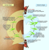
Figure 1
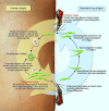
Figure 1. Life cycle of T. brucei.
HAT is caused by infection with the T.b. rhodesiense or T.b. gambiense subspecies. Over 20 species and subspecies of tsetse flies transmit these parasites, but only approximately 1 per 1,000 flies has the mature salivary gland infection that is necessary to transmit the parasite to humans. Trypanosomes enter the fly when it takes a meal of parasite-containing blood from an infected human or animal and, over a period of four weeks, undergo morphological and physiological transformations in the alimentary tract and in the salivary glands, where they become infective. During a blood meal on the mammalian host, an infected tsetse fly injects parasites into skin tissue. The parasites pass via the lymphatic system into the bloodstream, which carries them throughout the body. The parasites continue to replicate by binary fission. In late-stage disease the parasites invade the CNS and reside in the cerebrospinal fluid and intercellular spaces. Figure modified with permission from Alexander J. da Silva and Melanie Moser, Centers for Disease Control Public Health Image Library.
Figure 2
Figure 2. Life cycle of T. cruzi.
Persistent infection with T. cruzi causes Chagas disease. The parasite is transmitted to humans by infected blood-sucking Triatominae insects, which deposit trypomastigotes in their feces during feeding. The trypomastigotes enter the wound and invade nearby cells, within which they differentiate into intracellular amastigotes that multiply by binary fission. The amastigotes differentiate into trypomastigotes, which are released into the bloodstream and infect cells of multiple organs and tissues, including the heart, gut, CNS, smooth muscle, and adipose tissue and once again become amastigotes. The Triatominae insects become infected when they take a parasite-containing blood meal from an infected human or animal. The trypomastigotes undergo morphological and physiological transformations in the midgut of the vector and differentiate into infective trypomastigotes in the hindgut. Image modified with permission from Alexander J. da Silva and Melanie Moser, Centers for Disease Control Public Health Image Library.
Figure 3
Figure 3. Life cycle of Leishmania spp.
Infection with different species of Leishmania causes distinct forms of leishmaniasis. For example, VL is caused by infection with either L. infantum or L. donovani; whereas CL is caused by infection with any one of several different Leishmania spp. Sandflies are the vectors that transmit the disease-causing protozoan parasites, injecting infective promastigotes when they take a blood meal. The parasites invade mammalian macrophages by receptor-mediated endocytosis, where they transform into amastigotes that multiply by binary fission. Sandflies become infected by ingesting infected cells when they take a meal of parasite-containing blood from an infected human or animal. The amastigotes transform into promastigotes and develop in the gut into metacyclic promastigotes that are infective to humans. Figure modified with permission from Alexander J. da Silva and Melanie Moser, Centers for Disease Control Public Health Image Library.

References

    1. Burri, C., and Brun, R. 2003. Human African trypanosomiasis. InManson’s tropical diseases. G.C. Cook and A.I. Zumla, editors. 21st edition. W.B. Saunders/Elsevier. Edinburgh, United Kingdom. 1303–1323.
    1. [No authors listed]. . Human African trypanosomiasis (sleeping sickness): epidemiological update. Wkly. Epidemiol. Rec. 2006;81:71–80. - PubMed
    1. Radwanska M., et al. The serum resistance-associated gene as a diagnostic tool for the detection of Trypanosoma brucei rhodesiense. Am. J. Trop. Med. Hyg. 2002;67:684–690. - PubMed
    1. Radwanska M., et al. Novel primer sequences for polymerase chain reaction-based detection of Trypanosoma brucei gambiense. Am. J. Trop. Med. Hyg. 2002;67:289–295. - PubMed
    1. Magnus E., Vervoort T., Van Meirvenne N. A card-agglutination test with stained trypanosomes (C.A.T.T.) for the serological diagnosis of T. B. gambiense trypanosomiasis. Ann. Soc. Belg. Med. Trop. 1978;58:169–176. - PubMed

Publication types

MeSH terms