. 2008 May;118(5):1846-57.
doi: 10.1172/JCI32503.

An antiproliferative BMP-2/PPARgamma/apoE axis in human and murine SMCs and its role in pulmonary hypertension

Affiliations

An antiproliferative BMP-2/PPARgamma/apoE axis in human and murine SMCs and its role in pulmonary hypertension

Georg Hansmann et al. J Clin Invest. 2008 May.

Abstract

Loss-of-function mutations in bone morphogenetic protein receptor II (BMP-RII) are linked to pulmonary arterial hypertension (PAH); the ligand for BMP-RII, BMP-2, is a negative regulator of SMC growth. Here, we report an interplay between PPARgamma and its transcriptional target apoE downstream of BMP-2 signaling. BMP-2/BMP-RII signaling prevented PDGF-BB-induced proliferation of human and murine pulmonary artery SMCs (PASMCs) by decreasing nuclear phospho-ERK and inducing DNA binding of PPARgamma that is independent of Smad1/5/8 phosphorylation. Both BMP-2 and a PPARgamma agonist stimulated production and secretion of apoE by SMCs. Using a variety of methods, including short hairpin RNAi in human PASMCs, PAH patient-derived BMP-RII mutant PASMCs, a PPARgamma antagonist, and PASMCs isolated from PPARgamma- and apoE-deficient mice, we demonstrated that the antiproliferative effect of BMP-2 was BMP-RII, PPARgamma, and apoE dependent. Furthermore, we created mice with targeted deletion of PPARgamma in SMCs and showed that they spontaneously developed PAH, as indicated by elevated RV systolic pressure, RV hypertrophy, and increased muscularization of the distal pulmonary arteries. Thus, PPARgamma-mediated events could protect against PAH, and PPARgamma agonists may reverse PAH in patients with or without BMP-RII dysfunction.


Figures
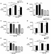
Figure 1
Figure 1. Antiproliferative effects of BMP-2 (A, C, D, and F), the PPARγ agonist rosiglitazone (Rosi; B), and apoE (E) on PDGF-BB–induced proliferation of human (A, B, C, and E) and murine (D and F) PASMCs.
PASMCs were seeded at 2.5 × 10⁴ cells per well of a 24-well plate in 500 μl of growth medium and allowed to adhere overnight. The cells were washed with PBS prior to the addition of starvation media (0.1% FBS) and incubated for 24 hours (murine PASMCs) or 48 hours (HPASMCs) and then stimulated with PDGF-BB (20 ng/ml) for 72 hours. BMP-2 (10 ng/ml), rosiglitazone (1 μM), and recombinant human apoE (1–10 μM) were added to quiescent cells 30 minutes prior to PDGF-BB stimulation. The PPARγ antagonist GW9662 (GW; 1 μM) was added 24 hours prior to the addition of BMP-2. Cells were finally washed twice with PBS, trypsinized, and counted in a hemacytometer (4 counts per well). Cell numbers in controls at time points 0 (CON) and 72 hours were not significantly different. A: shLacZi, HPASMCs transfected with short hairpin LacZi pLentivirus 6 (control); shBMP-RIIi, HPASMCs transfected with short hairpin pLentivirus 6 BMP-RIIi (i.e., BMP-RII–deficient PASMCs). D: Littermates, littermate control PASMCs; SMC PPARγ–/–, PASMCs isolated from SM22α Cre PPARγflox/flox mice. F: C57BL/6, control murine PASMCs; apoE–/–, PASMCs isolated from apoE-deficient mice. Bars represent mean ± SEM (n = 3 in A, D, and F; n = 4 in B and C; n = 6 in E; n = 12 in controls of A). *P < 0.05; **P < 0.01; ***P < 0.001 as indicated; ANOVA with Bonferroni’s multiple comparison test.
Figure 2
Figure 2. PDGF-BB (A and C) and BMP-2 (B and D) have opposing effects in HPASMCs on protein levels of phospho-ERK/total ERK (A and B), PPARγ DNA binding in nuclear extracts (upper panels in C and D), and PPARγ protein in nuclear and cytoplasmic extracts (lower panels in C and D).
Cells were stimulated with PDGF-BB (20 ng/ml) or BMP-2 (10 ng/ml) as described in the legend for Figure 1. In separate experiments, we determined that neither of the solvents (DMSO, sterile water; both 1:10,000) influenced the results. Western immunoblotting and PPARγ DNA binding assays are described in Methods. For the PPARγ DNA binding assay, bars represent median ± SEM of triplicate measurements of 1 representative experiment of 2 (C) and 3 (D) independent experiments with similar results. For protein levels in cell fractions, bars represent mean ± SEM (n = 3–4). *P < 0.05; **P < 0.01 versus control; ANOVA with Dunnett’s post-hoc test.
Figure 3
Figure 3. BMP-2 and rosiglitazone inhibit PDGF-BB–mediated ERK phosphorylation (A and C), and concomitant BMP-2 and PDGF-BB stimulation increases PPARγ DNA binding (B), in HPASMCs.
Cells were preincubated with BMP-2 (10 ng/ml) for 30 minutes (A and B) or rosiglitazone (1 μM) for 24 hours (C), followed by PDGF-BB (20 ng/ml) stimulation for 10 minutes (A and B) or 5–60 minutes. (C) Western immunoblotting and PPARγ DNA binding assays are described in Methods and Figure 2. For protein levels in cell fractions (A) or cell lysates (C), bars represent mean ± SEM (n = 3 each). In C, all samples are compared with the DMSO control. For the PPARγ DNA binding assay (B), bars represent median ± SEM of triplicate measurements of 1 representative experiment of 3 independent experiments with similar results. *P < 0.05; **P < 0.01 versus control; ANOVA with Dunnett’s post-hoc test.
Figure 4
Figure 4. Antiproliferative effects of BMP-2 and the PPARγ agonist rosiglitazone on PDGF-BB–induced proliferation of human wild-type and BMP-RII mutant PASMCs.
Control PASMCs were isolated from surgical resection specimens derived from patients undergoing lobectomy or pneumonectomy for suspected lung tumor. Additional peripheral pulmonary arteries (<1–2 mm external diameter) were obtained from a patient undergoing heart-lung transplantation for FPAH and known to harbor a mutation (W9X) in BMP-RII. The nature of the BMP-RII mutation, cell isolation, culture techniques, and cell counts are described in Methods and in Figure 1. HPASMCs were incubated for 48 hours in starvation media (0.1% FBS) and then stimulated with PDGF-BB (20 ng/ml) for 72 hours. BMP-2 (10 ng/ml) or rosiglitazone (1 μM) were added to quiescent cells 30 minutes prior to PDGF-BB stimulation. Bars represent mean ± SEM (n = 3). **P < 0.01; ***P < 0.001 as indicated; ANOVA with Bonferroni’s multiple comparison test. The number of PDGF-BB–stimulated cells was significantly higher than that of untreated control cells (P < 0.001).
Figure 5
Figure 5. BMP-2 and the PPARγ agonist rosiglitazone induce apoE in PASMCs.
(A) apoE protein expression in cell lysates (left) and apoE protein secretion in supernatant (right) induced by BMP-2 (10 ng/ml, 24 hours) and rosiglitazone (1 μM, 24 hours) were detected by immunoblotting as described in Methods (for cell lysates, densitometric values were corrected for equal loading using α-tubulin). For apoE secretion, the media of 3–4 cell culture flasks per condition were pooled and concentrated for the blots shown (representative of 2 independent experiments with similar results). (B) BMP-2–induced (10 ng/ml, 24 hours) upregulation of apoE in murine control PASMCs was reduced by half in PASMCs harvested from SM22α Cre PPARγflox/flox mice. PASMCs were isolated from 5 littermate control and 5 SM22α Cre PPARγflox/flox mice as described in Methods. PASMCs from each genotype were then pooled and subcultured prior to stimulation with BMP-2. The blot is representative of 2 independent experiments with similar results. For apoE protein levels in cell lysates (A), bars represent mean ± SEM (n = 3). *P < 0.05; **P < 0.01 versus control; unpaired 2-tailed t test.
Figure 6
Figure 6. Mice with targeted deletion of PPARγ in SMCs maintain BMP-2–induced pSmad1/5/8 signaling.
(A and B) Genotyping of mice with targeted deletion of PPARγ in SMCs. (A) PCR reactions showing gain of a 300-bp knockout transcript and almost complete loss of the 700-bp wild-type transcript in PASMCs and aorta from SM22α Cre PPARγflox/flox mice. In the lung, which contains SMCs but also many other cell types, both transcripts are found in SM22α Cre PPARγflox/flox mice, whereas only the wild-type transcript is detected in littermate control mice. (B) Western immunoblotting of PASMC lysates isolated from both littermate and SM22α Cre PPARγflox/flox (SMC PPARγ–/–) mice (n = 2 each) showed no detectable PPARγ protein expression when compared with control cells. (C) Both littermate control and SMC PPARγ–/– PASMCs were stimulated with BMP-2 (10 ng/ml) for 5–60 minutes as described in the legend for Figure 1, and phospho-Smad 1/5/8 protein expression was detected by immunoblotting as described in Methods (densitometric values were corrected for equal loading using α-tubulin). Data for 1 of 2 representative experiments with similar results are shown.
Figure 7
Figure 7. PAH in mice with targeted deletion of PPARγ in SMCs.
Thirteen- to 15-week-old mice underwent RV catheterization, followed by organ harvest. (A) RVSP measurements, as described in Methods. (B) Right ventricular hypertrophy (RVH), measured as ratio of the weight of the RV to that of the LV plus septum (RV/LV+S), as described in Methods. (C) Muscularization of alveolar wall arteries (Musc. Arteries Alv. Wall), as described in Methods. (D) Representative photomicrographs of lung tissue (stained by Movat pentachrome) of 15-week-old mice showing a typical nonmuscular peripheral alveolar artery in a littermate control mouse. (E) A similar section in the SM22α Cre PPARγflox/flox (SMC PPARγ–/–) mouse shows an alveolar wall artery surrounded by a rim of muscle. (FI) Immunohistochemistry in serial lung tissue sections from littermate control (CON) and SMC PPARγ–/– mice stained for α-SMA (F and G) and proliferating cell nuclear antigen (PCNA; H and I). Arrows in I indicate enhanced PCNA staining in PASMCs. See also Table 1. Bars represent mean ± SEM (n = 5). ***P < 0.001 versus control; unpaired 2-tailed t test.
Figure 8
Figure 8. Model: A novel antiproliferative BMP-2/PPARγ/apoE axis protects against PAH.
This schema incorporates the findings described in our article and the literature to date as discussed. (A) BMP-2 inhibits SMC proliferation via PPARγ and apoE. apoE impairs PDGF-BB/MAPK signaling by binding to LDL receptor–related protein (LRP), thereby initiating endocytosis and degradation of the LRP/PDGFR-β/PDGF-B complex. PPARγ induces LRP and other growth-inhibitory/proapoptotic genes in SMCs and inhibits cell-cycle and other growth-promoting genes such as telomerase, cyclin D1, and retinoblastoma protein. Moreover, PPARγ induces phosphatases that can directly inactivate phospho-ERK. (B) BMP-RII dysfunction promotes SMC proliferation and survival in PAH. Heightened PDGF-BB signaling leading to SMC proliferation is a key clinical feature of PAH. Deficiency of both apoE and LRP enhances mitogenic PDGF-BB/MAPK signaling. Loss-of-function mutations in the BMP-RII gene will decrease endogenous PPARγ activity, leading to unopposed MAPK signaling, SMC proliferation and survival, and ultimately development of PAH.TF, transcription factor. (C) PPARγ agonists can rescue BMP-RII dysfunction and reverse PAH. PPARγ agonists such as rosiglitazone or pioglitazone might reverse SMC proliferation and vascular remodeling in PAH patients with or without BMP-RII dysfunction via induction of apoE and other growth-inhibitory/proapoptotic genes (as indicated) and through repression of growth-promoting genes (not shown).

References

    1. Merklinger S.L., Jones P.L., Martinez E.C., Rabinovitch M. Epidermal growth factor receptor blockade mediates smooth muscle cell apoptosis and improves survival in rats with pulmonary hypertension. Circulation. 2005;112:423–431. doi: 10.1161/CIRCULATIONAHA.105.540542. - DOI - PubMed
    1. Schermuly R.T., et al. Reversal of experimental pulmonary hypertension by PDGF inhibition. . J. Clin. Invest. 2005;115:2811–2821. doi: 10.1172/JCI24838. - DOI - PMC - PubMed
    1. Lane K.B., et al. Heterozygous germline mutations in BMPR2, encoding a TGF-beta receptor, cause familial primary pulmonary hypertension. The International PPH Consortium. Nat. Genet. 2000;26:81–84. doi: 10.1038/79226. - DOI - PubMed
    1. Deng Z., et al. Familial primary pulmonary hypertension (gene PPH1) is caused by mutations in the bone morphogenetic protein receptor-II gene. . Am. J. Hum. Genet. 2000;67:737–744. doi: 10.1086/303059. - DOI - PMC - PubMed
    1. Newman J.H., et al. Genetic basis of pulmonary arterial hypertension: current understanding and future directions. J. Am. Coll. Cardiol. 2004;43:33S–39S. doi: 10.1016/j.jacc.2004.02.028. - DOI - PubMed

Publication types

MeSH terms