. 2008 Apr;27(4):825-33.
doi: 10.1002/jmri.21330.

Estrogen receptor and breast MR imaging features: a correlation study

Affiliations

Estrogen receptor and breast MR imaging features: a correlation study

Jeon-Hor Chen et al. J Magn Reson Imaging. 2008 Apr.

Abstract

Purpose: To compare the MRI features between estrogen receptor (ER) positive and negative breast cancers.

Materials and methods: Breast MRI of 90 consecutive patients confirmed with invasive ductal carcinoma (IDC), 51 ER positive and 39 ER negative, were analyzed. The tumor morphology and dynamic contrast-enhanced (DCE) kinetics were evaluated based on the American College of Radiology (ACR) Breast Imaging Reporting and Data System (BI-RADS) MRI lexicon and compared. Enlarged axillary lymph nodes on MRI and choline (Cho) detection using MR spectroscopy (MRS) were also analyzed and compared. For patients receiving axillary node dissection the pathological nodal status was also compared.

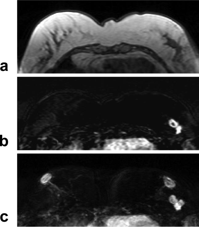
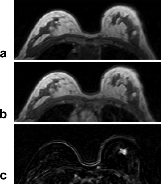
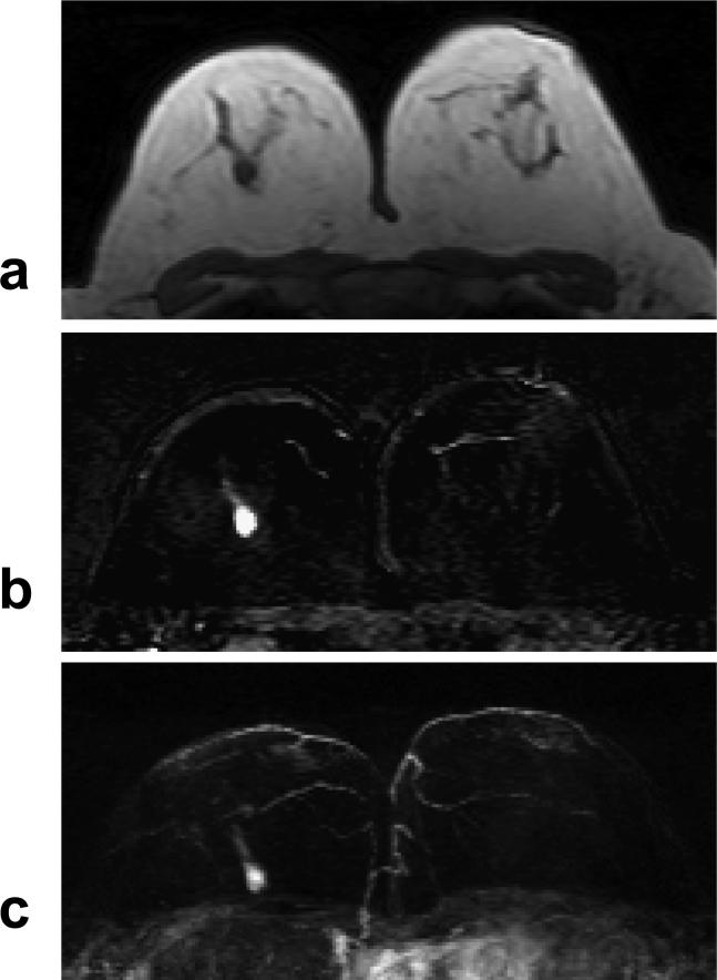
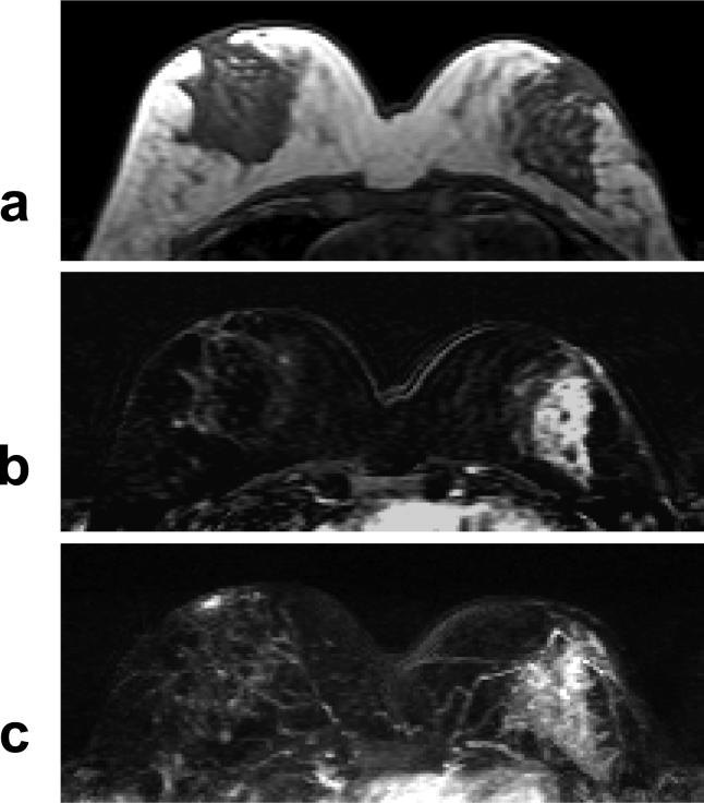
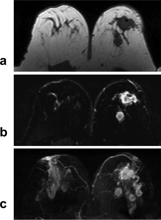
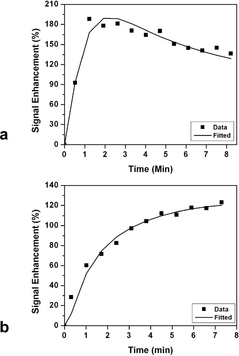
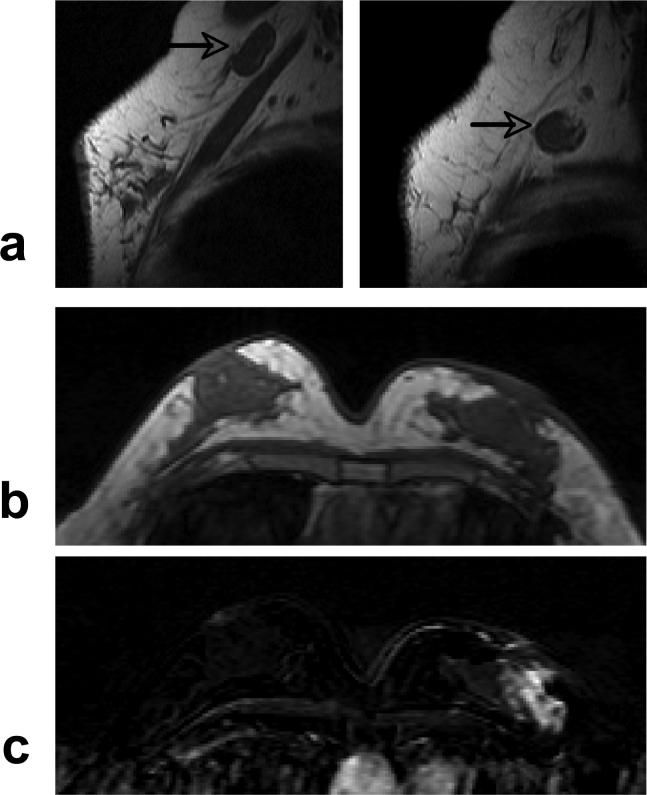
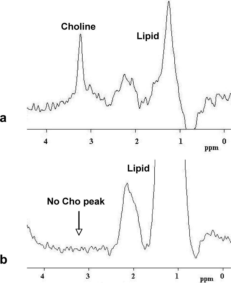
Results: ER negative breast cancer had bigger tumors compared to ER positive cancer (3.6 +/- 2.0 cm vs. 1.8 +/- 1.3 cm, P < 0.00005). ER negative cancer was more likely to exhibit nonmass type enhancements compared to ER positive cancer (P < 0.005). Enlarged axillary lymph nodes were more frequently identified on MRI in ER negative compared to ER positive patients (P < 0.05). After excluding patients undergoing neoadjuvant chemotherapy, auxiliary lymph node status did not show significant difference between ER positive and ER negative cancer on MRI and pathology. ER negative cancer was more likely to show the malignant type enhancement kinetics (P = 0.15), rim enhancement (P = 0.15), and Cho detection on MRS (P = 0.23) compared to ER positive cancer, but it did not reach a level of statistical significance.

Conclusion: ER negative breast cancer was more aggressive, with larger tumor size, more non-mass-type enhancement lesions, and a higher percentage showing enlarged axillary nodes on MRI. These features might be related to its poorer cellular differentiation and/or a higher angiogenesis.


Figures

Figure 1
Figure 1
A 47 year-old woman with ER positive IDC. a: Pre-contrast T1 weighted image shows a hypointense lesion in the left breast. b: Post-contrast subtraction image at 1-min after injection shows a lesion with rim-enhancement, and another connected lesion. c: Maximal intensity projection (MIP) image shows three connected enhanced lesions, approximately 2 cm. The nipples in both breasts are enhanced. The MR spectrum measured this patient is shown in Figure 8b.
Figure 2
Figure 2
A 63 year-old woman with ER positive IDC. a: Pre-contrast T1 weighted image does not show suspicious lesions. b: Post-contrast enhanced image taken at 5-min after injection. c: Post-contrast subtraction image taken at 5-min clearly shows a 1-cm enhanced lesion in the left breast. The DCE kinetics measured from this lesion is shown in Figure 7b.
Figure 3
Figure 3
A 55 year-old woman with ER negative IDC. a: Pre-contrast T1 weighted image shows a hypointense mass in the right breast. b: Post-enhanced subtraction image shows a strongly enhanced 1-cm lesion. c: Maximal intensity projection (MIP) image shows a solitary lesion. The nipples in both breasts are not enhanced. The DCE kinetics measured this lesion is shown in Figure 7a.
Figure 4
Figure 4
A 40 year-old woman with ER negative IDC. a: Pre-contrast T1 weighted image does not show suspicious lesion. b: Post-contrast subtraction image shows a non-mass type lesion with regional or segmental enhancements, without clearly-defined borders. c: Maximal intensity projection (MIP) image also demonstrates a non-mass type lesion.
Figure 5
Figure 5
A 47 year-old woman with ER negative IDC. a: Pre-contrast T1 weighted image shows 2 hypointense mass lesions in the left breast. b: Contrast enhanced subtraction image shows two enhanced lesions with rim-enhancement pattern. c: Maximal intensity projection (MIP) image shows 4 enhanced lesions in the left breast, with unspecific fibroglandular enhancements in the right breast. The MRS spectrum measured from the largest mass is shown in Figure 8a.
Figure 6
Figure 6
a: An example of malignant type DCE kinetics with rapid initial enhancement followed by wash-out in the late phase. This was measured from the lesion shown in Figure 3. b: An example of benign type DCE kinetics with continuous enhancements, measured from the lesion shown in Figure 2.
Figure 7
Figure 7
A 51 year-old woman with ER negative IDC. a: Pre-contrast sagittal view T1 weighted images from two imaging slices show two enlarged lymph nodes (arrows), one oval and one round, with complete loss of fatty hilum. b: Pre-contrast axial view T1 weighted image does not show suspicious lesion. c: Post-contrast subtraction image shows a non-mass type lesion with regional enhancements.
Figure 8
Figure 8
a: An example of MRS spectrum showing an identifiable Choline peak at 3.2 ppm. This spectrum was measured from the largest lesion shown in Figure 5. b: An example of MRS spectrum without an identifiable Choline peak around 3.2 ppm. This spectrum was measured from the lesion shown in Figure 1.

References

    1. Yasui Y, Potter JD. The shape of age-incidence curves of female breast cancer by hormone-receptor status. Cancer Causes Control. 1999;10:431–437. - PubMed
    1. Bernoux A, de Cremoux P, Lainé-Bidron C, Martin EC, Asselain B, Magdelénat H. Estrogen receptor negative and progesterone receptor positive primary breast cancer: pathological characteristics and clinical outcome. Institut Curie Breast Cancer Study Group. Breast Cancer Res Treat. 1998;49:219–225. - PubMed
    1. Putti TC, El-Rehim DM, Rakha EA, et al. Estrogen receptor-negative breast carcinomas: a review of morphology and immunophenotypical analysis. Mod Pathol. 2005;18:26–35. - PubMed
    1. Maki DD, Grossman RI. Patterns of disease spread in metastatic breast carcinoma: influence of estrogen and progesterone receptor status. AJNR Am J Neuroradiol. 2000;21:1064–1066. - PMC - PubMed
    1. Sanna G, Franceschelli L, Rotmensz N, et al. Brain metastases in patients with advanced breast cancer. Anticancer Res. 2007;27:2865–2869. - PubMed

Publication types