. 2008 Jun;46(6):508-17.
doi: 10.1002/mrc.2201.

Characterization of nonderivatized plant cell walls using high-resolution solution-state NMR spectroscopy

Affiliations

Characterization of nonderivatized plant cell walls using high-resolution solution-state NMR spectroscopy

Daniel J Yelle et al. Magn Reson Chem. 2008 Jun.

Erratum in

Abstract

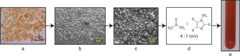
A recently described plant cell wall dissolution system has been modified to use perdeuterated solvents to allow direct in-NMR-tube dissolution and high-resolution solution-state NMR of the whole cell wall without derivatization. Finely ground cell wall material dissolves in a solvent system containing dimethylsulfoxide-d(6) and 1-methylimidazole-d(6) in a ratio of 4:1 (v/v), keeping wood component structures mainly intact in their near-native state. Two-dimensional NMR experiments, using gradient-HSQC (heteronuclear single quantum coherence) 1-bond (13)C--(1)H correlation spectroscopy, on nonderivatized cell wall material from a representative gymnosperm pinus taeda (loblolly pine), an angiosperm Populus tremuloides (quaking aspen), and a herbaceous plant Hibiscus cannabinus (kenaf) demonstrate the efficacy of the system. We describe a method to synthesize 1-methylimidazole-d(6) with a high degree of perdeuteration, thus allowing cell wall dissolution and NMR characterization of nonderivatized plant cell wall structures.


Figures

Figure 1
Figure 1
A diagram illustrating the nonderivatized dissolution of plant cell walls. Pinus taeda is shown here as an example: a, shavings produced via conventional planer; b, cryogenically mixer-milled; c, planetary ball-milled; d, dissolution method; e, NMR tube of cell walls in solution.
Figure 2
Figure 2
Proton spectra of various stages in NMI deuteration: a, 1-methylimidazole; b, 1-methyl-d3-imidazole; c, ring deuteration as per Hardacre et al.[27]; d, ring deuteration as per Esaki et al.[28]; e, ring deuteration with combination of methods.
Figure 3
Figure 3
Key to the chemical structures found in the spectra in Figs 4–7. The structures are: (A) β-aryl ether in cyan, (B) phenylcoumaran in green, (C) resinol in purple, (D) dibenzodioxocin in red, (X1) cinnamyl alcohol endgroups in magenta, (G) guaiacyl in dark blue, (S) syringyl in fuchsia, and (NMI) 1-methylimidazole in gray. Other structures include: lignin methoxyl in brown, H-2/C-2 & H-3/C-3 correlations for the acetylated structure of β-d-ManpI in maroon, H-2/C-2 & H-3/C-3 correlations for the acetylated structure of β-d-XylpI in chartreuse, polysaccharide anomerics in orange, and structures currently not assigned or unresolved in black.
Figure 4
Figure 4
HSQC spectra from nonderivatized plant cell walls: a, pine; b, aspen; and c, kenaf at 500 MHz. Note that the spectra cover the range for all the cell wall components – cellulose, hemicelluloses, and lignin. All contour colors can be matched to their respective structures in Fig. 3. Expanded regions are shown in Figs 5–7.
Figure 5
Figure 5
Aliphatic region of the whole plant cell wall HSQC spectra: a, pine; b, aspen; and c, kenaf. The black contours are currently unresolved, mostly because of heavy overlapping in these regions. All contour colors and labels can be matched to their respective structure in Fig. 3. As noted in the text, AβG is any syringyl or guaiacyl coupled to a guaiacyl β-aryl ether, AβS is any syringyl or guaiacyl coupled to a syringyl β-aryl ether, and AβγAc is a tentative assignment for a β-aryl ether linkage with a γ-acetate. 2-O-Ac-β-d-Manp, H-2/C-2 correlation for the acetylated structure of β-D-ManpI; 3-O-Ac-β-d-Manp, H-3/C-3 correlation for the acetylated structure of β-d-ManpI[32]; 2-O-Ac-β-d-Xylp, H-2/C-2 correlation for the acetylated structure of β-d-XylpI; 3-O-Ac-β-d-Xylp, H-3/C-3 correlation for the acetylated structure of β-d-XylpI[30,31].
Figure 6
Figure 6
Aromatic region of the whole plant cell wall HSQC spectra: a, pine; b, aspen; and c, kenaf. The guaiacyl and syringyl contours are a combination of free phenolic and etherified aromatic units. Contour colors and labels can be matched to their respective structure in Fig. 3.
Figure 7
Figure 7
Anomeric region of the whole plant cell wall HSQC spectra: a, pine; b, aspen; and c, kenaf. Assignments are as follows: β-d-GlcpI, internal β-d-glucopyranoside units (cellulose)[–33]; β-d-ManpI, internal β-d-mannopyranoside units[32,33] ; β-d-XylpI, internal β-d-xylopyranoside units[30]; α-d-GalpT, terminal α-d-galactopyranoside units[32,33]; β-d-GalpT, terminal β-d-galactopyranoside units[33]; α-l-ArafT, terminal α-l-arabinofuranoside units[31,32,50]; 2-O-Ac-β-d-Manp, anomeric correlation for the acetylated H-2/C-2 structure of β-d-ManpI; 3-O-Ac-β-d-Manp, anomeric correlation for the acetylated H-3/C-3 structure of β-d-ManpI[32]; 2-O-Ac-β-d-Xylp, anomeric correlation for the acetylated H-2/C-2 structure of β-d-XylpI; 3-O-Ac-β-d-Xylp, anomeric correlation for the acetylated H-3/C-3 structure of β-d-Xylp I[30,31]; 4-O-MeGlcA, 4-O-methyl-α-d-glucuronic acid[30]; α and β-d-GlcpR, reducing ends of glucopyranoside[32,33]; β-d-ManpR, reducing end of mannopyranoside[32]; α and β-d-GalpR, reducing ends of galactopyranoside[33]; α and β-d-XylpR, reducing ends of xylopyranoside.[30].

References

    1. Barron PF, Frost RL, Doimo L, Kennedy MJ. J. Macromol. Sci.-Chem. 1985;A22:303.
    1. Sterk H, Sattler W, Esterbauer H. Carbohydr. Res. 1987;164:85.
    1. Bardet M, Emsley L, Vincendon M. Solid State Nucl. Magn. Reson. 1997;8:25. - PubMed
    1. Johnson CE, Smernik RJ, Siccama TG, Kiemle DK, Xu Z, Vogt DJ. Can. J. For. Res. 2005;35:1821.
    1. Lu F, Ralph J. Plant J. 2003;35:535. - PubMed

Publication types

MeSH terms

LinkOut - more resources