

Role of endogenous sleep-wake and analgesic systems in anesthesia
- PMID: 18383504
- PMCID: PMC4924624
- DOI: 10.1002/cne.21685
Role of endogenous sleep-wake and analgesic systems in anesthesia
Abstract
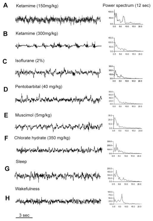
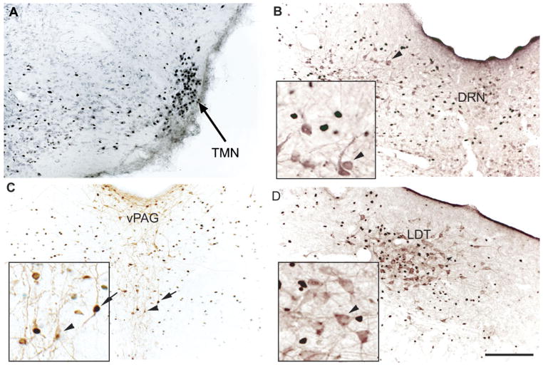
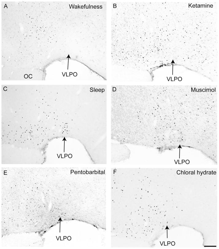
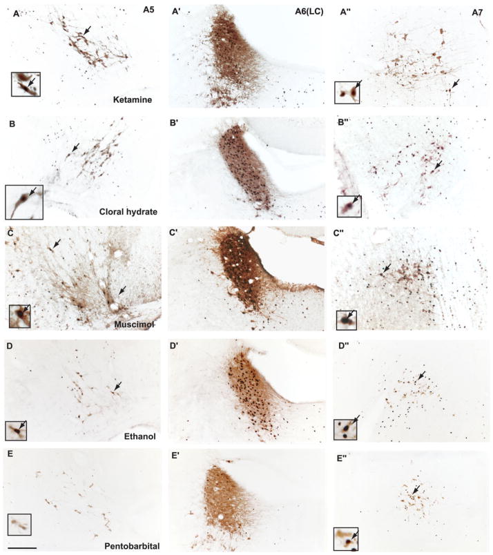
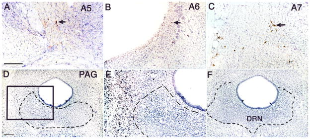
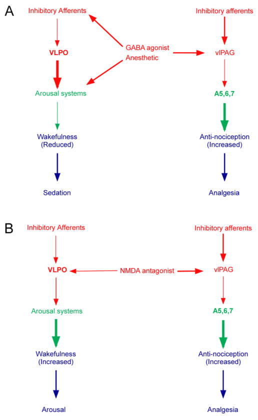
Classical anesthetics of the gamma-aminobutyric acid type A receptor (GABA(A))-enhancing class (e.g., pentobarbital, chloral hydrate, muscimol, and ethanol) produce analgesia and unconsciousness (sedation). Dissociative anesthetics that antagonize the N-methyl-D-aspartate (NMDA) receptor (e.g., ketamine, MK-801, dextromethorphan, and phencyclidine) produce analgesia but do not induce complete loss of consciousness. To understand the mechanisms underlying loss of consciousness and analgesia induced by general anesthetics, we examined the patterns of expression of c-Fos protein in the brain and correlated these with physiological effects of systemically administering GABAergic agents and ketamine at dosages used clinically for anesthesia in rats. We found that GABAergic agents produced predominantly delta activity in the electroencephalogram (EEG) and sedation. In contrast, anesthetic doses of ketamine induced sedation, followed by active arousal behaviors, and produced a faster EEG in the theta range. Consistent with its behavioral effects, ketamine induced Fos expression in cholinergic, monoaminergic, and orexinergic arousal systems and completely suppressed Fos immunoreactivity in the sleep-promoting ventrolateral preoptic nucleus (VLPO). In contrast, GABAergic agents suppressed Fos in the same arousal-promoting systems but increased the number of Fos-immunoreactive neurons in the VLPO compared with waking control animals. All anesthetics tested induced Fos in the spinally projecting noradrenergic A5-7 groups. 6-hydroxydopamine lesions of the A5-7 groups or ibotenic acid lesions of the ventrolateral periaqueductal gray matter (vlPAG) attenuated antinociceptive responses to noxious thermal stimulation (tail-flick test) by both types of anesthetics. We hypothesize that neural substrates of sleep-wake behavior are engaged by low-dose sedative anesthetics and that the mesopontine descending noradrenergic cell groups contribute to the analgesic effects of both NMDA receptor antagonists and GABA(A) receptor-enhancing anesthetics.
Figures

Similar articles
-
Paradoxical anesthesia: Sleep-like EEG during anesthesia induced by mesopontine microinjection of GABAergic agents.Exp Neurol. 2021 Sep;343:113760. doi: 10.1016/j.expneurol.2021.113760. Epub 2021 May 15. Exp Neurol. 2021. PMID: 34000248
-
Effects of propofol on sleep architecture and sleep-wake systems in rats.Behav Brain Res. 2021 Aug 6;411:113380. doi: 10.1016/j.bbr.2021.113380. Epub 2021 May 24. Behav Brain Res. 2021. PMID: 34033853
-
The sedative component of anesthesia is mediated by GABA(A) receptors in an endogenous sleep pathway.Nat Neurosci. 2002 Oct;5(10):979-84. doi: 10.1038/nn913. Nat Neurosci. 2002. PMID: 12195434
-
[Neurochemical mechanisms of sleep regulation].Glas Srp Akad Nauka Med. 2009;(50):97-109. Glas Srp Akad Nauka Med. 2009. PMID: 20666118 Review. Serbian.
-
Underlying brain mechanisms that regulate sleep-wakefulness cycles.Int Rev Neurobiol. 2010;93:1-21. doi: 10.1016/S0074-7742(10)93001-8. Int Rev Neurobiol. 2010. PMID: 20969999 Review.
Cited by
-
General Anesthetic Conditions Induce Network Synchrony and Disrupt Sensory Processing in the Cortex.Front Cell Neurosci. 2016 Apr 14;10:64. doi: 10.3389/fncel.2016.00064. eCollection 2016. Front Cell Neurosci. 2016. PMID: 27147963 Free PMC article.
-
Divergent Neural Activity in the VLPO During Anesthesia and Sleep.Adv Sci (Weinh). 2023 Jan;10(2):e2203395. doi: 10.1002/advs.202203395. Epub 2022 Dec 3. Adv Sci (Weinh). 2023. PMID: 36461756 Free PMC article.
-
Different Alterations of Hippocampal and Reticulo-Thalamic GABAergic Parvalbumin-Expressing Interneurons Underlie Different States of Unconsciousness.Int J Mol Sci. 2023 Apr 5;24(7):6769. doi: 10.3390/ijms24076769. Int J Mol Sci. 2023. PMID: 37047741 Free PMC article.
-
Brainstem Influence on Thalamocortical Oscillations during Anesthesia Emergence.Front Syst Neurosci. 2017 Sep 14;11:66. doi: 10.3389/fnsys.2017.00066. eCollection 2017. Front Syst Neurosci. 2017. PMID: 28959192 Free PMC article.
-
Animal models of sleep disorders.Comp Med. 2013 Apr;63(2):91-104. Comp Med. 2013. PMID: 23582416 Free PMC article. Review.
References
-
- Abi-Saab WM, D’Souza DC, Moghaddam B, Krystal JH. The NMDA antagonist model for schizophrenia: promise and pitfalls. Pharmacopsychiatry Suppl. 1998;2:104–109. - PubMed
-
- Aston-Jones G. Brain structures and receptors involved in alertness. Sleep Med Suppl. 2005;1:S3–7. - PubMed
-
- Bajic D, Proudfit HK, Van Bockstaele EJ. Periaqueductal gray neurons monosynaptically innervate extranuclear noradrenergic dendrites in the rat pericoerulear region. J Comp Neurol. 2000;427:649–662. - PubMed
-
- Bajic D, Van Bockstaele EJ, Proudfit HK. Ultrastructural analysis of ventrolateral periaqueductal gray projections to the A7 catecholamine cell group. Neuroscience. 2001;104:181–197. - PubMed
-
- Barbaro NM, Hammond DL, Fields HL. Effects of intrathecally administered methysergide and yohimbine on microstimulation-produced antinociception in the rat. Brain Res. 1985;343:223–229. - PubMed
Publication types
MeSH terms
Substances
Grants and funding
LinkOut - more resources
Full Text Sources
Medical
