

Imaging second messenger dynamics in developing neural circuits
- PMID: 18383551
- PMCID: PMC2930489
- DOI: 10.1002/dneu.20619
Imaging second messenger dynamics in developing neural circuits
Abstract
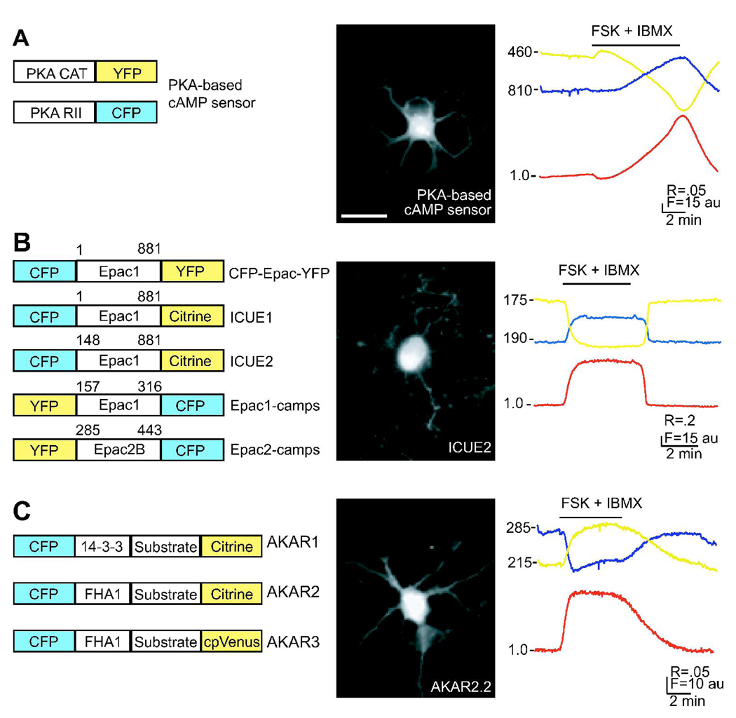
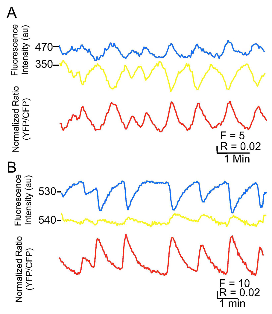
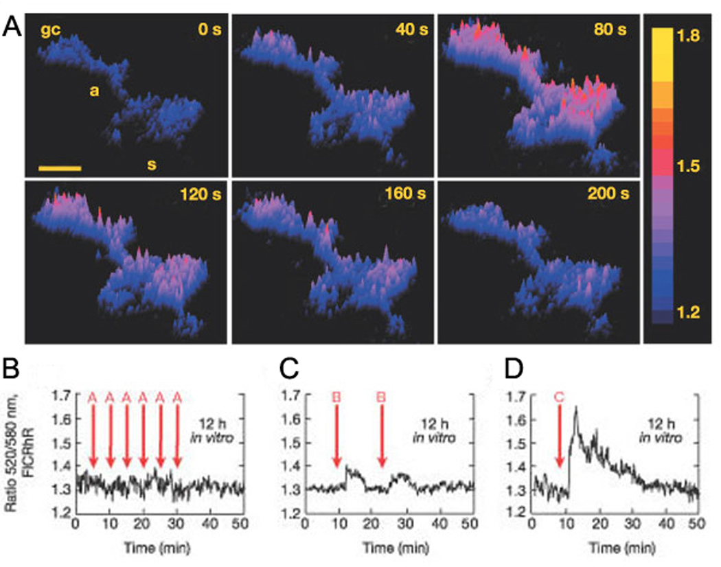
A characteristic feature of developing neural circuits is that they are spontaneously active. There are several examples, including the retina, spinal cord, and hippocampus, where spontaneous activity is highly correlated among neighboring cells, with large depolarizing events occurring with a periodicity on the order of minutes. One likely mechanism by which neurons can "decode" these slow oscillations is through activation of second messenger cascades that either influence transcriptional activity or drive posttranslational modifications. Here, we describe recent experiments where imaging has been used to characterize slow oscillations in the cAMP/PKA second messenger cascade in retinal neurons. We review the latest techniques in imaging this specific second messenger cascade, its intimate relationship with changes in intracellular calcium concentration, and several hypotheses regarding its role in neurodevelopment.
Figures
References
-
- Adams SR, Harootunian AT, Buechler YJ, Taylor SS, Tsien RY. Fluorescence ratio imaging of cyclic AMP in single cells. Nature. 1991;349:694–697. - PubMed
-
- Adelsberger H, Garaschuk O, Konnerth A. Cortical calcium waves in resting newborn mice. Nat Neurosci. 2005;8:988–990. Epub 2005 Jul 2010. - PubMed
-
- Allen MD, Zhang J. Subcellular dynamics of protein kinase A activity visualized by FRET-based reporters. Biochemical And Biophysical Research Communications. 2006;348:716–721. - PubMed
-
- Bacskai BJ, Hochner B, Mahaut-Smith M, Adams SR, Kaang BK, Kandel ER, Tsien RY. Spatially resolved dynamics of cAMP and protein kinase A subunits in Aplysia sensory neurons. Science. 1993;260:222–226. - PubMed
Publication types
MeSH terms
Grants and funding
LinkOut - more resources
Full Text Sources
Medical
