. 2008 Aug;10(8):1622-33.
doi: 10.1111/j.1462-5822.2008.01152.x. Epub 2008 Apr 1.

Antibody action after phagocytosis promotes Cryptococcus neoformans and Cryptococcus gattii macrophage exocytosis with biofilm-like microcolony formation

Affiliations

Antibody action after phagocytosis promotes Cryptococcus neoformans and Cryptococcus gattii macrophage exocytosis with biofilm-like microcolony formation

Mauricio Alvarez et al. Cell Microbiol. 2008 Aug.

Abstract

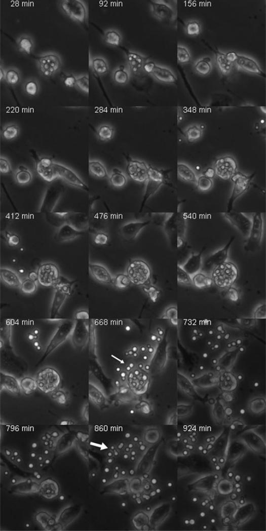
Antibody-mediated phagocytosis was discovered over a century ago but little is known about antibody effects in phagolysosomes. We explored the consequences of antibody-mediated phagocytosis for two closely related human pathogenic fungal species, Cryptococcus neoformans and Cryptococcus gattii, of which C. neoformans encompasses two varieties: neoformans and grubii. The interaction between C. neoformans varieties grubii and neoformans and host cells has been extensively studied, but that of C. gattii and macrophages remains largely unexplored. Like C. neoformans, antibody-mediated phagocytosis of C. gattii cells was followed by intracellular replication, host cell cytoplasmic polysaccharide accumulation and phagosomal extrusion. Both C. gattii and C. neoformans cells exited macrophages in biofilm-like microcolonies where the yeast cells were aggregated in a polysaccharide matrix that contained bound antibody. In contrast, complement-opsonized C. neoformans variety grubii cells were released from macrophages dispersed as individual cells. Hence, both antibody- and complement-mediated phagocytosis resulted in intracellular replication but the mode of opsonization affected the outcome of exocytosis. The biofilm-like microcolony exit strategy of C. neoformans and C. gattii following antibody opsonization reduced fungal cell dispersion. This finding suggests that antibody agglutination effects persist in the phagosome to entangle nascent daughter cells and this phenomenon may contribute to antibody-mediated protection.


Figures

Fig. 1
Fig. 1
IgM-mediated complement-independent phagocytosis of C. gattii via CR3 and CR4. Phagocytosis assays with various concentration of IgM (12A1) were performed and a dose-dependent opsonization of C. gattii and subsequent phagocytosis was observed (black bars). Blockage of CR with anti-CR ablates phagocytosis of C. gattii (grey bars with asterisk on top). Line bars represent data carried out in triplicates, ± SD.
Fig. 2
Fig. 2
Intracellular polysaccharide shedding by C. gattii cells. Organized left to right are phase, fluorescence and merged phase and fluorescence images taken at 40×. A. Intracellular shedding of cryptococcal polysaccharide from C. gattii cells into J774.16 cells. Top: 5.0 s exposure, bottom: 4.0 s exposure. B. J774.16 cells lacking intracellular cryptococcal cells showed no fluorescence, even when overexposed (20.0 s).
Fig. 3
Fig. 3
C. gattii forms microcolony following macrophage exit. Following extrusion or lysis of macrophages, C. gattii exits in microcolony form as shown in (A) where the thick arrow points to the forming microcolony while the thin one indicates the macrophage that extruded. The yeast cells appear to be closely apposed to one another in an apparent polysaccharide matrix, as shown in a 3D composite (E) of combined Z-steps from (C) and (D) to recognize the polysaccharide capsules. In (E) the arrow points to polysaccharide residues and indicates the bottom end of the sample where the coverslip is located. The yeast cells continue to reproduce in such form, adding to the diameter of the microcolony. (B) Differential interference contrast (DIC) image. (A) was captured at 10× while (B–E) were captured at 63× with oil.
Fig. 4
Fig. 4
C. neoformans gets extruded as dispersed cells following complement opsonized phagocytosis. C. neoformans strain H99 was opsonized with complement, incubated for 4 h with macrophage-like cells and taken for time-lapse imaging as indicated in Experimental procedures. Following phagocytosis and intracellular residence the yeast cells were extruded as dispersed cells out to the extracellular media as the white arrows point to. Images were collected at 10×.
Fig. 5
Fig. 5
C. gattii remains in a microcolony following phagosome extrusion. Following 20 h incubation of C. gattii with macrophage-like cells, careful collection of the extracellular media demonstrates clumps of C. gattii microcolonies can be found (A). Overnight incubation of C. gattii in feeding media indicates that no microcolonies form (B). Overnight incubation of C. gattii in sabouraud dextrose broth indicates that no microcolonies form. Overnight incubation of C. gattii with mAb 18B7 (100 mg ml-1) shows clumping formation (D), whereas overnight incubation with 20% pooled human sera does not. Image in (A) was collected at 48× and images in (B–E) were collected at 15×. Difference in media colour is not relevant to results and is due to a difference in lighting.
Fig. 6
Fig. 6
Monoclonal antibody (mAb) 18B7 remains attached to yeast cells post extrusion. Yeast cells were opsonized with mAb 18B7 and phagocytosis was carried out as indicated in the experimental procedures section. Extruded C. gattii strain NIH 198 remained stained with fluorescein-labelled goat anti-mouse IgG while new buds after extrusion appeared not stained (A). Note that in these images the macrophages are not lysed and hence the intracellular yeast does not stain with the secondary antibody. Likewise, C. neoformans strain H99 was remained opsonized by mAb post extrusion (B). Macrophage-like cells that were incubated with and endocytosed mAb demonstrated the presence of the endocytosed mAb after 1 h incubation (C). Controls in which no primary mAb was added demonstrated no staining (data not shown). The panels are composed of DIC (left), FITC (middle) and overlaid images (right). Confocal images were collected at 63× with oil.

References

    1. Alvarez M, Casadevall A. Phagosome extrusion and host-cell survival after Cryptococcus neoformans phagocytosis by macrophages. Curr Biol. 2006;16:2161–2165. - PubMed
    1. Alvarez M, Casadevall A. Cell-to-cell spread and massive vacuole formation after Cryptococcus neoformans infection of murine macrophages. BMC Immunol. 2007;8:16. - PMC - PubMed
    1. Baro T, Torres-Rodriguez JM, De Mendoza MH, Morera Y, Alia C. First identification of autochthonous Cryptococcus neoformans var. gattii isloated from goats with predominantly severe pulmonary disease in Spain. J Clin Microbiol. 1998;36:458–461. - PMC - PubMed
    1. Campbell LT, Fraser JA, Nichols CB, Dietrich FS, Carter D, Heitman J. Clinical and environmental isolates of Cryptococcus gattii from Australia that retain sexual fecundity. Eukaryot Cell. 2005;4:1410–1419. - PMC - PubMed
    1. Casadevall A, Perfect J. Cryptococcus Neoformans. American Society for Microbiology Press; Washington, DC: 1998.

Publication types

LinkOut - more resources