Toward improving therapeutic regimens for Bacillus endophthalmitis
- PMID: 18385066
- PMCID: PMC2531288
- DOI: 10.1167/iovs.07-1303
Toward improving therapeutic regimens for Bacillus endophthalmitis
Abstract
Purpose: Bacillus cereus causes the most virulent and refractory form of endophthalmitis. The authors analyzed the effectiveness of early treatment with vancomycin or gatifloxacin, with or without dexamethasone, for experimental B. cereus endophthalmitis.
Methods: Rabbit eyes were injected intravitreally with 100 colony-forming units of B. cereus. At 2, 4, or 6 hours after infection, eyes were injected intravitreally with 0.1 mL gatifloxacin (0.3%), vancomycin (1.0%), either antibiotic plus dexamethasone, dexamethasone alone (1.0%), or PBS. Eyes were analyzed by electroretinography, bacterial quantitation, and antibiotic penetration analysis. Drug toxicity toward Müller cells, retinal pigment epithelium, and cones was also analyzed.
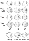
Results: Eyes treated at 2 hours with vancomycin or gatifloxacin, with or without dexamethasone, maintained higher ERG amplitudes than the dexamethasone alone and PBS control groups. Eyes treated with antibiotic plus dexamethasone at 6 hours had reduced retinal function compared to antibiotic treatment alone. With the exception of vancomycin with or without dexamethasone at 6 hours, all antibiotic treatments sterilized eyes. Only gatifloxacin reached aqueous concentrations greater than the minimal inhibitory concentration for B. cereus when measured at 8 hours. Neither gatifloxacin nor vancomycin was toxic to retinal cells in vitro.
Conclusions: Early intravitreal injection of vancomycin or gatifloxacin improved the therapeutic outcome of B. cereus endophthalmitis. The addition of dexamethasone to antibiotic treatment did not provide a therapeutic benefit over antibiotics alone and appeared to reduce the antibiotic efficacy of vancomycin 6 hours after infection. In this model, delay in treatment past 6 hours significantly reduced the potential for salvaging useful vision.
Figures


Similar articles
-
The efficacy of intravitreal vancomycin and dexamethasone in the treatment of experimental bacillus cereus endophthalmitis.Curr Eye Res. 2008 Sep;33(9):761-8. doi: 10.1080/02713680802344690. Curr Eye Res. 2008. PMID: 18798079
-
Efficacy of vitrectomy in improving the outcome of Bacillus cereus endophthalmitis.Retina. 2011 Sep;31(8):1518-24. doi: 10.1097/IAE.0b013e318206d176. Retina. 2011. PMID: 21606892 Free PMC article.
-
Effects of intravitreal corticosteroids in the treatment of Bacillus cereus endophthalmitis.Arch Ophthalmol. 2000 Jun;118(6):803-6. doi: 10.1001/archopht.118.6.803. Arch Ophthalmol. 2000. PMID: 10865318
-
Endophthalmitis caused by Gram-positive organisms with reduced vancomycin susceptibility: literature review and options for treatment.Br J Ophthalmol. 2016 Apr;100(4):446-52. doi: 10.1136/bjophthalmol-2015-307722. Epub 2015 Dec 23. Br J Ophthalmol. 2016. PMID: 26701686 Free PMC article. Review.
-
The cereus matter of Bacillus endophthalmitis.Exp Eye Res. 2020 Apr;193:107959. doi: 10.1016/j.exer.2020.107959. Epub 2020 Feb 4. Exp Eye Res. 2020. PMID: 32032628 Free PMC article. Review.
Cited by
-
Bacillus S-Layer-Mediated Innate Interactions During Endophthalmitis.Front Immunol. 2020 Feb 12;11:215. doi: 10.3389/fimmu.2020.00215. eCollection 2020. Front Immunol. 2020. PMID: 32117322 Free PMC article.
-
Evidence for and against intravitreous corticosteroids in addition to intravitreous antibiotics for acute endophthalmitis.Int Ophthalmol Clin. 2014 Spring;54(2):215-24. doi: 10.1097/IIO.0000000000000020. Int Ophthalmol Clin. 2014. PMID: 24613894 Free PMC article. Review.
-
Modeling intraocular bacterial infections.Prog Retin Eye Res. 2016 Sep;54:30-48. doi: 10.1016/j.preteyeres.2016.04.007. Epub 2016 May 3. Prog Retin Eye Res. 2016. PMID: 27154427 Free PMC article. Review.
-
C-X-C Chemokines Influence Intraocular Inflammation During Bacillus Endophthalmitis.Invest Ophthalmol Vis Sci. 2021 Nov 1;62(14):14. doi: 10.1167/iovs.62.14.14. Invest Ophthalmol Vis Sci. 2021. PMID: 34784411 Free PMC article.
-
Temporal retinal transcriptome and systems biology analysis identifies key pathways and hub genes in Staphylococcus aureus endophthalmitis.Sci Rep. 2016 Feb 11;6:21502. doi: 10.1038/srep21502. Sci Rep. 2016. PMID: 26865111 Free PMC article.
References
-
- Thompson ST, Parver LM, Enger CL, Meiler WF, Ligget PE. Infectious endophthalmitis after penetrating injuries with retained intraocular foreign bodies. Ophthalmology. 1993;100:1468–1474. - PubMed
-
- Meredith TA. Posttraumatic endophthalmitis. Arch Ophthalmol. 1999;117:520–521. - PubMed
-
- Chhabra S, Kunimoto DY, Kazi L, et al. Endophthalmitis after open globe injury: Microbiologic spectrum and susceptibilities of isolates. Am J Ophthalmol. 2007;142:852–854. - PubMed
-
- Davey RJ, Tauber WB. Posttraumatic endophthalmitis: The emerging role of Bacillus cereus infection. Rev Infect Dis. 1987;9:110–123. - PubMed
Publication types
MeSH terms
Substances
Grants and funding
LinkOut - more resources
Full Text Sources
Other Literature Sources
Medical
Miscellaneous

