

Activated inflammatory infiltrate in HSV-1-infected corneas without herpes stromal keratitis
- PMID: 18385067
- PMCID: PMC2367224
- DOI: 10.1167/iovs.07-1107
Activated inflammatory infiltrate in HSV-1-infected corneas without herpes stromal keratitis
Abstract
Purpose: To investigate herpes stromal keratitis (HSK) immunopathology by studying HSV-1-infected corneas that fail to develop HSK.
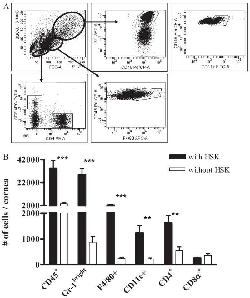
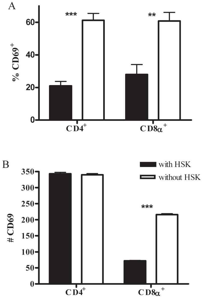
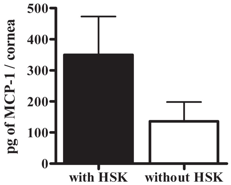
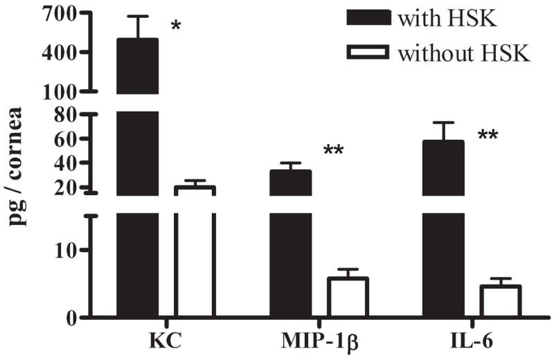
Methods: Plaque assay quantified HSV-1 in the tear film of infected mice. FACS analysis enumerated corneal leukocytic infiltrate and characterized infiltrate phenotypically after staining for activation and regulatory T cell (Treg) markers and for markers of antigen-presenting cell (APC) maturation. Treg cells were depleted in vivo using anti-CD25 mAb. Luminex analysis quantified the amount of cytokines and chemokines expressed in corneal tissue homogenate.
Results: Infected corneas without HSK exhibited a pronounced leukocytic infiltrate containing a significantly higher proportion and nearly identical absolute number of activated CD4+ T cells 15 days after infection when compared with those with HSK. Moreover, the frequency and absolute number of regulatory CD4+ T cells (Tregs) was lower in nondiseased corneas, and Treg depletion did not influence HSK incidence. The frequency of mature, immunogenic DCs and the ratio of mature DCs to CD4+ T cells were nearly identical in corneas with and without HSK. The authors observed a reduced population of neutrophils and reduced expression of neutrophil chemoattractants MIP-1beta and keratinocyte chemoattractant and the neutrophil-attracting cytokine IL-6 in corneas without HSK.
Conclusions: These findings demonstrate that HSV-1-infected corneas can retain clarity in the presence of a substantial secondary leukocytic infiltrate, that activated CD4+ T cells, while necessary, are not sufficient for HSK development, that susceptibility to HSK is not determined by Tregs, and that clinical disease correlates with the accumulation of a critical mass of neutrophils through chemoattraction.
Figures


Similar articles
-
CD8 T cells mediate transient herpes stromal keratitis in CD4-deficient mice.Invest Ophthalmol Vis Sci. 2006 Aug;47(8):3400-9. doi: 10.1167/iovs.05-0898. Invest Ophthalmol Vis Sci. 2006. PMID: 16877409 Free PMC article.
-
A novel p40-independent function of IL-12p35 is required for progression and maintenance of herpes stromal keratitis.Invest Ophthalmol Vis Sci. 2010 Jul;51(7):3591-8. doi: 10.1167/iovs.09-4368. Epub 2010 Mar 5. Invest Ophthalmol Vis Sci. 2010. PMID: 20207959 Free PMC article.
-
MCP-1 derived from stromal keratocyte induces corneal infiltration of CD4+ T cells in herpetic stromal keratitis.Mol Cells. 2008 Jul 31;26(1):67-73. Epub 2008 Jul 2. Mol Cells. 2008. PMID: 18594181
-
Immunological aspects of herpetic stromal keratitis.Semin Ophthalmol. 2008 Jul-Aug;23(4):221-7. doi: 10.1080/08820530802111390. Semin Ophthalmol. 2008. PMID: 18584559 Review.
-
Reviews for immune privilege in the year 2010: immune privilege and infection.Ocul Immunol Inflamm. 2010 Aug;18(4):237-43. doi: 10.3109/09273948.2010.501946. Ocul Immunol Inflamm. 2010. PMID: 20662654 Review.
Cited by
-
HSV-1 miR-H6 inhibits HSV-1 replication and IL-6 expression in human corneal epithelial cells in vitro.Clin Dev Immunol. 2012;2012:192791. doi: 10.1155/2012/192791. Epub 2012 Apr 9. Clin Dev Immunol. 2012. PMID: 22550533 Free PMC article.
-
Diphenyleneiodonium Treatment Inhibits the Development of Severe Herpes Stromal Keratitis Lesions.J Virol. 2022 Sep 14;96(17):e0101422. doi: 10.1128/jvi.01014-22. Epub 2022 Aug 10. J Virol. 2022. PMID: 35946937 Free PMC article.
-
NLRP3, NLRP12, and IFI16 Inflammasomes Induction and Caspase-1 Activation Triggered by Virulent HSV-1 Strains Are Associated With Severe Corneal Inflammatory Herpetic Disease.Front Immunol. 2019 Jul 16;10:1631. doi: 10.3389/fimmu.2019.01631. eCollection 2019. Front Immunol. 2019. PMID: 31367214 Free PMC article.
-
CD137 costimulation is associated with reduced herpetic stromal keratitis and with developing normal CD8+ T cells in trigeminal ganglia.J Gen Virol. 2022 Jun;103(6):001756. doi: 10.1099/jgv.0.001756. J Gen Virol. 2022. PMID: 35766977 Free PMC article.
-
Herpesvirus entry mediator on radiation-resistant cell lineages promotes ocular herpes simplex virus 1 pathogenesis in an entry-independent manner.mBio. 2015 Oct 20;6(5):e01532-15. doi: 10.1128/mBio.01532-15. mBio. 2015. PMID: 26489863 Free PMC article.
References
-
- Hendricks RL, Epstein RJ, Tumpey T. The effect of cellular immune tolerance to HSV-1 antigens on the immunopathology of HSV-1 keratitis. Invest Ophthalmol Vis Sci. 1989;30:105–115. - PubMed
-
- Shimeld C, Whiteland JL, Nicholls SM, Easty DL, Hill TJ. Immune cell infiltration in corneas of mice with recurrent herpes simplex virus disease. J Gen Virol. 1996;77(pt 5):977–985. - PubMed
-
- Tumpey TM, Cheng H, Yan XT, Oakes JE, Lausch RN. Chemokine synthesis in the HSV-1-infected cornea and its suppression by interleukin-10. J Leukoc Biol. 1998;63:486–492. - PubMed
-
- Yan XT, Tumpey TM, Kunkel SL, Oakes JE, Lausch RN. Role of MIP-2 in neutrophil migration and tissue injury in the herpes simplex virus-1-infected cornea. Invest Ophthalmol Vis Sci. 1998;39:1854–1862. - PubMed
Publication types
MeSH terms
Substances
Grants and funding
LinkOut - more resources
Full Text Sources
Research Materials
