Comparative Study
. 2008 Apr;49(4):1580-90.
doi: 10.1167/iovs.07-1110.

Retinal laminar architecture in human retinitis pigmentosa caused by Rhodopsin gene mutations

Affiliations
Comparative Study

Retinal laminar architecture in human retinitis pigmentosa caused by Rhodopsin gene mutations

Tomas S Aleman et al. Invest Ophthalmol Vis Sci. 2008 Apr.

Abstract

Purpose: To determine the underlying retinal micropathology in subclasses of autosomal dominant retinitis pigmentosa (ADRP) caused by rhodopsin (RHO) mutations.

Methods: Patients with RHO-ADRP (n = 17, ages 6-73 years), representing class A (R135W and P347L) and class B (P23H, T58R, and G106R) functional phenotypes, were studied with optical coherence tomography (OCT), and colocalized visual thresholds were determined by dark- and light-adapted chromatic perimetry. Autofluorescence imaging was performed with near-infrared light. Retinal histology in hT17M-rhodopsin mice was compared with the human results.

Results: Class A patients had only cone-mediated vision. The outer nuclear layer (ONL) thinned with eccentricity and was not detectable within 3 to 4 mm of the fovea. Scotomatous extracentral retina showed loss of ONL, thickening of the inner retina, and demelanization of RPE. Class B patients had superior-inferior asymmetry in function and structure. The superior retina could have normal rod and cone vision, normal lamination (including ONL) and autofluorescence of the RPE melanin; laminopathy was found in the scotomas. With Fourier-domain-OCT, there was apparent inner nuclear layer (INL) thickening in regions with ONL thinning. Retinal regions without ONL had a thick hyporeflective layer that was continuous with the INL from neighboring regions with normal lamination. Transgenic mice had many of the laminar abnormalities found in patients.

Conclusions: Retinal laminar abnormalities were present in both classes of RHO-ADRP and were related to the severity of colocalized vision loss. The results in human class B and the transgenic mice support the following disease sequence: ONL diminution with INL thickening; amalgamation of residual ONL with the thickened INL; and progressive retinal remodeling with eventual thinning.


Figures
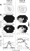
Figure 1
Figure 1
Classes of disease expression in RHO-ADRP exemplified by data from two patients. (A) Kinetic perimetry results from the right eye using two targets (V-4e and I-4e). (B) Static threshold perimetry results, dark-adapted (top) and light-adapted (bottom), are displayed as grayscale maps of rod and cone sensitivity loss. The scale has 16 levels of gray, representing 0- to 30-dB losses (right). The physiological blind spot is represented as a black square at 12° in the temporal field. N, nasal; T, temporal; I, inferior; and S, superior visual field. (C) Dark-adapted two-color (500 and 650 nm) vertical sensitivity profiles in the patients (symbols) compared with lower limits of normal (thick gray lines) for rod-mediated sensitivity to the 500-nm stimulus and for cone-mediated sensitivity to the 650-nm stimulus at the cone plateau. The photoreceptor mediation at each locus, based on the sensitivity difference between the two colors is given: R, rod-mediated; M, mixed rod- and cone-mediated; and C, cone-mediated.
Figure 2
Figure 2
Retinal laminar architecture in class A RHO-ADRP. (A) Cross-sectional OCT images along the vertical meridian through the fovea in a normal subject (age 25; top) compared with three patients (bottom) representing different ages and disease stages in class A RHO-ADRP. Brackets defining ONL and the inner retina are labeled (left) and a bracket showing total retinal thickness is at the right edge. Bars above the scans show psychophysically determined rod (blue bar: dark-adapted, 500-nm stimulus) and cone (red bar: light-adapted, 600-nm stimulus) sensitivity. Arrows: discernible ONL in F2,P1 and F2,P2. (*) Cystoid changes. I, inferior; S, superior retina. Calibration bar at left. Inset: schematic location of the scans. (B) Thickness of the overall retina and inner retina along the vertical meridian at eccentricities >2 mm in 9 class A patients grouped by age. Measurements in some patients are interrupted in regions with or adjacent to cystoid changes. Shaded areas: normal limits (mean ± 2SD) for retinal thickness (n = 27, ages 5–58) and inner retina (n = 14, ages 5–58). Insets: schematic location of the scans.
Figure 3
Figure 3
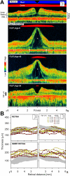
Retinal laminar architecture in class B RHO-ADRP. (A) Cross-sectional OCT images along the vertical meridian through the fovea in a normal subject (age 24; top) compared with two patients (bottom) representing class B patients. Brackets defining the ONL and the inner retina are labeled (left) and a bracket showing total retinal thickness is at the right edge. White bracket in the image obtained from F7,P1 delimits the segment with normal lamination. Bars above the scans indicate rod (blue) and cone (red) sensitivity (as in Fig. 2). I, inferior; S, superior. Left: calibration bar. Insets: schematic location of the scans. (B) Thickness of the overall retina, ONL, and inner retina along the vertical meridian in the eight patients. Shaded areas: normal limits (mean ± 2 SD) as in Fig. 2.
Figure 4
Figure 4
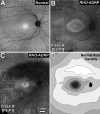
Topography of RPE disease in RHO-ADRP. (AC) AF imaging results obtained with near-infrared (NIR) excitation in a 24-year-old normal subject (A) and in patients from the two classes of RHO-ADRP (B, C). The intensity of the NIR-AF image of F8,P1 (C) is shown scaled by 1.75× compared with the normal subject (A) and F1,P1 (B) for better visualization of the regional features. (D) Map of mean rod photoreceptor density in the human retina (generated from data published in Ref. 48) for comparison with the NIR-AF images. Lighter intensities correspond to higher spatial densities (∼130,000–160,000 rods/mm−2) and darker intensities to lower densities. Black oval: optic nerve head.
Figure 5
Figure 5
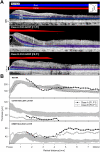
Detailed retinal structure of the inferior retina in RHO-ADRP examined by FD-OCT. (A) Cross-sectional FD-OCT along the vertical meridian from the fovea extending into the inferior retina in a normal subject (top) and two patients representing each class of RHO-ADRP (middle, bottom). Bars above the cross-sections indicate rod (blue) and cone (red) sensitivity (as in Fig. 2). Nuclear layers are labeled and highlighted (ONL, blue; INL and hyporeflective layer continuous with it: purple). Inset: schematic location of the scans. Epiretinal membranes were visible in both patients. (*) Cystoid changes. Left: calibration bar. (B) Overall retinal, ONL, and INL thicknesses along the vertical meridian in the inferior retina in both patients. Circles: retinal regions with two detectable nuclear layers; diamonds: regions with a single hyporeflective layer that is continuous with the INL from the more central retina. Shaded areas: normal limits (mean ± 2SD; n = 9, age range, 15–63).
Figure 6
Figure 6
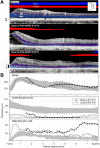
Detailed laminar structure of the superior retina in RHO-ADRP examined by FD-OCT. (A) Cross-sectional FD-OCT along the vertical meridian from the fovea extending into the superior retina in a normal subject (top) and two patients (middle, bottom; same subjects as Fig. 5) representing each class of RHO-ADRP. Bars above the cross-sections indicate rod (blue) and cone (red) sensitivity (as in Fig. 2). Nuclear layers are highlighted (ONL, blue; INL and hyporeflective layer continuous with it: purple). Inset: schematic location of the scans. Epiretinal membranes are visible in both patients. (*) Cystoid changes. Left: calibration bar. (B) Overall retinal thickness, ONL, and INL thicknesses along the vertical superior meridian in both patients. Symbols and shaded areas are as defined in Figure 5.
Figure 7
Figure 7
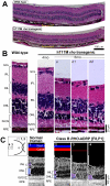
Comparison of histopathology of hT17M rho mutant mouse with results in a class B RHO-ADRP patient. (A) Low-magnification views of vertical retinal sections crossing the optic nerve from a 6-month-old hT17M rho transgenic mouse compared with an age-matched wild-type (WT) mouse. Yellow bracket: region examined at higher magnification in (B). (B) Magnified (40-μm-wide) retinal images taken at ∼400 to 500 μm from the optic nerve in a 4-month-old mouse (Ba) and in two 6-month-old (Bb1, Bb2) transgenic mice compared to a 4-month-old WT mouse. (Bb2, arrows) Clusters of remaining nuclei in the ONL. (C) Cross-sectional, 500-μm-long FD-OCT images obtained at 5 to 9 mm of eccentricity in the superior and temporal retina in a class B RHO-RP patient compared with a normal subject. Schematic to the left depicts retinal regions sampled in each cross section. Reflectivity profiles (white traces) are overlaid on the FD-OCT scans; signal features representing nuclear layers are shown adjacent to highlighted layers (ONL, blue; INL and hyporeflective layer continuous with it: purple). Bars above the images indicate rod (blue) and cone (red) (as in Fig. 2) sensitivity. (C3, arrows) Hyporeflectivities that may correspond to clumps of remnant photoreceptor nuclei.

References

    1. Gal A, Apfelstedt-Sylla E, Janecke AR, Zrenner E. Rhodopsin mutations in inherited retinal dystrophies and dysfunctions. Prog Retinal Eye Res. 1997;16(1):51–79.
    1. Kennan A, Aherne A, Humphries P. Light in retinitis pigmentosa. Trends Genet. 2005;21(2):103–110. - PubMed
    1. Hartong DT, Berson EL, Dryja TP. Retinitis pigmentosa. Lancet. 2006;368(9549):1795–1809. - PubMed
    1. Daiger SP, Bowne SJ, Sullivan LS. Perspective on genes and mutations causing retinitis pigmentosa. Arch Ophthalmol. 2007;125(2):151–158. - PMC - PubMed
    1. Palczewski K, Hofmann KP, Baehr W. Rhodopsin–advances and perspectives. Vision Res. 2006;46(27):4425–4426. - PubMed

Publication types

MeSH terms