. 2008 Jul 1;17(13):2047-57.
doi: 10.1093/hmg/ddn102. Epub 2008 Apr 1.

The loss of methyl-CpG binding protein 1 leads to autism-like behavioral deficits

Affiliations

The loss of methyl-CpG binding protein 1 leads to autism-like behavioral deficits

Andrea M Allan et al. Hum Mol Genet. .

Abstract

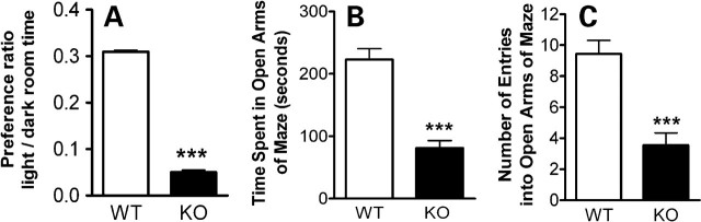
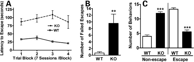
Methyl-CpG binding proteins (MBDs) are central components of DNA methylation-mediated epigenetic gene regulation. Alterations of epigenetic pathways are known to be associated with several neurodevelopmental disorders, particularly autism. Our previous studies showed that the loss of Mbd1 led to reduced hippocampal neurogenesis and impaired learning in mice. However, whether MBD1 regulates the autism-related cognitive functions remains unknown. Here we show that Mbd1 mutant (Mbd1(-/-)) mice exhibit several core deficits frequently associated with autism, including reduced social interaction, learning deficits, anxiety, defective sensory motor gating, depression and abnormal brain serotonin activity. Furthermore, we find that Mbd1 can directly regulate the expression of Htr2c, one of the serotonin receptors, by binding to its promoter, and the loss of Mbd1 led to elevated expression of Htr2c. Our results, therefore, demonstrate the importance of epigenetic regulation in mammalian brain development and cognitive functions. Understanding how the loss of Mbd1 could lead to autism-like behavioral phenotypes would reveal much-needed information about the molecular pathogenesis of autism.


Figures

Figure 1.
Figure 1.
Mbd1−/− mice have impaired social interaction. (A) Both Mbd1−/− (KO) and wild-type (WT) mice spent similar amounts of time interacting with the tent during the first 5 min. During the last 5 min, when a stranger mouse was in the tent (right panel), WT mice spent significantly more time in contact with the tent, demonstrating a normal level of socialization. KO mice, however, failed to display this interest in social interaction. (B) KO and WT mice exhibited no differences in the number (frequency) of visits to the tent, whether there was a novel object (first 5 min) or a novel mouse (last 5 min) inside the tent. Data are presented as mean ± SEM, n = 12; ***P < 0.0001.
Figure 2.
Figure 2.
Mbd1−/− mice have impaired sensorimotor gating as assessed by prepulse inhibition (PPI). (A and B) Mbd1−/− (KO) mice have reduced PI compared with WT littermate controls at both 120 ms ISI (A) and 300 ms ISI (B) across a range of prepulse intensities. (C) The tactile startle response was no different for WT and KO mice. Data are shown as mean percent startle at different prepulse amplitudes ± SEM (n = 7).
Figure 3.
Figure 3.
Mbd1−/−mice exhibited deficits in fear-conditioning learning tests. (A) Mbd1−/− (KO) mice had deficits in the cued fear conditioning test, which assessed amygdala-mediated learning; (B) KO mice had deficits in the contextual fear conditioning test, which assessed both hippocampus- and amygdala-mediated learning. *P < 0.05, n = 6, t-test.
Figure 4.
Figure 4.
Mbd1−/− mice exhibited increased anxiety. (A) In a light–dark preference test, Mbd1−/− (KO) mice spent significantly less time in the light room than their wild-type (WT) littermates. (B and C) In an elevated plus-maze test, KO mice spent significantly less time in open arms (B) and had significantly fewer entries into open arms (C) compared with WT littermate controls, while the total numbers of transitions between arms were no different (data not shown). Data presented are mean ± SEM, n = 9–12, *** indicates significance, P < 0.001.
Figure 5.
Figure 5.
Mbd1−/− mice exhibited enhanced susceptibility to depressive behaviors. (A and B) In a learned helplessness test of depression, Mbd1−/− (KO) mice exhibited significantly longer latency for escape across all four trial blocks (A) and a significantly greater number of failed escapes (B) than their wild-type (WT) littermates. In a forced swim test (C), KO mice exhibited significantly more non-escape-directed behaviors (floating, twitching and paddling) and significantly fewer escape-directed behaviors (swimming, climbing and thrashing) than WT littermates. Data presented are mean ± SEM, n = 9; ***P < 0.0001; **P < 0.01.
Figure 6.
Figure 6.
Mbd1 directly regulates the expression of serotonin receptor Htr2c. (A) Mbd1−/− mice (KO) displayed more wet-dog shake behaviors compared with their wild-type (WT) littermates, indicative of abnormal brain serotonin activity; (B) Real-time PCR analyses showing increased Htr2c mRNA in the MFC of KO mice; (C) Representative western blot analysis showing increased Htr2c protein in the KO MFC. (D and E) Agonist-mediated serotonin binding assay showing that KO mice exhibited increased serotonin binding to the Htr2c receptor, indicated by the higher KD value (D); however, Htr2c receptors in KO brains have lower binding affinity for serotonin, indicated by the higher Bmax value (E). Data are represented as mean ± SEM, n = 11–12. (F) Chromatin immunoprecipitation assay demonstrating that Mbd1 bound to the promoter region of Htr2c. (G) Chromatin immunoprecipitation assay demonstrating that Mbd1 bound to the intron 1 of Htr2c in WT brains, but not in KO brains. The amount of binding of Mbd1 to Htr2c intron 1 was not significantly different between male (KO1 and WT1) and female (KO2 and WT2) brains. (H and I), immunohistochemistry using an anti-β-gal antibody showing expression of Mbd1 in the MFC region. Scale bar = 50 µm. (H) Negative control using mouse IgG instead of primary antibody. (I) Anti-β-gal antibody. ***P < 0.0001. **P < 0.01; *P < 0.05.

References

    1. Muhle R., Trentacoste S.V., Rapin I. The genetics of autism. Pediatrics. 2004;113:e472–e486. - PubMed
    1. Liu J., Nyholt D.R., Magnussen P., Parano E., Pavone P., Geschwind D., Lord C., Iversen P., Hoh J., Ott J., et al. A genomewide screen for autism susceptibility loci. Am. J. Hum. Genet. 2001;69:327–340. - PMC - PubMed
    1. Yonan A.L., Alarcon M., Cheng R., Magnusson P.K., Spence S.J., Palmer A.A., Grunn A., Juo S.H., Terwilliger J.D., Liu J., et al. A genomewide screen of 345 families for autism-susceptibility loci. Am. J. Hum. Genet. 2003;73:886–897. - PMC - PubMed
    1. Holliday R., Pugh J.E. DNA modification mechanisms and gene activity during development. Science. 1975;187:226–232. - PubMed
    1. Riggs A.D. X inactivation, differentiation, and DNA methylation. Cytogenet. Cell. Genet. 1975;14:9–25. - PubMed

Publication types