. 2008 Mar;178(3):1533-45.
doi: 10.1534/genetics.107.082792.

Lgals6, a 2-million-year-old gene in mice: a case of positive Darwinian selection and presence/absence polymorphism

Affiliations

Lgals6, a 2-million-year-old gene in mice: a case of positive Darwinian selection and presence/absence polymorphism

Denis Houzelstein et al. Genetics. 2008 Mar.

Abstract

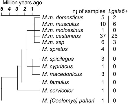
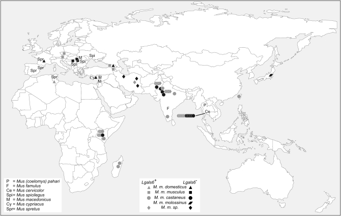
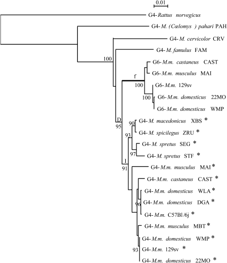
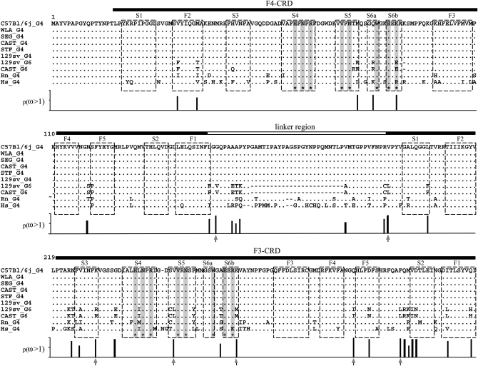
Duplications of genes are widely considered to be a driving force in the evolutionary process. The fate of such duplicated genes (paralogs) depends mainly on the early stages of their evolution. Therefore, the study of duplications that have already started to diverge is useful to better understand their evolution. We present here the example of a 2-million-year-old segmental duplication at the origin of the Lgals4 and Lgals6 genes in the mouse genome. We analyzed the distribution of these genes in samples from 110 wild individuals and wild-derived inbred strains belonging to eight mouse species from Mus (Coelomys) pahari to M. musculus and 28 laboratory strains. Using a maximum-likelihood method, we show that the sequence of the Lgals6 gene has evolved under the influence of strong positive selection that is likely to result in its neofunctionalization. Surprisingly, despite this selection pressure, the Lgals6 gene is present in some mouse species, but not all. Furthermore, even within the species and populations where it is present, the Lgals6 gene is never fixed. To explain this paradox, we propose different hypotheses such as balanced selection and neutral retention of ancient polymophism and we discuss this unexpected result with regard to known galectin properties and response to infections by pathogens.


Figures

F<sc>igure</sc> 1.—
Figure 1.—
Comparison of Lgals4 and Lgals6 genomic organization. (a) Genomic organization of the Lgals4 (top) and Lgals6 (bottom) genes. Exons are represented as boxes, numbered from 01 to 10. Note that, for clarity, we ascribe the same reference number to homologous exons in Lgals4 and Lgals6, i.e., the exons of both genes are numbered from 01 to 10 with exons 05 and 06 (shaded on Lgals4) missing from the Lgals6 gene. Scale bar is in base pairs. The Lgals4 and Lgals6 genes differ by a 1.8-kb deletion in Lgals6 shown here as an open triangle. The primer pair 1f-1r (numbered 1) amplifies a 305-bp fragment specific for the Lgals4 gene and an 82-bp fragment specific for the Lgals6 gene. The primer pair 2f-2r (numbered 2) amplifies a 142-bp fragment specific for the Lgals6 gene. The fragment containing the intronic sequences that were cloned and sequenced to build the phylogenetic tree is shown as a solid line (numbered 3.1 in Lgals4 and 3.2 in Lgals6, respectively). (b) Ethidium bromide-stained gel showing bands amplified from the primer pair 1f-1r (numbered 1) and 2f-2r (numbered 2) from 129sv genomic DNA. L, DNA ladder.
F<sc>igure</sc> 2.—
Figure 2.—
Presence/absence of the Lgals6 gene in Mus species and subspecies from which the wild-derived inbred strains were derived. ni, number of individuals from a given species or subspecies for which the presence of the Lgals6 gene was tested. Lgals6+, number of individuals from a given species or subspecies in which the Lgals6 gene has been detected. The Mus musculus ssp. branch regroups individuals of strains of still unsettled taxonomic status. The evolutionary tree has been modified from Guénet and Bonhomme (2003).
F<sc>igure</sc> 3.—
Figure 3.—
Geographical origin of the different individuals in which the presence of the Lgals6 gene was tested. Symbols shown as a line group individuals belonging to the same population. Lgals6+, an individual from a given species or subspecies in which the Lgals6 gene has been detected (shaded). Lgals6, an individual from a given species or subspecies in which the Lgals6 is absent (solid).
F<sc>igure</sc> 4.—
Figure 4.—
Phylogenetic tree of Lgals4 and Lgals6. This maximum-likelihood tree was reconstructed with 1234 sites of intronic sequences (input tree generated by BIONJ; HKY model including a Γ-correction with four categories of sites and ts:tv ratio estimated from the data). The numbers at nodes correspond to the percentage support of 1000 bootstrap replicates and percentages only >90% are shown. The intron 04 of sequences with an asterisk contains the SINE element. The branch f is postulated to be under positive selection and is considered as the foreground branch for branch-site models. D represents the gene duplication event; I, the insertion of the SINE element in the intron 04 of some Mus Lgals4 sequences.
F<sc>igure</sc> 5.—
Figure 5.—
Comparison of amino acid sequences of galectin-4 and galectin-6 sequences. Dashes represent gaps introduced for alignment: a genomic deletion in the Lgals6 gene removed two exons (05 and 06; see Figure 1), coding for part of the linker. Residues identical to those of the corresponding C57BL/6J galectin-4 are indicated by dots. Horizontal filled bars correspond to the F4- and F3-carbohydrate recognition domains (Houzelstein et al. 2004). The horizontal open bar corresponds to the linker region that is shorter in galectin-6 (G6) because of the deletion. Asterisks at the bottom of the sequences and corresponding shaded vertical bars mark the position of residues that interact with carbohydrates (Lobsanov et al. 1993). Open boxes indicate the β-strands. Vertical bars below the alignment show the Bayesian posterior probability of ω > 1 for each site. Arrows under vertical bars indicate sites with p(ω >1) > 0.95.

Similar articles

Cited by

References

    1. Adams, D. J., E. T. Dermitzakis, T. Cox, J. Smith, R. Davies et al., 2005. Complex haplotypes, copy number polymorphisms and coding variation in two recently divergent mouse strains. Nat. Genet. 37 532–536. - PubMed
    1. Aitman, T. J., R. Dong, T. J. Vyse, P. J. Norsworthy, M. D. Johnson et al., 2006. Copy number polymorphism in Fcgr3 predisposes to glomerulonephritis in rats and humans. Nature 439 851–855. - PubMed
    1. Bailey, J. A., Z. Gu, R. A. Clark, K. Reinert, R. V. Samonte et al., 2002. Recent segmental duplications in the human genome. Science 297 1003–1007. - PubMed
    1. Bailey, J. A., D. M. Church, M. Ventura, M. Rocchi and E. E. Eichler, 2004. Analysis of segmental duplications and genome assembly in the mouse. Genome Res. 14 789–801. - PMC - PubMed
    1. Beck, J. A., S. Lloyd, M. Hafezparast, M. Lennon-Pierce, J. T. Eppig et al., 2000. Genealogies of mouse inbred strains. Nat. Genet. 24 23–25. - PubMed

Publication types

Associated data