. 2008 Jun;82(11):5340-7.
doi: 10.1128/JVI.00135-08. Epub 2008 Apr 2.

Structural basis for the receptor binding specificity of Norwalk virus

Affiliations

Structural basis for the receptor binding specificity of Norwalk virus

Weiming Bu et al. J Virol. 2008 Jun.

Abstract

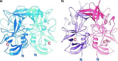
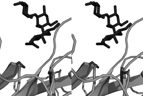
Noroviruses are positive-sense, single-stranded RNA viruses that cause acute gastroenteritis. They recognize human histo-blood group antigens as receptors in a strain-specific manner. The structures presented here were analyzed in order to elucidate the structural basis for differences in ligand recognition of noroviruses from different genogroups, the prototypic Norwalk virus (NV; GI-1) and VA387 (GII-4), which recognize the same A antigen but differ in that NV is unable to bind to the B antigen. Two forms of the receptor-binding domain of the norovirus coat protein, the P domain and the P polypeptide, that were previously shown to differ in receptor binding and P-particle formation properties were studied. Comparison of the structures of the NV P domain with and without A trisaccharide and the NV P polypeptide revealed no major ligand-induced changes. The 2.3-A cocrystal structure reveals that the A trisaccharide binds to the NV P domain through interactions with the residues Ser377, Asp327, His329, and Ser380 in a mode distinct from that previously reported for the VA387 P-domain-A-trisaccharide complex. Mutational analyses confirm the importance of these residues in NV P-particle binding to native A antigen. The alpha-GalNAc residue unique to the A trisaccharide is buried deeply in the NV binding pocket, unlike in the structures of A and B trisaccharides bound to VA387 P domain, where the alpha-fucose residue forms the most protein contacts. The A-trisaccharide binding mode seen in the NV P domain complex cannot be sterically accommodated in the VA387 P domain.


Figures

FIG. 1.
FIG. 1.
(a) Structure-based superimposition of the amino acid sequences of NV P domain and VA387 P domain. Identical residues are highlighted with a red background, while similar residues are in red type. Secondary structures present in the P polypeptide structures of NV and VA387 are indicated above and below the sequences, respectively; arrows represent β-strands, and coils represent helices (either 310 [η] or alpha [α]). Blue circles indicate residues involved in A-trisaccharide interaction with the NV P domain, and pink circles indicate residues involved in A-trisaccharide interaction with the VA387 P domain. Sequence alignment was done with ESPript (8), and secondary structure assignment was done with STRIDE (7). (b) Ribbon diagram showing one subunit of the NV P domain (generated with RIBBONS [4]). Strands (β) are shown in pink, α-helices (α) in cyan, 310 helices (η) in green, coils and turns in gray. Secondary structural elements are labeled as in panel a. The N terminus of the structure (Pro230) is indicated in blue, and a dotted arrow indicates the location of the S domain in the intact VP1 protein. The C terminus of the crystallographic model (Val516) is indicated, along with the residues not visible in the experimental density map (red sequence). (c) Superimposition of the NV (blue) and VA387 (pink) P domain structures. Green boxes highlight regions where the secondary structure is distinctly different.
FIG. 2.
FIG. 2.
Dimers of the NV P domain (a) and the VA387 P domain (2OBR.PDB) (3) (b). In each case the two subunits are shown in different colors. The N and C termini of the structures are indicated.
FIG. 3.
FIG. 3.
(a) Stereo view of the electron density for the A trisaccharide in the experimental Fo-Fc map calculated after the initial molecular replacement solution for the NV P-domain-A-trisaccharide complex structure was refined with only protein coordinates. The coordinates of the final refined A trisaccharide are also shown. The map is contoured at 1.5σ. (b) Ribbon diagram showing superimposition of the A-trisaccharide-bound NV P domain (blue) and the A-trisaccharide-bound VA387 P domain (pink; 2OBS.PDB) superimposed. The A trisaccharide in the NV complex is shown as a ball-and-stick model in blue, and the A trisaccharide in the VA387 complex is in red, demonstrating the different binding sites occupied by the receptor oligosaccharides. (c) Stereo view of the A-trisaccharide binding site in the NV P domain. Dashed lines indicate hydrogen bonds, “W” indicates solvent molecules, and the apostrophe in Ser338′ indicates an interaction with the noncognate subunit. The image was generated with PyMOL (PyMOL Graphics System; DeLano Scientific, San Carlos, CA).
FIG. 4.
FIG. 4.
Schematic representations of contacts between the A trisaccharide and the NV P domain seen in the two noncrystallographically related subunits of the NV P domain dimer (a and b) and the contacts between the A trisaccharide and the VA387 P domain identified in the previously published crystal structure (2OBS.PDB) (3) (c). Dashed lines indicate hydrogen bonds, and “W” indicates solvent molecules. The apostrophe in Ser338′ indicates an interaction with the noncognate subunit. Obb and Nbb refer to contacts with the protein backbone. H-bond distances are indicated.
FIG. 5.
FIG. 5.
HBGA-binding assays of wild-type and mutant P particles with single amino acid mutations at the A-trisaccharide-binding site. The x axes show the protein concentrations of the P particles, while the y axes indicate measurements of optical density at 450 (OD450) nm. Data are averages from triplicate experiments. O, A, and N, type O (containing H antigen), A, and nonsecretor saliva, respectively.
FIG. 6.
FIG. 6.
The structures of the NV P domain-A trisaccharide complex and the VA387 P domain-A trisaccharide complex (2OBS.PDB) were superimposed. Shown here, as a stereo view, is the A trisaccharide from the NV P domain complex and that from the VA387 protein, demonstrating steric clashes that prevent an NV-type carbohydrate binding mode for the VA387 P domain.

References

    1. Bertolotti-Ciarlet, A., L. J. White, R. Chen, B. V. Prasad, and M. K. Estes. 2002. Structural requirements for the assembly of Norwalk virus-like particles. J. Virol. 764044-4055. - PMC - PubMed
    1. Brunger, A. T., P. D. Adams, G. M. Clore, W. L. DeLano, P. Gros, R. W. Grosse-Kunstleve, J. S. Jiang, J. Kuszewski, M. Nilges, N. S. Pannu, R. J. Read, L. M. Rice, T. Simonson, and G. L. Warren. 1998. Crystallography & NMR system: a new software suite for macromolecular structure determination. Acta Crystallogr. D 54(Pt. 5)905-921. - PubMed
    1. Cao, S., Z. Lou, M. Tan, Y. Chen, Y. Liu, Z. Zhang, X. C. Zhang, X. Jiang, X. Li, and Z. Rao. 2007. Structural basis for the recognition of blood group trisaccharides by norovirus. J. Virol. 815949-5957. - PMC - PubMed
    1. Carson, M. 1987. Ribbon models of macromolecules. J. Mol. Graphics 5103-106.
    1. Chakravarty, S., A. M. Hutson, M. K. Estes, and B. V. Prasad. 2005. Evolutionary trace residues in noroviruses: importance in receptor binding, antigenicity, virion assembly, and strain diversity. J. Virol. 79554-568. - PMC - PubMed

Publication types

MeSH terms