. 2008 Jun;294(6):F1323-35.
doi: 10.1152/ajprenal.00401.2007. Epub 2008 Apr 2.

Signaling pathways modulated by fish oil in salt-sensitive hypertension

Affiliations

Signaling pathways modulated by fish oil in salt-sensitive hypertension

Montserrat M Diaz Encarnacion et al. Am J Physiol Renal Physiol. 2008 Jun.

Abstract

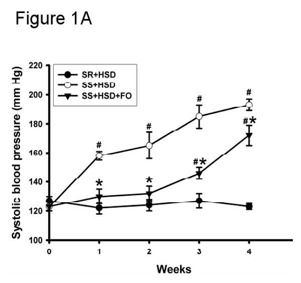
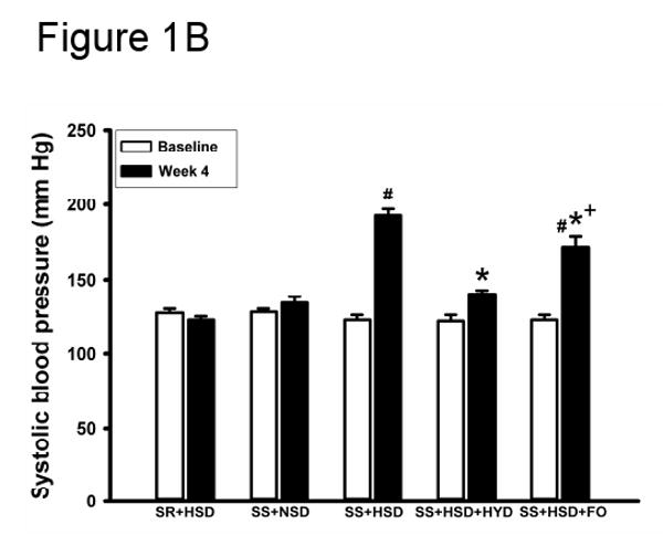
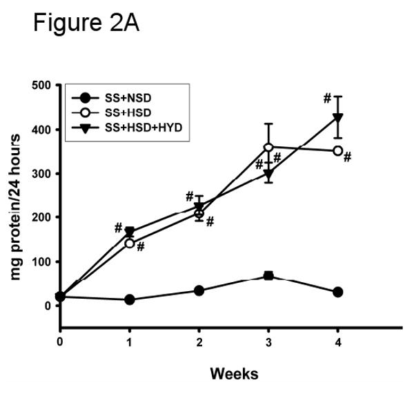
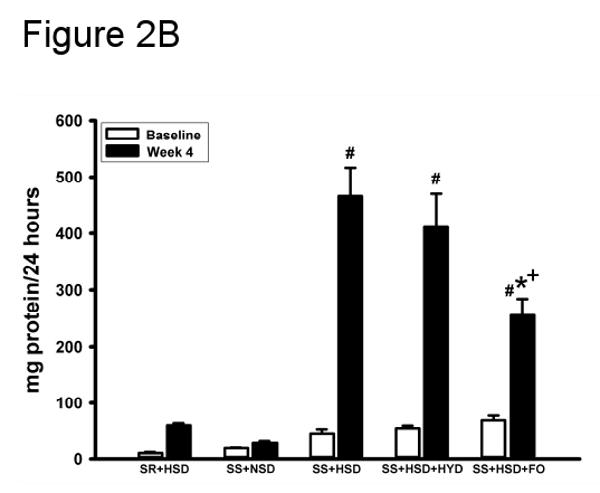
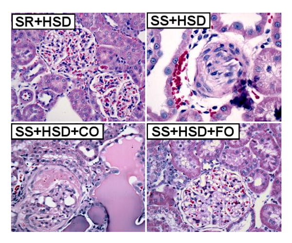
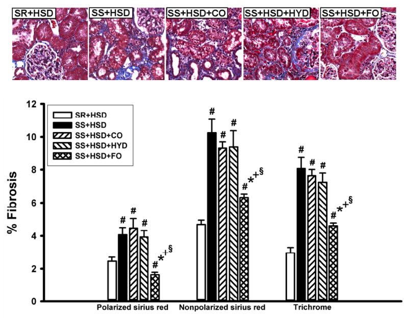
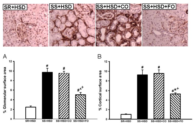
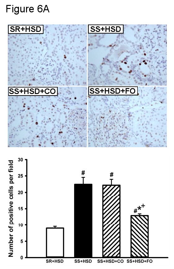
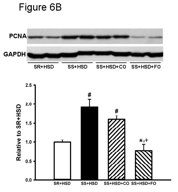
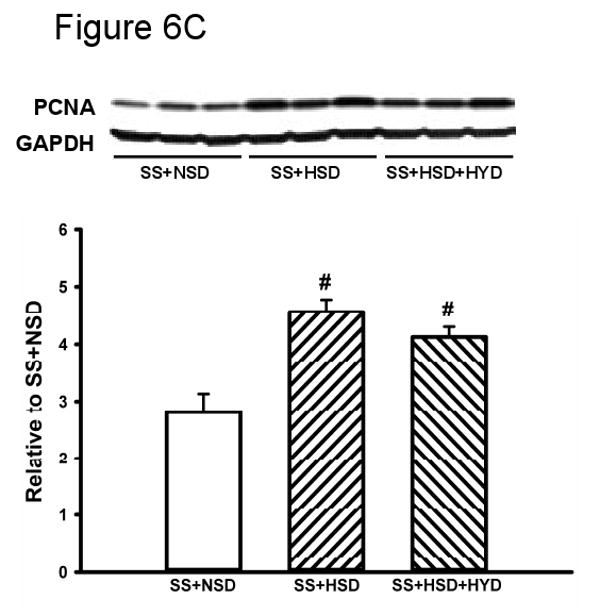
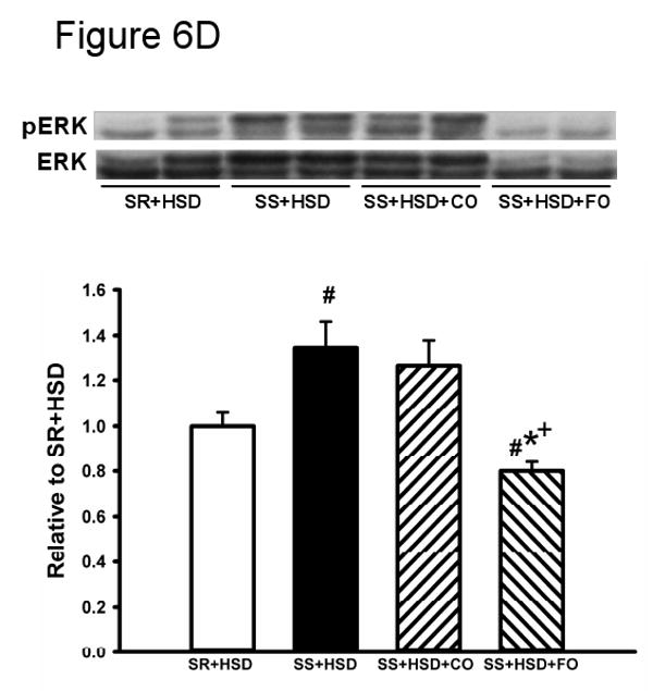
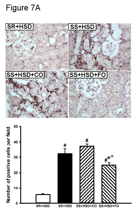
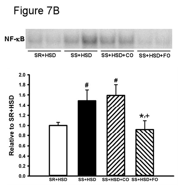
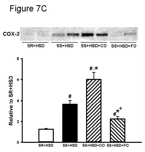
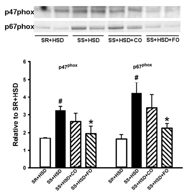
Although many studies have indicated that fish oil (FO) improves cardiovascular risk factors and reduces histopathological manifestations of injury in experimental renal injury models, potential mechanisms underlying this protective effect have not been adequately defined. The objective of this study was to identify potential signaling pathways that confer protection in the Dahl rat model of salt-sensitive hypertension. Male Dahl salt-sensitive rats (n = 10/group) were provided with formulated diets containing 8% NaCl, 20% protein, and 25% FO or 25% corn oil (CO) for 28 days. FO reduced blood pressure (-11% at 4 wk; P < 0.05), urine protein excretion (-45% at 4 wk; P < 0.05), plasma cholesterol and triglyceride levels (-54%, P < 0.001; and -58%, P < 0.05), and histopathological manifestations of renal injury, including vascular hypertrophy, segmental and global glomerular sclerosis, interstitial fibrosis, and tubular atrophy. Interstitial inflammation was significantly reduced by FO (-32%; P < 0.001), as assessed by quantitative analysis of ED1-positive cells in sections of the renal cortex. FO reduced tubulointerstitial proliferative activity, as assessed by Western blot analysis of cortical homogenates for PCNA (-51%; P < 0.01) and quantitative analysis of Mib-1-stained sections of the renal cortex (-42%; P < 0.001). Decreased proliferative activity was associated with reduced phospho-ERK expression (-37%; P < 0.005) and NF-kappaB activation (-42%; P < 0.05). FO reduced cyclooxygenase (COX)-2 expression (-63%; P < 0.01) and membrane translocation of the NADPH oxidase subunits p47(phox) and p67(phox) (-26 and -34%; P < 0.05). We propose that FO ameliorates renal injury in Dahl salt-sensitive rats through the inhibition of ERK, decreased NF-kappaB activation, inhibition of COX-2 expression, and decreased NADPH oxidase activation.


Figures

Figure 1
Figure 1. Fish oil decreases salt-sensitive hypertension in Dahl SS rats
(A) Systolic blood pressure was measured in SR and SS rats at baseline and at weekly intervals during administration of high-salt diet alone (HSD, 8% NaCl; SR+HSD and SS+HSD) or supplemented with fish oil (FO, 25%; SS+HSD+FO). Values are mean ± SEM (n=10). #P<0.05 vs. SR+HSD, *P<0.05 vs. SS+HSD. (B) Rats were treated with normal-salt diet (NSD, 0.45% NaCl; SS+NSD, n=3), high-salt diet alone (SR+HSD and SS+HSD, n=10), or high-salt diet supplemented with hydralazine (HYD, 5 mg/kg/d; SS+HSD+HYD, n=6) or fish oil (SS+HSD+FO, n=10). Blood pressure was measured at baseline and after 4 weeks on diet. Values are mean ± SEM. #P<0.05 vs. SS+NSD, *P<0.05 vs. SS+HSD, +P<0.05 vs. SS+HSD+HYD.
Figure 2
Figure 2. Fish oil reduces proteinuria in Dahl SS rats
(A) 24 hour urine protein excretion was assessed at baseline and at weekly intervals in SS rats fed diets containing normal salt (NSD, 0.45% NaCl; SS+NSD, n=3), high salt (HSD, 8% NaCl; SS+HSD, n=10), or high salt with hydralazine (HYD, 5 mg/kg/d; SS+HSD+HYD, n=6). Values are mean ± SEM. #P<0.05 vs. SS+HSD. (B) 24 hour urine protein was measured at baseline and after 4 weeks in rats on normal-salt diet (SS+NSD, n=3), high-salt diet alone (SR+HSD and SS+HSD, n=10), or high-salt diet supplemented with hydralazine (SS+HSD+HYD, n=6) or fish oil (SS+HSD+FO, n=10). Values are mean ± SEM. #P<0.05 vs. SS+NSD, *P<0.05 vs. SS+HSD, +P<0.05 vs. SS+HSD+HYD.
Figure 3
Figure 3. Fish oil decreases histopathologic changes brought about by a high-salt diet in Dahl SS rats
Representative H&E stains of renal tissue from rats after 4 weeks on high-salt diet alone (SR+HSD and SS+HSD), or supplemented with corn oil (SS+HSD+CO) or fish oil (SS+HSD+FO) as indicated.
Figure 4
Figure 4. Fish oil reduces interstitial fibrosis in Dahl SS rats on a high-salt diet
SR and SS rats were maintained for 4 weeks on high-salt diet alone (SR+HSD and SS+HSD), or supplemented with corn oil (SS+HSD+CO), hydralazine (SS+HSD+HYD), or fish oil (SS+HSD+FO) as indicated. Histologic sections of renal tissue were stained with Sirius red or Masson's trichrome; staining was quantitated, relative to cortical surface area, with the MetaVue Image Analysis System. Values are mean ± SEM (n=10). #P<0.05 vs. SR+HSD, *P<0.05 vs. SS+HSD, +P<0.05 vs. SS+HSD+CO, §P<0.05 vs. SS+HSD+HYD. Images = representative trichrome stains.
Figure 5
Figure 5. Fish oil decreases salt-dependent α-SMA expression in Dahl SS rats
Glomerular (A) and interstitial (B) staining for α-SMA in kidneys of SR and SS rats maintained for 4 weeks on high-salt diet alone (SR+HSD and SS+HSD), or supplemented with corn oil (SS+HSD+CO) or fish oil (SS+HSD+FO) as indicated. Staining was analyzed with the MetaVue Image Analysis System. Values are mean ± SEM (n=10). #P<0.05 vs. SR+HSD, *P<0.05 vs. SS+HSD, +P<0.05 vs. SS+HSD+CO. Images = representative α-SMA stains.
Figure 6
Figure 6. Fish oil has an antiproliferative effect in Dahl SS rats on a high-salt diet
(A) Mib-1 stains of SR and SS rats after 4 weeks on high-salt diet alone (SR+HSD and SS+HSD), or supplemented with corn oil (SS+HSD+CO) or fish oil (SS+HSD+FO) as indicated. Values are mean ± SEM (n=10). (B) Western blot of PCNA expression in renal cortical homogenates from above rats, normalized to GAPDH. Values are mean ± SEM (n=10). (C) Comparison of PCNA expression in SS rats on normal-salt diet (SS+NSD, n=3), high-salt diet alone (SS+HSD, n=10) or supplemented with hydralazine (SS+HSD+HYD, n=6), normalized to GAPDH. Values are mean ± SEM. (D) Western blot of pERK expression in renal cortical homogenates from rats in (A), normalized to total ERK. Values are mean ± SEM (n=10). For (A), (B), and (D): #P<0.05 vs. SR+HSD, *P<0.05 vs. SS+HSD, +P<0.05 vs. SS+HSD+CO. For (C): #P<0.05 vs. SS+NSD. Insets = representative Mib-1 stains (A), PCNA Western blots (B, C), and pERK Western blot (D).
Figure 7
Figure 7. Fish oil reduces salt-induced inflammation in Dahl SS rats
SR and SS rats were maintained for 4 weeks on high-salt diet alone (SR+HSD and SS+HSD), or supplemented with corn oil (SS+HSD+CO) or fish oil (SS+HSD+FO) as indicated. (A) ED1 stains of kidney sections. (B) NF-κB EMSA of renal nuclear extracts. (C) COX-2 expression of renal microsomal fractions by Western blot analysis. Values are mean ± SEM (n=10); #P<0.05 vs. SR+HSD, *P<0.05 vs. SS+HSD, +P<0.05 vs. SS+HSD+CO. Insets = representative ED1 stain (A), NF-κB EMSA (B), and COX-2 Western blot (C).
Figure 8
Figure 8. Fish oil prevents membrane translocation of NADPH oxidase subunits p47phox and p67phox in Dahl SS rats on a high-salt diet
Western blots for p47phox and p67phox in renal microsomal fractions of SR or SS rats maintained on high-salt diet alone (SR+HSD and SS+HSD), or supplemented with corn oil (SS+HSD+CO) or fish oil (SS+HSD+FO) as indicated. Values are mean ± SEM (n=10); #P<0.05 vs. SR+HSD, *P<0.05 vs. SS+HSD. Inset = representative Western blots.
Figure 9
Figure 9. Fish oil does not induce HO-1 expression in hypertensive Dahl SS rats
Western blot for HO-1 in renal microsomal fractions of SR and SS rats on high-salt diet alone (SR+HSD and SS+HSD), or supplemented with corn oil (SS+HSD+CO) or fish oil (SS+HSD+FO) as indicated. Values are mean ± SEM (n=10); #P<0.05 vs. SR+HSD, *P<0.05 vs. SS+HSD. Inset = representative Western blot.

Similar articles

Cited by

References

    1. Abraham N, Lutton J, Drummond G, Kappas A. The biological significance and physiological role of heme oxygenase. Cell Physiol Biochem. 1996;6:129–168.
    1. Adam O. Dietary fatty acids and immune reactions in synovial tissue. Eur J Med Res. 2003;8:381–387. - PubMed
    1. Adler S, Huang H. Oxidant stress in kidneys of spontaneously hypertensive rats involves both oxidase overexpression and loss of extracellular superoxide dismutase. Am J Physiol Renal Physiol. 2004;287:F907–913. - PubMed
    1. Agarwal A, Balla J, Alam J, Croatt AJ, Nath KA. Induction of heme oxygenase in toxic renal injury: a protective role in cisplatin nephrotoxicity in the rat. Kidney Int. 1995;48:1298–1307. - PubMed
    1. Agarwal A, Nick HS. Renal response to tissue injury: lessons from heme oxygenase-1 GeneAblation and expression. J Am Soc Nephrol. 2000;11:965–973. - PubMed

Publication types

MeSH terms