. 2008 Jun;18(6):847-58.
doi: 10.1101/gr.075903.107. Epub 2008 Apr 2.

Nonrecurrent MECP2 duplications mediated by genomic architecture-driven DNA breaks and break-induced replication repair

Affiliations

Nonrecurrent MECP2 duplications mediated by genomic architecture-driven DNA breaks and break-induced replication repair

Marijke Bauters et al. Genome Res. 2008 Jun.

Abstract

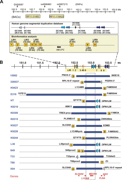
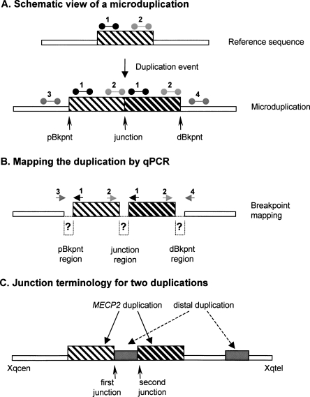
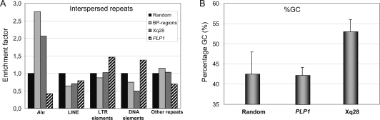
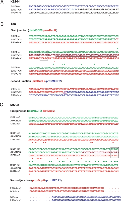
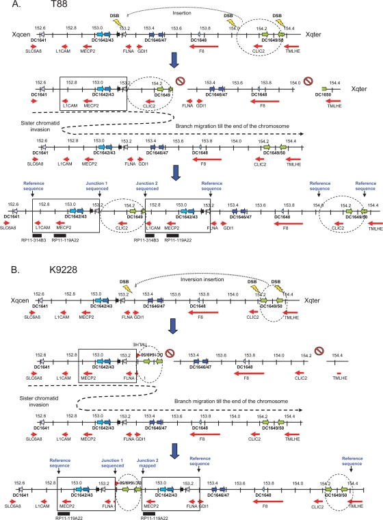
Recurrent submicroscopic genomic copy number changes are the result of nonallelic homologous recombination (NAHR). Nonrecurrent aberrations, however, can result from different nonexclusive recombination-repair mechanisms. We previously described small microduplications at Xq28 containing MECP2 in four male patients with a severe neurological phenotype. Here, we report on the fine-mapping and breakpoint analysis of 16 unique microduplications. The size of the overlapping copy number changes varies between 0.3 and 2.3 Mb, and FISH analysis on three patients demonstrated a tandem orientation. Although eight of the 32 breakpoint regions coincide with low-copy repeats, none of the duplications are the result of NAHR. Bioinformatics analysis of the breakpoint regions demonstrated a 2.5-fold higher frequency of Alu interspersed repeats as compared with control regions, as well as a very high GC content (53%). Unexpectedly, we obtained the junction in only one patient by long-range PCR, which revealed nonhomologous end joining as the mechanism. Breakpoint analysis in two other patients by inverse PCR and subsequent array comparative genomic hybridization analysis demonstrated the presence of a second duplicated region more telomeric at Xq28, of which one copy was inserted in between the duplicated MECP2 regions. These data suggest a two-step mechanism in which part of Xq28 is first inserted near the MECP2 locus, followed by breakage-induced replication with strand invasion of the normal sister chromatid. Our results indicate that the mechanism by which copy number changes occur in regions with a complex genomic architecture can yield complex rearrangements.


Figures

Figure 1.
Figure 1.
Bioinformatics analysis of the breakpoint regions and mapping of the duplications in the 16 patients. (A) Schematic overview of the repeat structures that coincide with duplication breakpoints in a 1-Mb region flanking MECP2 (152.55–153.55 Mb). Position and size of interchromosomal (DC1640 and DC1641) and intrachromosomal (DC1642, DC1643, DC1644, DC1645, DC1646, and DC1647) segmental duplications are depicted as reported in the Human genome segmental duplication database. In the Bioinformatics analysis box, LIR1 to LIR9 represent the breakpoint regions enriched for interspersed repeats with their lengths in brackets. The position of the BAC clones RP11-314B3 (152.63 Mb) and RP11-119A22 (152.90 Mb) used for FISH analysis is shown, as well as the position of DXS8087 (152.54 Mb) and the SNPs rs4898460 (152.83 Mb) and rs5987215 (153.05 Mb), used to investigate the origin of the duplications. (B) Duplication breakpoint mapping in the 16 patients (indicated on the left). Horizontal blue bars represent duplicated regions at Xq28. Size and location of each duplication were determined by iterative rounds of qPCR. Positions and sizes of all duplications were different. The colocalization of the breakpoint regions with the repeat structures, if present, is indicated with the name of the last duplicated qPCR primer set mentioned next to it (primer sequences can be found in Supplemental Table S1). The genes for which mutations are known to result in XLMR that also map in this region are shown at the bottom. The commonly duplicated region is boxed and includes only one XLMR gene, MECP2. Except for E316, all duplications lie within a 1.2-Mb region.
Figure 2.
Figure 2.
Terminology used for Xq28 duplication breakpoint mapping and cloning. (A) Schematic of a duplicated segment in the genome. The reference genomic sequence (top), with the region that is duplicated in the lower scheme, is indicated as a thin-striped box. qPCR primer pairs used for duplication size mapping are shown above this box at the proximal (1) and distal (2) side of this region. When duplicated in tandem (bottom), the proximal position where the duplication starts is called the proximal breakpoint (pBkpnt). Similarly, the most distal position where the duplication stops is called the distal breakpoint (dBkpnt). The position where the first copy is followed by the second one is the junction. In qPCR analysis, primer pairs 1 as well as 2 will yield a double dose while the primer pairs for 3 and 4 will give a single copy (relative to the reference genome). (B) For mapping purposes the pBkpnt and dBkpnt regions lie in between the last “normal” forward (3) and the first “duplicated” reverse primer (1), and the last “duplicated” forward (2) and the first “normal” reverse primer (4), respectively. To amplify over the junction, the first “duplicated” forward primer of the proximal breakpoint (1) was combined with the last “duplicated” reverse primer of the distal breakpoint (2). The region in between both primer pairs is called the junction region. Normal sequences are represented by thin open boxes; duplicated sequence by thin-striped and thick-striped boxes, which represent the first and second copy, respectively. (C) Junction terminology for the two duplications.
Figure 3.
Figure 3.
Interspersed repeat analysis of the MECP2 region. (A) Comparison of several regions on the X chromosome for potential differential enrichment in an interspersed repeat class (Alu, LINE, LTR elements, DNA elements, other repeats). (Random) Random DNA sequence on chromosome X (total length: 1140 kb); (BP-regions) sum of all breakpoint regions (total length of sequence is 113 kb); (Xq28) Xq28 region ranging from 152.5 to 153.6 Mb chopped into 100-kb fragments (total length, 1133 kb); (PLP1) BAC/PAC clones covering 231 kb of the PLP1 region. The Alu repeat content of all breakpoint regions (BP-regions) was significantly enriched compared with other analyzed sequences on the X chromosome. The other classes of interspersed repeats did not show significant differences. (B) GC content of the different genomic regions.
Figure 4.
Figure 4.
Alignment of the sequenced junctions in patients K9244, T88, and K9228 with the reference genome sequence. Proximal and distal reference sequences are printed in normal font and italics, respectively, and colored differently. All junctions are underlined and in the merge color of proximal and distal reference sequence. Asterisks represent sequence mismatches. (A) K9244: Proximal (top) and distal (bottom) sequence were aligned against the junction sequence (middle), demonstrating the tandem orientation of the duplication. A microhomology of 5 bp (CCTCT) is found at the junction between the distal and proximal sequences, characteristic for NHEJ. (B) T88: Proximal (top) and distal (bottom) sequence of the first and second junction were aligned against the respective junction sequences (middle). The first junction occurs within an AluY present at both dBkpnt and pBkpnt of the MECP2 and the distal duplication, respectively. There is a perfect match of 39 bp. The Alu pentanucleotide motif (CCAGC) is boxed. These data are consistent with homology-assisted NHEJ. A microhomology of 2 bp (GT) is found at the second junction, in agreement with a collapse of the BIR-fork at this locus and search for microhomology on the sister chromatid. (C) K9228: Alignment of the reference sequence against the respective junction sequences (middle). The first junction occurs within an AluJo at the dBkpnt of the MECP2 duplication and an AluSg at the dBkpnt of the distal duplication. The homology-assisted NHEJ at this junction results in the insertion of the distal duplication in between the MECP2 duplications in an inverted orientation. The 23-bp junction terminates in the Alu pentanucleotide motif (boxed). Boundaries of the PCR-fragment containing the second junction were sequenced and revealed sequences of the pBkpnt of the MECP2 duplication at one side (PCR-forw) while at the other end (PCR-rev) sequences of LCR DC1649 were obtained, coinciding with the pBkpnt of the distal duplication. Dots represent predicted reference sequence.
Figure 5.
Figure 5.
Proposed model for the recombination/repair mechanism originating both duplications in patients T88 and K9228. Illustration of the Xq28 region from 152.45 Mb to 154.65 Mb. The LCRs are represented by differently colored block arrows and genes by red block arrows. The MECP2 duplication is boxed while the additional distal duplication is circled (broken line). (A) Patient T88. Several DSBs occur simultaneously resulting in an excised fragment that is attached through Alu homology, in a direct orientation to the DSB generated proximally from FLNA. Repair of the remaining DSB is performed by BIR in which the BIR fork invaded the DNA duplex of the sister chromatid at a proximal location with microhomology of a few bp, and DNA synthesis proceeded to the telomere. (B) Patient K9228. Similar recombination/repair event in K9228 with minor differences. Several DSBs occur simultaneously resulting in an excised fragment at LCRs DC1649 and DC1650. This fragment is then rejoined, via Alu homology, in an inverted way to the DSB generated at GDI1. Then, the BIR fork invaded the sister chromatid at a more proximal locus, and DNA synthesis continued to the end of the X chromosome.

Similar articles

Cited by

References

    1. Aradhya S., Woffendin H., Bonnen P., Heiss N.S., Yamagata T., Esposito T., Bardaro T., Poustka A., D'Urso M., Kenwrick S., Woffendin H., Bonnen P., Heiss N.S., Yamagata T., Esposito T., Bardaro T., Poustka A., D'Urso M., Kenwrick S., Bonnen P., Heiss N.S., Yamagata T., Esposito T., Bardaro T., Poustka A., D'Urso M., Kenwrick S., Heiss N.S., Yamagata T., Esposito T., Bardaro T., Poustka A., D'Urso M., Kenwrick S., Yamagata T., Esposito T., Bardaro T., Poustka A., D'Urso M., Kenwrick S., Esposito T., Bardaro T., Poustka A., D'Urso M., Kenwrick S., Bardaro T., Poustka A., D'Urso M., Kenwrick S., Poustka A., D'Urso M., Kenwrick S., D'Urso M., Kenwrick S., Kenwrick S., et al. Physical and genetic characterization reveals a pseudogene, an evolutionary junction, and unstable loci in distal Xq28. Genomics. 2002;79:31–40. - PubMed
    1. Bagnall R.D., Ayres K.L., Green P.M., Giannelli F., Ayres K.L., Green P.M., Giannelli F., Green P.M., Giannelli F., Giannelli F. Gene conversion and evolution of Xq28 duplicons involved in recurring inversions causing severe hemophilia A. Genome Res. 2005;15:214–223. - PMC - PubMed
    1. Balciuniene J., Feng N., Iyadurai K., Hirsch B., Charnas L., Bill B.R., Easterday M.C., Staaf J., Oseth L., Czapansky-Beilman D., Feng N., Iyadurai K., Hirsch B., Charnas L., Bill B.R., Easterday M.C., Staaf J., Oseth L., Czapansky-Beilman D., Iyadurai K., Hirsch B., Charnas L., Bill B.R., Easterday M.C., Staaf J., Oseth L., Czapansky-Beilman D., Hirsch B., Charnas L., Bill B.R., Easterday M.C., Staaf J., Oseth L., Czapansky-Beilman D., Charnas L., Bill B.R., Easterday M.C., Staaf J., Oseth L., Czapansky-Beilman D., Bill B.R., Easterday M.C., Staaf J., Oseth L., Czapansky-Beilman D., Easterday M.C., Staaf J., Oseth L., Czapansky-Beilman D., Staaf J., Oseth L., Czapansky-Beilman D., Oseth L., Czapansky-Beilman D., Czapansky-Beilman D., et al. Recurrent 10q22-q23 deletions: A genomic disorder on 10q associated with cognitive and behavioral abnormalities. Am. J. Hum. Genet. 2007;80:938–947. - PMC - PubMed
    1. Bauters M., Van Esch H., Marynen P., Froyen G., Van Esch H., Marynen P., Froyen G., Marynen P., Froyen G., Froyen G. X chromosome array-CGH for the identification of novel X-linked mental retardation genes. Eur. J. Med. Genet. 2005;48:263–275. - PubMed
    1. Deeb S.S., Kohl S., Kohl S. Genetics of color vision deficiencies. Dev. Ophthalmol. 2003;37:170–187. - PubMed

Publication types

Substances