. 2008 Apr 2;28(14):3631-43.
doi: 10.1523/JNEUROSCI.0453-08.2008.

Cyclin-dependent kinase 5 phosphorylation of human septin SEPT5 (hCDCrel-1) modulates exocytosis

Affiliations

Cyclin-dependent kinase 5 phosphorylation of human septin SEPT5 (hCDCrel-1) modulates exocytosis

Niranjana D Amin et al. J Neurosci. .

Abstract

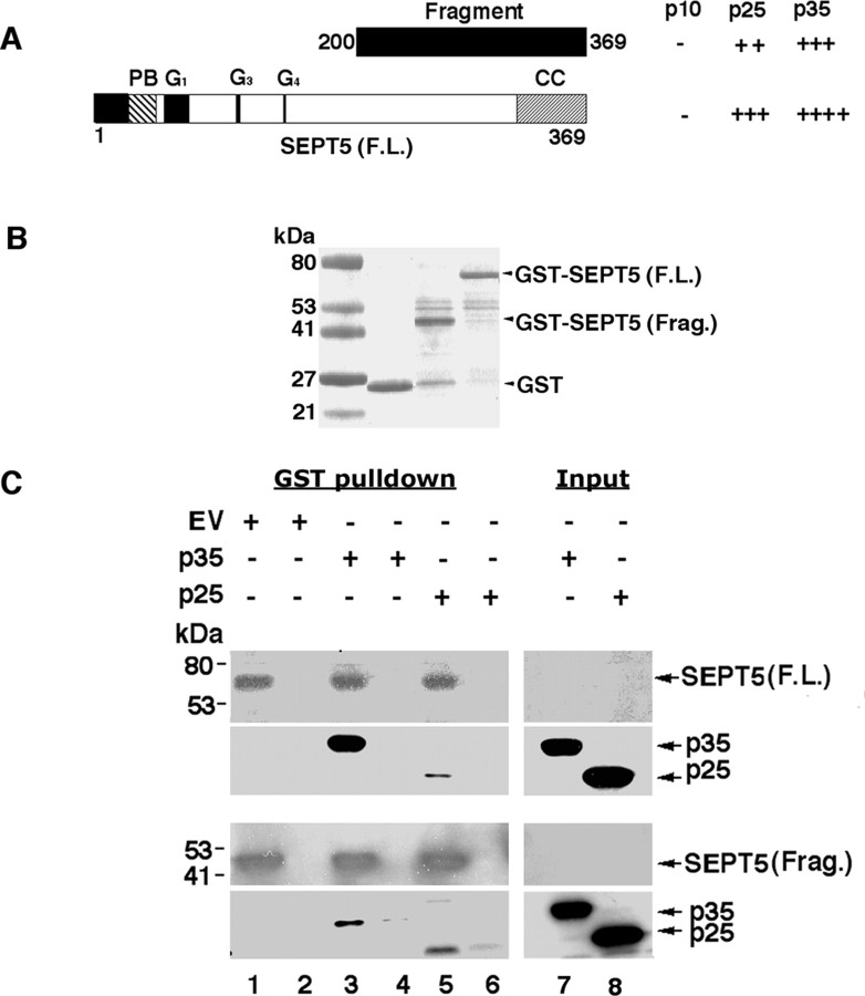
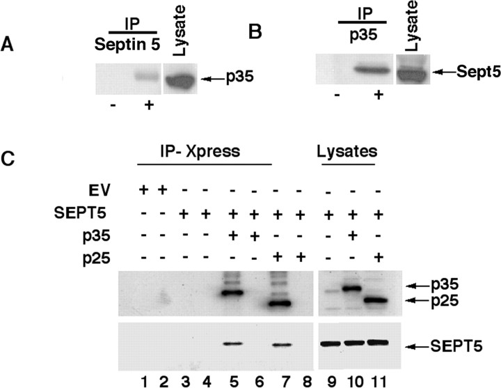
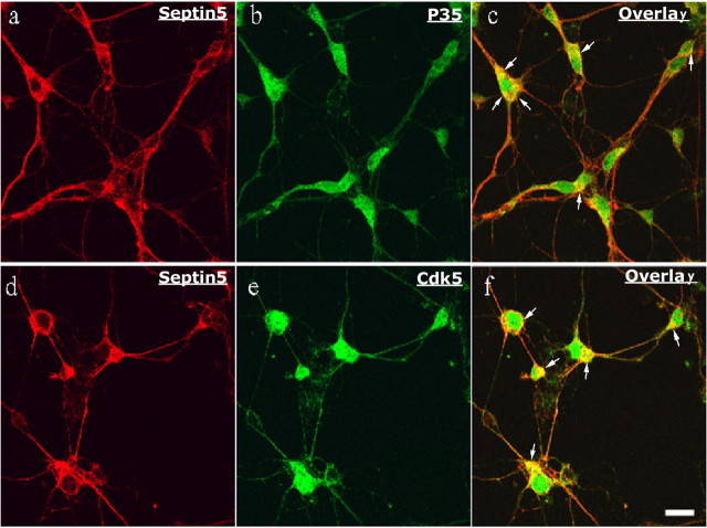
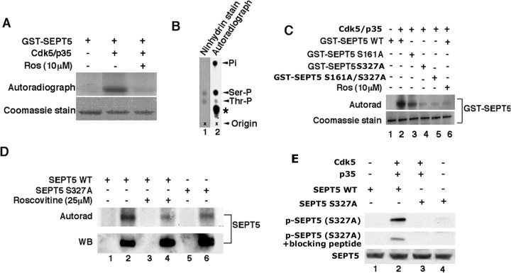
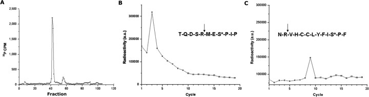
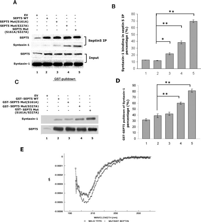
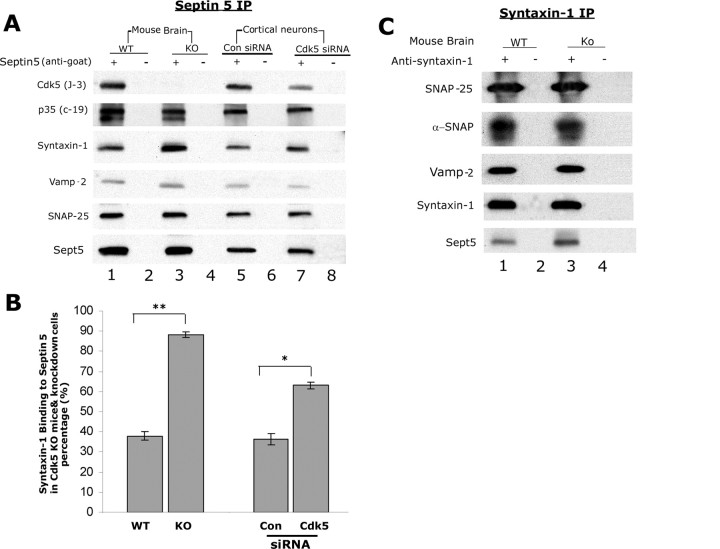
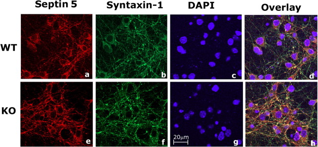
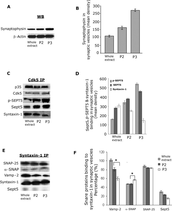
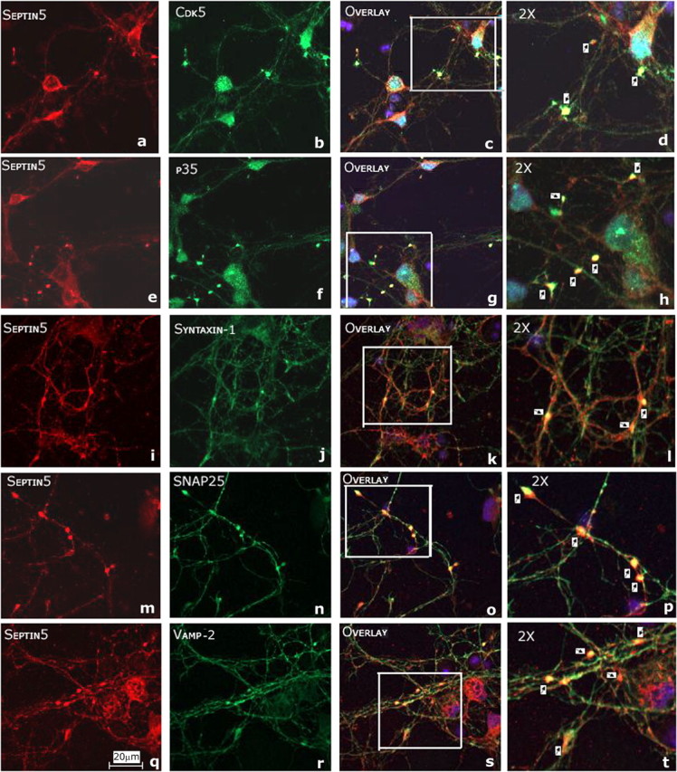
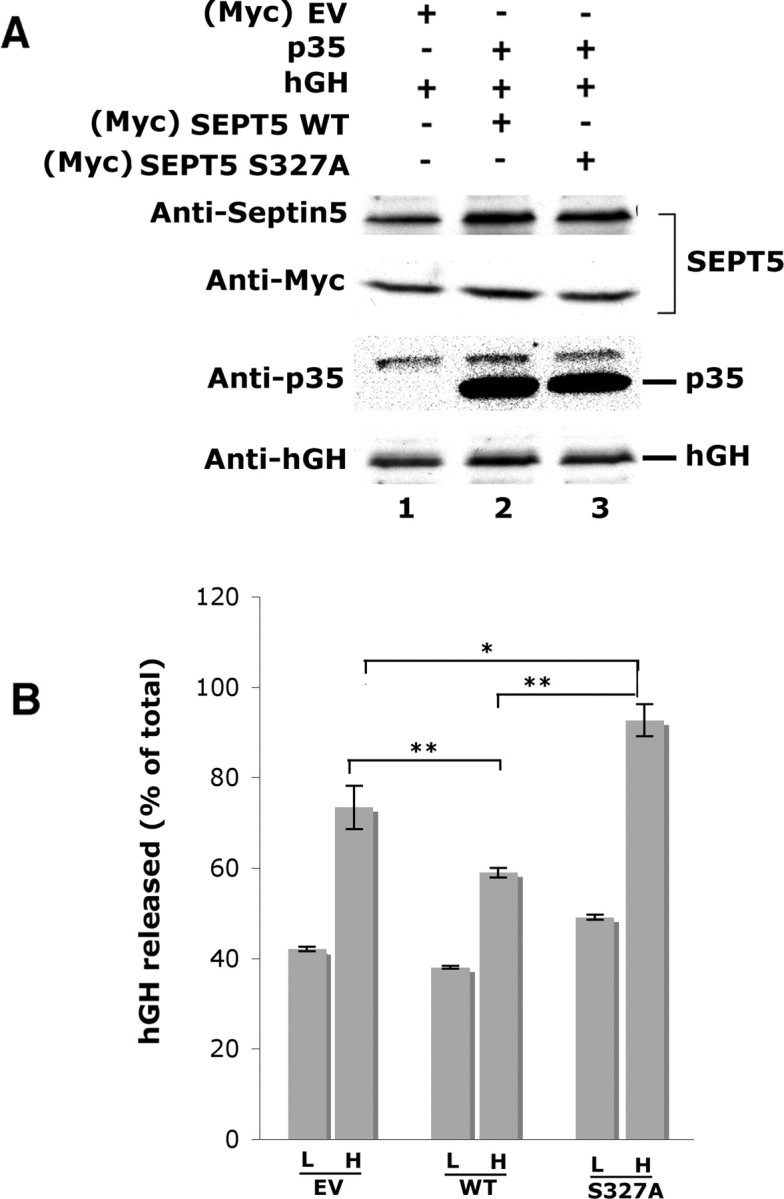
Cyclin-dependent kinase 5 (Cdk5) is predominantly expressed in the nervous system, where it is involved in neuronal migration, synaptic transmission, and survival. The role of Cdk5 in synaptic transmission is mediated by regulating the cellular functions of presynaptic proteins such as synapsin, Munc18, and dynamin 1. Its multifunctional role at the synapse is complex and probably involves other novel substrates. To explore this possibility, we used a yeast two-hybrid screen of a human cDNA library with p35 as bait and isolated human septin 5 (SEPT5), known also as hCDCrel-1, as an interacting clone. Here we report that p35 associates with SEPT5 in GST (glutathione S-transferase)-pull-down and coimmunoprecipitation assays. We confirmed that Cdk5/p35 phosphorylates SEPT5 in vitro and in vivo and identified S327 of SEPT5 as a major phosphorylation site. A serine (S)-to-alanine (A) 327 mutant of SEPT5 bound syntaxin more efficiently than SEPT5 wild type. Additionally, coimmunoprecipitation from synaptic vesicle fractions and Cdk5 wild-type and knock-out lysates showed that phosphorylation of septin 5 by Cdk5/p35 decreases its binding to syntaxin-1. Moreover, mutant nonphosphorylated SEPT5 potentiated regulated exocytosis more than the wild type when each was expressed in PC12 cells. These data suggest that Cdk5 phosphorylation of human septin SEPT5 at S327 plays a role in modulating exocytotic secretion.


Figures

Figure 1.
Figure 1.
SEPT5 interacts with p35 in yeast two-hybrid screen and GST-pull-down assays. A, SEPT5, a protein of 369 amino acid residues, contains a polybasic region (PB), G1, G3, and G4 GTP-binding sites in the GBD (GTP-binding domain), and a coiled-coiled domain (CC) at the C terminus. The SEPT5 fragment that interacted with p25 in the yeast two-hybrid screen is shown as a black box above the SEPT5 F.L. comprising amino acid residues 200–369. Yeast cotransformed with p35 or with different domains of p35 (p10, p25) and SEPT5 full length as well as the SEPT5 fragment (in activation domain vector pGADT7) were grown in media lacking tryptophan, leucine, and histidine containing 25 mm 3-aminotriazole. β-Galactosidase assays were performed to test interactions and were judged by comparing the color generation of the positive control. Interaction strength is indicated as follows: ++++, very strong; +++, medium interaction; ++, weak interaction. B, Coomassie blue stain shows expression of GST, GST-SEPT5 F.L., and GST-SEPT5 Frag. C, SEPT5 interacts with p35 and p25 in vitro in GST-pull-down assays. GST, GST-SEPT5 F.L., and GST-SEPT5 Frag. proteins were used as bait with CHO cell lysates transfected with empty vector (EV) (lanes 1, 2), p35 (lanes 3, 4), and p25 (lanes 5, 6). Samples were separated by SDS-PAGE and then probed with monoclonal septin 5 (SP18) antibody (first and third panels) and polyclonal p35 (C19) antibody (second and forth panels). Lanes 7 and 8 show the CHO cell lysates transfected with p35 and p25, respectively, as control. Lanes 2, 4, and 6 show the pull-down assay with only GST protein.
Figure 2.
Figure 2.
Septin 5 coimmunoprecipitates p35 from rat brain and transfected CHO cell lysates. A, Postnatal day 8 (PN8) rat brain lysate was immunoprecipitated with mouse monoclonal septin 5 antibody, and p35 was immunodetected in Western blots with polyclonal p35 antibody. B, Rat brain lysate as above was immunoprecipitated with monoclonal p35 antibody, and the expression of septin 5 was confirmed using polyclonal septin 5 antibody. The presence of p35 and septin 5 in rat brain lysate is shown using their respective antibodies as a positive control. The + and − signs are presence and absence of immunoprecipitating antibody, respectively. C, CHO cell lysates transfected with empty vector (EV) (lanes 1, 2), SEPT5 alone (lanes 3, 4), p35/SEPT5 (lanes 5, 6) and p25/SEPT5 (lanes 7, 8) were immunoprecipitated with monoclonal anti-Xpress antibody. Western blots show the expression of p35 and p25 with polyclonal p35 antibody (top), and SEPT5 was detected using polyclonal septin 5 antibody (bottom). Lanes 2, 4, 6, and 8 mark the absence of immunoprecipitating antibody. Lanes 9, 10, and 11 show the expression of p35, p25, and SEPT5 in the whole-cell lysates, using their corresponding antibodies.
Figure 3.
Figure 3.
p35 and Cdk5 colocalize with septin 5 in rat cortical neurons. a–f, 7-DIC E-18 rat cortical neurons were immunostained to detect endogenous Sept5 with p35 (a–c) and Cdk5 (d–f). Sept5 was immunostained using monoclonal septin 5 antibody and visualized with Texas Red (a, d), p35 was immunostained using polyclonal p35 antibody (b), and Cdk5 was immunostained using Cdk5 (C8) antibody (e), and signals were visualized using Oregon Green. The overlay shows the colocalization in yellow (c, f). Scale bar, 20 μm. Arrows point to colocalization sites.
Figure 4.
Figure 4.
Phosphorylation of SEPT5 by Cdk5/p35. GST-SEPT5 protein was expressed and purified as described previously. Active Cdk5 immunoprecipitated from rat brain lysate using C8 antibody or purchased from Millipore were used for phosphorylation assays. A, SEPT5 is phosphorylated by Cdk5/p35. Active Cdk5 immunoprecipitated from rat brain lysates phosphorylated GST-SEPT5 and was inhibited by 10 μm roscovitine, a specific Cdk5 inhibitor. B, Phosphoamino acid analysis of SEPT5 phosphorylated by active Cdk5/p35. The autoradiogram shows most incorporation of 32P into S residues (lane 2). The asterisk in lane 2 denotes the band of undigested material. Pi refers to inorganic phosphate. Ninhydrin staining of the unlabeled S phosphate and threonine phosphate mixed with the 5.7N HCl hydrolysate of [γ-32P]ATP SEPT5 is also shown (lane 1). C, Serine 327 of SEPT5 is phosphorylated by Cdk5/p35. Kinase assays were performed with GST-SEPT5 WT (lane 2) and individually with GST-SEPT5 mutants (S161A, lane 3; S327/A, lane 4; S161A/S327A, lane 5) as substrates and active Cdk5/p35 as an enzyme. Bottom, Coomassie stain; top, the autoradiogram of the respective proteins. Lanes 1 and 6 show the phosphorylation of GST-SEPT5 WT without and with Cdk5/p35 plus roscovitine, respectively. D, Phosphorylation of SEPT5 by Cdk5/p35 in cortical neurons. Primary rat cortical neurons were transfected with SEPT5 WT and SEPT5 S327A mutant and metabolically labeled with [32P]-orthophosphate in the absence or presence of 25 μm roscovitine. SEPT5 was immunoprecipitated with polyclonal septin 5 antibody with equal amounts of cell lysates, and Western blots were prepared. Top, Autoradiograph; bottom, the corresponding immunoblot to show the SEPT5 expression level with monoclonal septin 5 antibody. The results in lanes 2, 4, and 6 show the IPs in the presence of septin 5 antibody, and lanes 1, 3, and 5 show the IPs in the absence of septin 5 antibody. SEPT5 WT phosphorylation is shown in lane 2. Lane 4 shows the SEPT5 WT phosphorylation in the presence of 25 μm roscovitine, and SEPT5 mutant (S327A) phosphorylation is shown in lane 6. This experiment is an example of three independent experiments. E, Phosphorylation of SEPT5 by Cdk5/p35 in HEK 293 cells. HEK 293 cells were transfected with empty vector (EV), SEPT5 WT, and SEPT5 mutant S327/A in the presence or absence of Cdk5/p35. The lysates were immunoprecipitated with monoclonal septin 5 antibody, and the blots were immunodetected with site-specific polyclonal Ser(P)327 antibody (top), Ser(P)327 antibody plus blocking peptide (middle), and polyclonal septin 5 antibody (bottom). The results of transfected HEK 293 cells are shown as SEPT5 WT alone (lane 1); SEPT5 WT plus Cdk5 and p35 (lane 2); SEPT5 mutant (S327A) plus Cdk5 and p35 (lane 3); and SEPT5 mutant (S327A) alone (lane 4).
Figure 5.
Figure 5.
Serine 327 is the principal site on SEPT5 that is phosphorylated by Cdk5/p35. GST-SEPT5 was phosphorylated in vitro by Cdk5/p35 with [γ-32P]ATP, subjected to SDS-PAGE, and transferred to a PVDF membrane. The radioactive band was removed and digested with trypsin. A, The tryptic fragments were run through HPLC and the radioactivity assayed in each fraction. The fraction 42, showing peak activity, and the minor fraction at 55–58 were collected and subjected to Edman degradation sequence analysis. Sequence mapping was based on a procedure (Guszczynski et al., 2006) in which sequence cycles are collected as spots on filter papers in a microtiter plate and assayed for radioactivity. B, In the third cycle, the 32P signal is derived from a serine 3 residue from the N-terminal end of the tryptic peptide. The insert sequence shows the tryptic digestion site (arrow), and note that the phosphorylated S (*) at the third residue from the N-terminal end is S327. C, At the ninth cycle where activity is significantly lower, the 32P signal marks a serine 9 residue from the N-terminal end of the tryptic peptide. The sequence in the insert, representing the only SEPT5 sequence satisfying this criterion, is shown with the tryptic digest site at the arrow, and the S* (S161), the ninth residue from the N-terminal end of the peptide.
Figure 6.
Figure 6.
Mutation of SEPT5 (S327A) enhances binding to syntaxin-1. A, SEPT5 WT and SEPT5 mutants (S161A, S327A, and S161A/S327A) bind with syntaxin-1. 3-DIC cortical neurons were transfected with wild-type SEPT5 or SEPT5 mutants, S161A, S327A, and the double mutant S161A/S327A. Twenty-four hours after transfection, SEPT5 was immunoprecipitated from equal amounts of lysate with a polyclonal septin 5 antibody, and Western blots were prepared. The presence of SEPT5 was confirmed using monoclonal SEPT5 antibody (first panel). Syntaxin-1 was immunodetected with monoclonal syntaxin-1 antibody (second panel). The third and fourth panels show the presence of SEPT5 and syntaxin-1 in the whole-cell lysates. Robust binding of syntaxin-1 is shown by the single mutant S327A and double mutant S161A/S327A in lanes 4 and 5 of the second panel. B, The bar graph shows the percentage of the syntaxin-1 binding to SEPT5 in the septin 5 IP. Data represent the mean of three separate experiments, and error bars represent the ±SEM. *p < 0.01, **p < 0.001, values indicate significant differences from WT. C, Nonphosphorylated SEPT5 mutants bind syntaxin-1 more effectively than the Cdk5/p35 phosphorylated wild type in GST-pull-down assays. Equal amounts of GST-SEPT5 WT or GST-SEPT5 mutant (S161A, S327A, and S161A/S327A) proteins on glutathione-Sepharose beads were phosphorylated in the presence of active Cdk5/p35 in the kinase reaction buffer with cold ATP and used as bait with rat brain cortical neuron lysates. Samples were run by SDS-PAGE and immunoblotted with syntaxin-1 antibody (top) and reprobed with monoclonal septin 5 antibody (bottom). The interaction of syntaxin-1 with phosphorylated SEPT5 WT or SEPT5 mutants (S161A, S327A, and S161A/S327A) is shown in lanes 2, 3, 4, and 5, respectively. Lane 1 shows the GST-pull-down with GST protein alone. D, The bar graph shows the percentage of the syntaxin-1 bound to SEPT5 in the GST-SEPT5 pull-down. Data represent the mean of three separate experiments, and error bars represent the ±SEM. **p < 0.001 indicates significant difference. The bar graph shows mean density measurement of the syntaxin-1 binding in GST-SEPT5 pull-down. Results are expressed as mean ± SEM of three separate experiments. E, CD spectroscopy of the wild-type and S327A SEPT5 fusion proteins. The spectra were scanned from 190 to 250 nm and represented as mean residue ellipticity. All the spectra are averages of four accumulations.
Figure 7.
Figure 7.
Endogenous phosphorylation of septin 5 by Cdk5/p35 reduced binding to syntaxin-1. A, Lysates of Cdk5 WT and Cdk5 KO embryonic (E16) mouse brains and lysates from siRNA transfected (E18) rat cortical neurons were immunoprecipitated with polyclonal septin 5 antibody, and precipitates were analyzed by SDS-PAGE and Western blotting. Blots were then developed with antibodies specific to the proteins indicated to the left. Endogenous syntaxin binding to Sept5 is reduced by Sept5 phosphorylation in both cases. Exposures for SNAP-25 and Vamp-2 were considerably longer than for septin 5 and syntaxin-1, suggesting that the former SNARE proteins bound to septin 5 indirectly via direct association with syntaxin-1. B, The bar graph shows the percentage of the syntaxin-1 binding to Sept5 in septin 5 IP. Data represent the mean of three separate experiments, and error bars represent the ±SEM. *p < 0.01, significantly different from control; **p < 0.001, significantly different from WT. C, Cdk5 WT and KO mouse brain lysates were each immunoprecipitated with monoclonal syntaxin-1 antibody, and the blots were analyzed with antibodies specific to the proteins indicated to the left. Here, in contrast to A, exposure times were the same for all proteins.
Figure 8.
Figure 8.
Septin 5 colocalizes with syntaxin-1 in putative synapses of Cdk5 WT and Cdk5 KO mouse cortical neurons. a–h, 10-DIC E15 mouse cortical neurons were immunostained to detect endogenous Sept5 with syntaxin-1 in Cdk5 WT (a–d), and Cdk5 KO (e–h). Sept5 was immunostained using polyclonal septin 5 antibody and visualized with Texas Red (a, e); syntaxin-1 was immunostained using monoclonal syntaxin-1 antibody (b, f) and was visualized using Oregon Green. The overlay shows the colocalization in yellow (d, h). Scale bar, 20 μm.
Figure 9.
Figure 9.
Cdk5/p35 phosphorylation of septin 5 in a rat brain synaptic vesicle fraction decreases syntaxin-1 binding. Cortices from 10 adult rat brains were isolated, homogenized, and processed for an enriched synaptic vesicle fraction using a Sigma kit (SV0100) based on a procedure published by Roz et al. (2002). Three fractions were used for immunoprecipitation studies, the initial whole extract, a P2 crude synaptosome fraction, and P3, purified synaptic vesicles (see Materials and Methods). The course of the purification was monitored by Western blots for synaptophysin, a key synaptic vesicle protein. A, Western blots for synaptophysin in each fraction showed a progressive increase in expression as seen in the histogram in B. Actin was used as a measure of protein loading. C, A Cdk5 monoclonal antibody (J3) was used to immunoprecipitate the three fractions, and the immunoprecipitates were probed with polyclonal antibodies to p35, Cdk5, phospho-SEPT5 [Ser(P)327], septin 5, and syntaxin-1 (H-221). As Sept5 phosphorylation increased in the vesicle enriched fractions, syntaxin-1 binding to the Cdk5 complex declined significantly. D, A histogram of a quantitative analysis of three experiments showing the increasing levels of phospho-SEPT5 and Sept5 expression in the more purified fractions, P2 and P3, and the accompanying decline in syntaxin-1 expression. E, Using a monoclonal syntaxin-1 antibody (HPC-1), each fraction was immunoprecipitated and probed with rabbit polyclonal antibodies to syntaxin-1, SNAP-25, and Vamp-2 and a polyclonal antibody to septin 5. F, A histogram analysis of percentage binding to syntaxin-1 in three experiments showed in the P3 fraction a weak binding of Sept5 to syntaxin-1, a significant increase in binding of α-SNAP to syntaxin-1, and a decreased binding of Vamp-2 to syntaxin-1 in P3 (*p < 0.01). SNAP-25 binding to syntaxin-1 remained relatively unchanged during the purification.
Figure 10.
Figure 10.
Septin 5 colocalization with SNARE proteins in putative synaptic sites in long-term (2.5 weeks) cultures of rat cortical neurons. a–t, E18 rat cortical neurons were immunostained to detect endogenous Sept5 with Cdk5 (a–d), p35 (e–h), syntaxin-1 (i–l), SNAP-25 (m–p), and Vamp-2 (q–t). Sept5 was immunostained using monoclonal septin 5 antibody and visualized with Texas Red (a, e, i, m, q); Cdk5, p35, syntaxin-1, SNAP-25, and Vamp-2 were immunostained using their respective polyclonal antibodies (b, f, j, n, r) and were visualized using Oregon Green. The synaptic colocalizations are shown in yellow in the magnified inserts in the overlays (d, h, l, p, t, arrows). Scale bar, 20 μm.
Figure 11.
Figure 11.
Secretion of human growth hormone in PC12 cells is enhanced by overexpression of the nonphosphorylated SEPT5 (S327A) mutant. A, Expression constructs encoding hGH were cotransfected into PC12 cells with empty vector (EV), SEPT5 WT, and SEPT5 mutant (S327A) along with p35. Lysates of transfected PC12 cells were run on the gel and immunodetected using respective antibodies. The first panel shows the expression of total septin 5 in the lysates, whereas the second panel shows the transfected SEPT5 as immunodetected with Myc antibody. The presence of p35 was probed with polyclonal p35 antibody and hGH was detected using mouse monoclonal anti hGH antibody. B, Culture media with 2.2 mm Ca2+ in the presence of low (L) and high (H) K+ were collected, and the hGH secreted was assayed by hGH-linked immunosorbent assay. hGH released is calculated as a percentage of total with EV, with SEPT5 WT, and SEPT5 S327A. Each value shown represents the mean ± SEM from multiple (n = 9) experiments. Statistically significant differences are depicted (*p < 0.05; **p < 0.01; Student's t test).

References

    1. Amin ND, Albers W, Pant HC. Cyclin-dependent kinase 5 (cdk5) activation requires interaction with three domains of p35. J Neurosci Res. 2002;67:354–362. - PubMed
    1. Ballif BA, Villen J, Beausoleil SA, Schwartz D, Gygi SP. Phosphoproteomic analysis of the developing mouse brain. Mol Cell Proteomics. 2004;3:1093–1101. - PubMed
    1. Beites CL, Xie H, Bowser R, Trimble WS. The septin CDCrel-1 binds syntaxin and inhibits exocytosis. Nat Neurosci. 1999;2:434–439. - PubMed
    1. Beites CL, Campbell KA, Trimble WS. The septin Sept5/CDCrel-1 competes with alpha-SNAP for binding to the SNARE complex. Biochem J. 2005;385:347–353. - PMC - PubMed
    1. Chae T, Kwon YT, Bronson R, Dikkes P, Li E, Tsai LH. Mice lacking p35, a neuronal specific activator of Cdk5, display cortical lamination defects, seizures, and adult lethality. Neuron. 1997;18:29–42. - PubMed

Publication types

MeSH terms