. 2008 Apr 2;28(14):3668-82.
doi: 10.1523/JNEUROSCI.5553-07.2008.

Presynaptic calcium channel localization and calcium-dependent synaptic vesicle exocytosis regulated by the Fuseless protein

Affiliations

Presynaptic calcium channel localization and calcium-dependent synaptic vesicle exocytosis regulated by the Fuseless protein

A Ashleigh Long et al. J Neurosci. .

Abstract

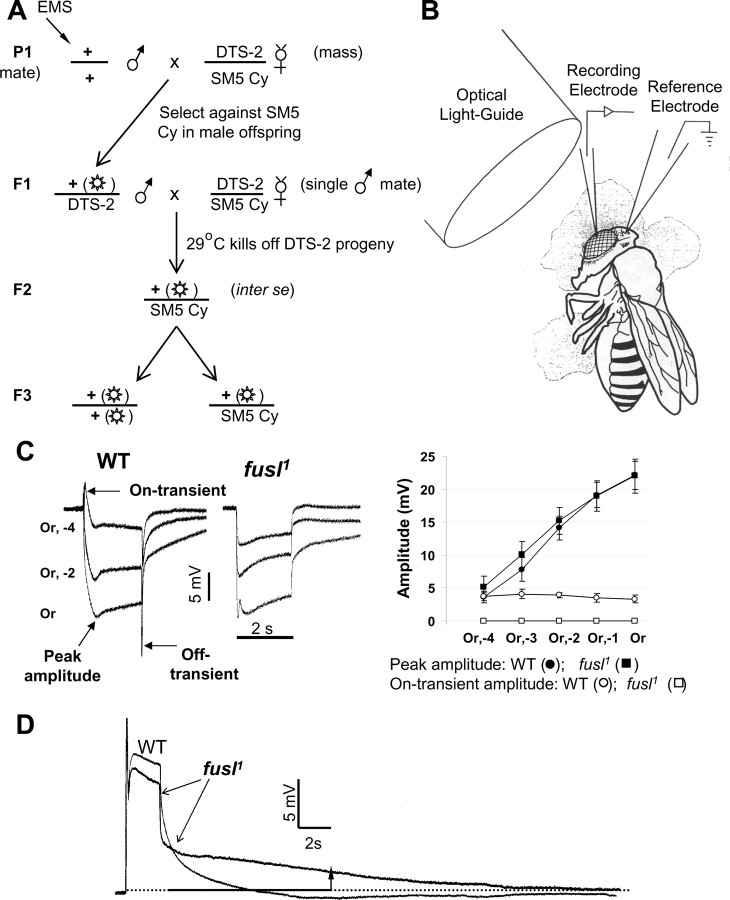
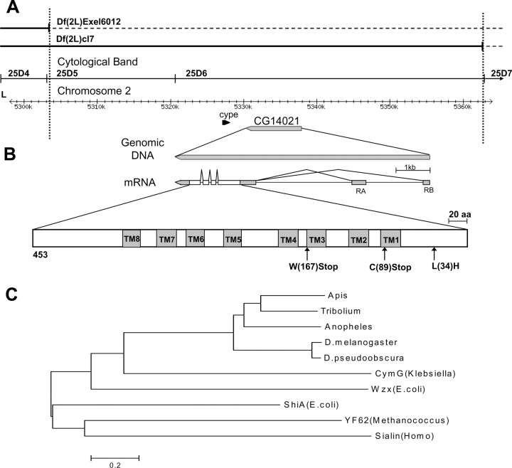
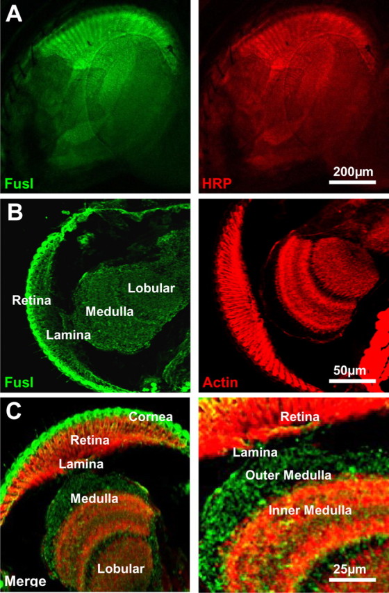
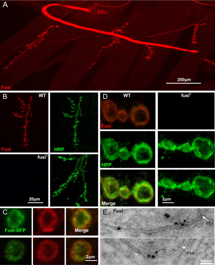
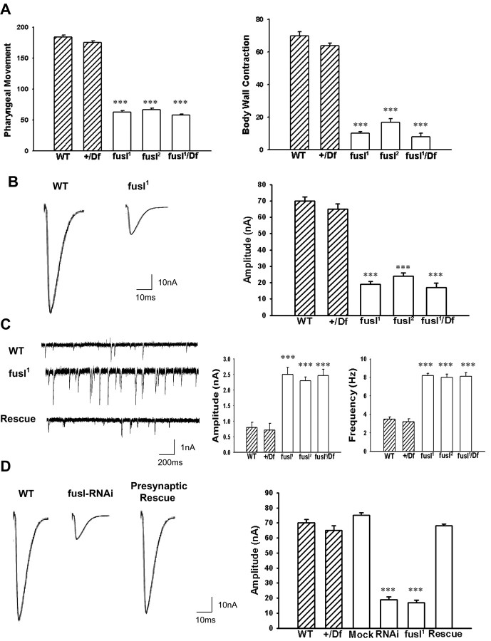
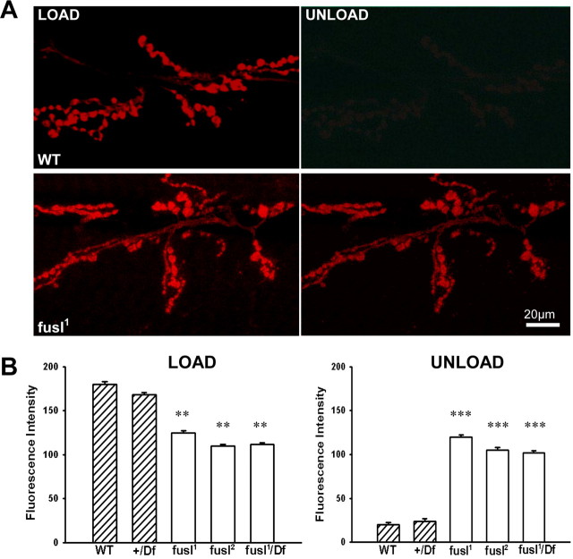
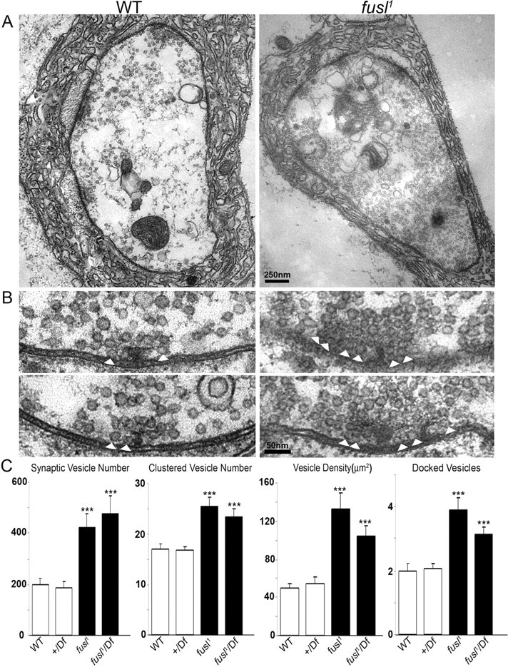
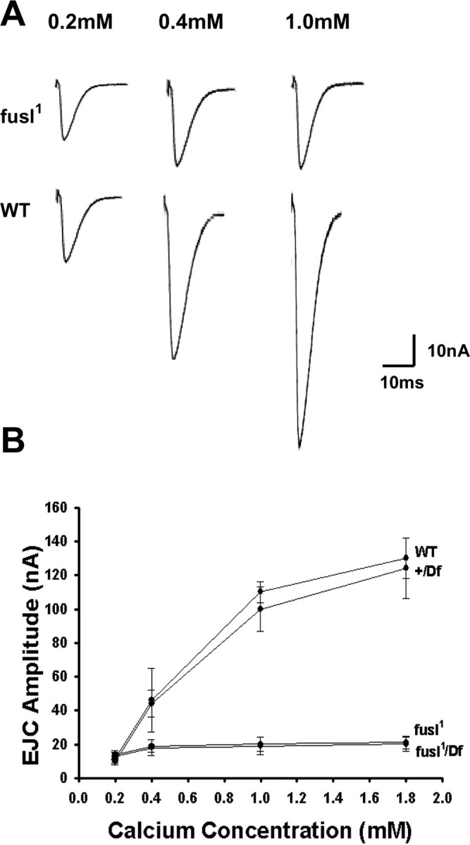
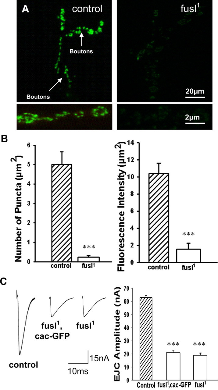
A systematic forward genetic Drosophila screen for electroretinogram mutants lacking synaptic transients identified the fuseless (fusl) gene, which encodes a predicted eight-pass transmembrane protein in the presynaptic membrane. Null fusl mutants display >75% reduction in evoked synaptic transmission but, conversely, an approximately threefold increase in the frequency and amplitude of spontaneous synaptic vesicle fusion events. These neurotransmission defects are rescued by a wild-type fusl transgene targeted only to the presynaptic cell, demonstrating a strictly presynaptic requirement for Fusl function. Defects in FM dye turnover at the synapse show a severely impaired exo-endo synaptic vesicle cycling pool. Consistently, ultrastructural analyses reveal accumulated vesicles arrested in clustered and docked pools at presynaptic active zones. In the absence of Fusl, calcium-dependent neurotransmitter release is dramatically compromised and there is little enhancement of synaptic efficacy with elevated external Ca(2+) concentrations. These defects are causally linked with severe loss of the Cacophony voltage-gated Ca(2+) channels, which fail to localize normally at presynaptic active zone domains in the absence of Fusl. These data indicate that Fusl regulates assembly of the presynaptic active zone Ca(2+) channel domains required for efficient coupling of the Ca(2+) influx and synaptic vesicle exocytosis during neurotransmission.


Figures

Figure 1.
Figure 1.
Phototransduction mutagenesis identifies a novel mutant specifically defective in synaptic transmission. A, Second chromosome mutagenesis scheme. EMS mutagenized males were single-mated in a second generation cross to track each mutant line independently. The DTS-2 mutation allowed only animals carrying the mutagenized second chromosome over balancer to survive among F2 offspring. Crossing inter SE produced offspring (F3) that are either homozygous or heterozygous for the mutagenized second chromosome. These animals were screened by ERG recording. B, Diagram of ERG recording. The fly was mounted in low-melting-point wax, with a recording electrode in the cornea and a reference electrode in head. The light stimulus (300 W halogen lamp) was led to eye using an optical light guide. C, The fusl mutants show a specific loss of ERG synaptic transients. Representative ERG traces (left) elicited from WT and fusl using 2 s orange light stimuli. Responses at three different light intensities are shown superimposed. Synaptic on- and off-transients are absent in fusl mutants. Right, Peak ERG and synaptic on-transient amplitudes (as defined in the left image) plotted against stimulus intensity for WT (N = 8) and fusl (N = 9). Error bars indicate SEM. D, Representative photoreceptor intracellular recordings from WT and fusl, shown superimposed. The receptor potentials have indistinguishable amplitudes (WT, 23.8 ± 2.7 mV; fusl, 22.5 ± 2.1 mV) and time (N = 7). However, the fusl response is slowly decaying: WT has completely returned to baseline [9.8 ± 0.9 s (N = 7) after stimulus termination], whereas the mutant response still displays a 2.0 ± 0.8 mV (N = 7) depolarization (arrow).
Figure 2.
Figure 2.
Maps of the fusl genomic region, gene, and gene product. A, The mutant genomic region. The fusl mutation is uncovered by the Df(2L)Exel6012 deficiency (top line), but complemented by Df(2L)cl7 (second line). The third line indicates cytogenetic boundaries and the fourth line shows the nucleotide coordinates. B, The CG14021 gene and its gene products. The gene generates two transcripts (RA, RB) by alternative splicing that differ in the location of the noncoding first exon but contain identical coding sequence. Untranslated regions are shown in gray. The predicted protein is 453 aa and contains eight predicted transmembrane domains. Mutations found in the coding regions of CG14021 in three characterized fusl alleles are shown at the bottom. C, Related proteins from multiple species were used to construct the phylogenetic tree. Protein sequences are identified from top to bottom by species, protein name, function (if known), and accession numbers, as follows: Apis, Apis mellifera (XM_623675.2); Tribolium, Tribolium castaneum (XM_968828.1); Anoph, Anopheles gambiae (XM_317156.2); D. mel., Drosophila melanogaster CG14021 (NM_135079.4); D. pseudo, Drosophila pseudoobscura (XM_001356784.1); Cym G, Klebsiella oxytoca, cyclodextrin transport (Q48397); Wzx, Escherichia coli, transbilayer movement of a trisaccharide-lipid intermediate (P27834); Shi A, Escherichia coli, shikimate transporter (P76350); YF62, Methanococcus jannaschii, putative membrane protein (Q58957); Sialin, Homo sapiens, transport of sialic acid in lysosomes (Q9UGH0).
Figure 3.
Figure 3.
Fusl protein expression in the visual system. A, Whole-mount preparation of the adult Drosophila eye, colabeled with anti-Fuseless (green; left) and anti-HRP (red; right), a neuronal plasma membrane marker. Scale bar, 200 μm. B, C, Cryosections of the adult visual system colabeled with anti-Fusl (green; left) and actin phalloidin (red; right). The sections shows retina, lamina, and medulla, as indicated. Scale bars: B, C, left, 50 μm; C, right, 25 μm.
Figure 4.
Figure 4.
Fusl protein localizes in the presynaptic plasma membrane. A, Very low magnification image of the neuromusculature. Anti-Fusl immunoreactivity observed only in presynaptic nerve and NMJ synapses. Scale bar, 200 μm. B, Low magnification images of Fusl staining within a single NMJ synaptic arbor. Neuronal membranes labeled with anti-HRP (green) compared with anti-Fusl (red). The WT control is show on top, and the fusl1; fusl1 mutant on the bottom. Scale bar, 20 μm. C, High magnification images of individual NMJ synaptic boutons in control (left) and mutant (right). Fusl is restricted to the plasma membrane. Scale bar, 2 μm. D, The transgenic Fusl-GFP fusion protein colocalizes with anti-HRP in synaptic boutons, revealing a peripheral, plasma membrane associated localization. Scale bar, 2 μm. E, Fusl-GFP distribution by immuno-EM. Anti-GFP with an electron-dense gold (15 nm immunogold) label visualized by TEM. Gold label (arrows) observed in and immediately adjacent to the presynaptic plasma membrane (PM) in the NMJ bouton. Scale bar, 50 nm.
Figure 5.
Figure 5.
Loss of Fusl critical impairs movement and NMJ synaptic function. A, Defective larval movement in fusl mutants. Movements were quantified over a 1 min period for both pharynx contraction (left) and body wall peristalsis (right). All fusl mutants (open bars) display significantly reduced movements compared with both controls (hatched bars). p < 0.001; N = 10 animals for all genotypes shown. B, Impaired NMJ synaptic transmission in fusl mutants. Representative traces of EJCs (left), evoked by 0.5 Hz motor nerve stimulation in control and fusl mutant. Current recordings were made in TEVC mode at −60 mV from muscle 6 (segment 3). Right, Quantified mean EJC amplitude for controls (hatched bars) and mutants (open bars). p < 0.001; N = 10 animals for each genotype. C, Mutants display increased spontaneous neurotransmission events. Representative mEJCs (left) traces in control (top), mutant (middle), and mutant with one copy of wild-type fusl transgene (bottom). The fusl mutant displays a higher frequency of mEJCs with obviously elevated amplitudes. Quantification of mEJCs reveals a threefold increase in mean mEJC amplitude (left) and more than twofold increase in mean mEJC frequency (right). p < 0.001; N = 10 animals for all genotypes. D, RNAi knockdown of CG14021 mimics the fusl mutant transmission defect, and targeted presynaptic expression of a wild-type CG14021 transgene rescues the fusl mutant transmission defect. Representative EJC traces (left) from WT control, dsRNAi injected animal, and presynaptic transgenic rescue of fusl mutant (elav-GAL4; UAS-CG14021 in the fusl1/fusl1 background). Right, Quantification of mean EJC amplitude in controls (hatched bars), mock and dsRNAi injected animals, fusl mutant alone and with transgenic CG14021 rescue. Sample size of 10 animals for all genotypes represented (p < 0.001). Error bars indicate SEM.
Figure 6.
Figure 6.
Lipophilic dye imaging of the synaptic vesicle cycle reveals a significant impairment in the endo-exo cycling pool in fusl mutants. A, Representative images of the NMJ synapse after FM1-43 loading and unloading. Preparations were incubated for 2 min in 90 mm [K+] saline to depolarize the synapse in the presence of FM1-43, washed in Ca2+-free saline to arrest SV cycling, and then imaged for the endocytosed dye (load). Synapses were then again depolarized for 2 min in 90 mm [K+] saline in the absence of FM1-43, washed in Ca2+-free saline, and then imaged for the loss of dye via exocytosis (unload). Scale bar, 20 μm. B, Quantification of FM1-43 fluorescence intensity in NMJ boutons after dye loading and unloading. Null fusl mutants display a significant impairment of endo-exo cycling (p < 0.01), with a dramatic reduction in dye exocytosis (p < 0.001). Sample size: 10 animals, 20 NMJs per genotype. Error bars indicate SEM.
Figure 7.
Figure 7.
Ultrastructural analyses reveal accumulation of clustered and docked synaptic vesicles at presynaptic active zones in fusl mutant. A, Representative TEM images of WT control (left) and fusl mutant (right) NMJ boutons. The mutant has normal bouton size, morphology, and postsynaptic subsynaptic reticulum. Normal active zones are visible in both panels as electron-dense synaptic membranes and T-bars. Null fusl mutant synapses display an obvious increase of synaptic vesicles throughout the terminal. Scale bar, 250 nm. B, High magnification images of active zones. In control animals (left), the clustered area (250 nm radius from T-bar center) has ∼15 vesicles localized around the T-bar and ∼2 docked vesicles (white arrowheads) contacting the presynaptic plasma membrane adjacent to the T-bar. In fusl mutants (right), there is a dense aggregation of clustered vesicles and clear increase in the number of docked vesicles. Scale bar, 50 nm. C, Quantitative analysis of ultrastructural phenotypes, including total vesicle number, clustered vesicle number (<250 nm from T-bar), vesicle density, and docked vesicle number (<20 nm from AZ). The white bars represent control animals, and the black bars represent mutant animals. The bouton profile sample size is >17 for each parameter; four animals were assayed for each genotype. Significance is indicated as follows: ***p < 0.001. Error bars indicate SEM.
Figure 8.
Figure 8.
Loss of the Ca2+ sensitivity of neurotransmission. A, Representative EJC records from fusl mutants (top row) and WT controls (bottom row) in a range of external [Ca2+] as follows: 0.2 mm (left), 0.5 mm (center), and 1.0 mm (right). Null fusl mutants display insensitivity to increasing [Ca2+], with only small increases in EJC amplitude. B, Quantification of EJC amplitude as a function of [Ca2+]. Mean EJC amplitudes are shown for 0.2, 0.4, 1.0, and 1.8 mm [Ca2+] for two controls (WT, Df/+) and two mutants (fusl1, fusl1/Df). Sample size: 10 animals per genotype for each data point. Error bars indicate SEM.
Figure 9.
Figure 9.
Presynaptic voltage-gated Ca2+ channels lost in fusl mutants. A transgenic line of the Cacophony α-1 pore subunit of the presynaptic voltage-gated Ca2+ channel fused to GFP (UAS-Cac-GFP) driven by the elav-GAL4 neural driver in the presynaptic NMJ terminal in the control or fusl1 null backgrounds. A, Representative images of control (left) and fusl1 mutant (right) expressing Cac-GFP. Low magnification images of the NMJ (top) and high magnification images of synaptic boutons (bottom). Scale bars: Top, 20 μm; bottom, 2 μm. B, Quantitation of the number of Cac-GFP puncta (left) and the Cac-GFP fluorescent intensity density (right). Control and mutant are very highly significantly different (***p < 0.0001). C, Representative EJC traces of elav-GAL4; UAS-Cac-GFP in a wild-type background (control), a fusl1 homozygous background (fusl1, Cac-GFP), and the fusl1 mutant alone (fusl1). Right, Quantification of mean EJC amplitudes. There was no significant difference between fusl1, Cac-GFP, and fusl1 mutants, but there was a significant p < 0.0001 difference between mutants and control. N = 10 animals for each genotype. Error bars indicate SEM.

Similar articles

Cited by

References

    1. Ambudkar IS. TRPC1: a core component of store-operated calcium channels. Biochem Soc Trans. 2007;35:96–100. - PubMed
    1. Andrews HK, Zhang YQ, Trotta N, Broadie K. Drosophila sec10 is required for hormone secretion but not general exocytosis or neurotransmission. Traffic. 2002;3:906–921. - PubMed
    1. Aravamudan B, Broadie K. Synaptic Drosophila UNC-13 is regulated by antagonistic G-protein pathways via a proteasome-dependent degradation mechanism. J Neurobiol. 2003;54:417–438. - PubMed
    1. Aravamudan B, Fergestad T, Davis WS, Rodesch CK, Broadie K. Drosophila UNC-13 is essential for synaptic transmission. Nat Neurosci. 1999;2:965–971. - PubMed
    1. Becherer U, Moser T, Stuhmer W, Oheim M. Calcium regulates exocytosis at the level of single vesicles. Nat Neurosci. 2003;6:846–853. - PubMed

Publication types

MeSH terms

LinkOut - more resources