. 2008 Apr 2;28(14):3689-96.
doi: 10.1523/JNEUROSCI.0375-08.2008.

Long-distance retrograde effects of botulinum neurotoxin A

Affiliations

Long-distance retrograde effects of botulinum neurotoxin A

Flavia Antonucci et al. J Neurosci. .

Abstract

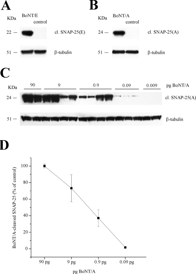
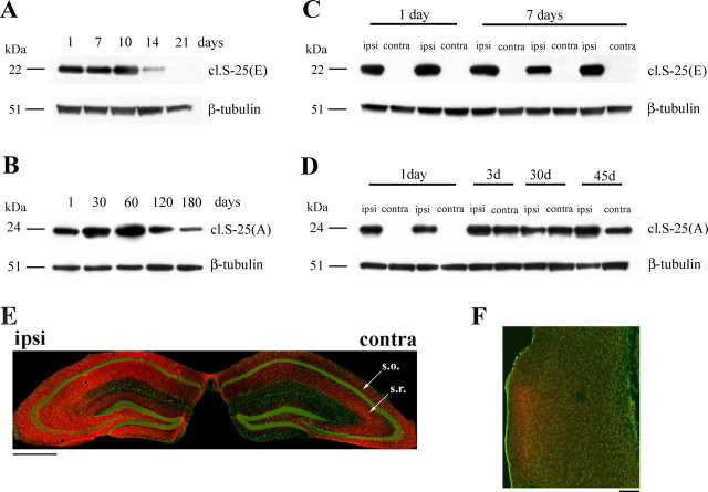
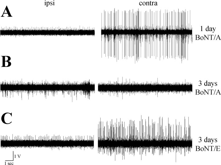
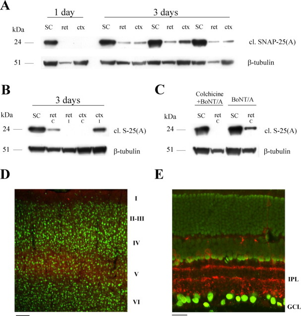
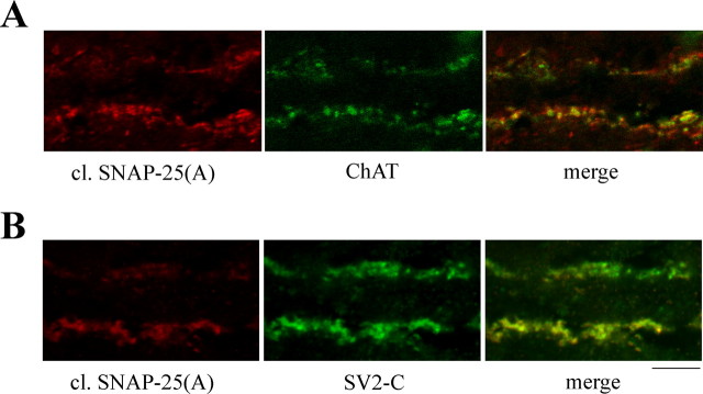
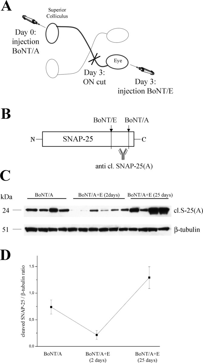
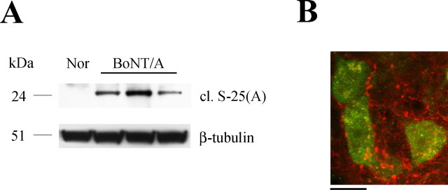
Botulinum neurotoxins (designated BoNT/A-BoNT/G) are bacterial enzymes that block neurotransmitter release by cleaving essential components of the vesicle fusion machinery. BoNT/A, which cleaves SNAP-25 (synaptosomal-associated protein of 25 kDa), is extensively exploited in clinical medicine to treat neuromuscular pathologies, facial wrinkles, and various types of pain. It is widely assumed that BoNT/A remains at the synaptic terminal and its effects are confined to the injection site. Here we demonstrate that catalytically active BoNT/A is retrogradely transported by central neurons and motoneurons and is then transcytosed to afferent synapses, in which it cleaves SNAP-25. SNAP-25 cleavage by BoNT/A was observed in the contralateral hemisphere after unilateral BoNT/A delivery to the hippocampus. Appearance of cleaved SNAP-25 resulted in blockade of hippocampal activity in the untreated hemisphere. Injections of BoNT/A into the optic tectum led to the appearance of BoNT/A-truncated SNAP-25 in synaptic terminals within the retina. Cleaved SNAP-25 also appeared in the facial nucleus after injection of the toxin into rat whisker muscles. Experiments excluded passive spread of the toxin and demonstrated axonal migration and neuronal transcytosis of BoNT/A. These findings reveal a novel pathway of BoNT/A trafficking in neurons and have important implications for the clinical uses of this neurotoxin.


Figures

Figure 1.
Figure 1.
Characterization of the antibodies against cleaved SNAP-25. A, Immunoblotting for BoNT/E-cleaved SNAP-25 [cl. SNAP-25(E)] on protein extracts from the mouse hippocampus. A 22 kDa band is detected in the sample injected with BoNT/E but not in a control uninjected hippocampus (control). β-Tubulin, Internal standard. B, Immunoblotting for BoNT/A-cleaved SNAP-25 [cl. SNAP-25(A)] on protein extracts from the mouse hippocampus. A 24 kDa band is detected in the sample injected with BoNT/A but not in a control uninjected hippocampus (control). β-Tubulin, Internal standard. C, Immunoblotting for BoNT/A-cleaved SNAP-25 [cl. SNAP-25(A)] on hippocampal extracts after injection of different doses of toxin. D, Quantification of the levels of BoNT/A-damaged SNAP-25 versus amounts of injected toxin. Values are normalized to the data obtained with 90 pg of BoNT/A. Error bars indicate SE and, when not seen, are within the symbol.
Figure 2.
Figure 2.
Retrograde propagation of BoNT/A effects in the limbic system. A, Immunoblotting for BoNT/E-cleaved SNAP-25 [cl. S-25(E)] on protein extracts from the injected hippocampus at different times after BoNT/E injection. BoNT/E effects are extinguished by 21 d. Fifteen micrograms of protein were loaded per lane. β-Tubulin, Internal standard. B, Persistence of BoNT/A-truncated SNAP-25 in the injected hippocampus. Levels of BoNT/A-cleaved SNAP-25 [cl.S-25(A)] remain high for up to 120 d after injection and decrease at 180 d. C, Unilateral BoNT/E administration results in SNAP-25 cleavage in the injected (ipsi) but not contralateral (contra) hemisphere. D, Appearance of BoNT/A-truncated SNAP-25 on the side contralateral to the injection starting from 3 d after toxin delivery. E, Hippocampal coronal section from a mouse unilaterally injected with BoNT/A 3 d earlier. Staining for cleaved SNAP-25 (red) is evident in both hemispheres. Green, Neuronal counterstaining (NeuN antibody). Arrows indicate stratum oriens (s.o.) and radiatum (s.r.). Scale bar, 500 μm. F, Section through the entorhinal cortex ipsilateral to the side of intrahippocampal BoNT/A injection. Labeling for cleaved SNAP-25 (red) is evident in superficial layers. Neuronal counterstaining is in green. Note that other areas of the cortex are not stained, ruling out passive spread. Dorsal is up and lateral is to the left. Scale bar, 100 μm.
Figure 3.
Figure 3.
Unilateral, intrahippocampal infusion of BoNT/A results in bilateral blockade of activity within 3 d. A, B, Representative examples of extracellular recordings of spiking activity from CA1, performed 1 d (A) and 3 d (B) after unilateral administration of BoNT/A into the dorsal hippocampus. Note that spontaneous discharges in the contralateral side are strongly suppressed 3 d after BoNT/A. Ipsi, Injected side; contra, contralateral side. C, Extracellular recordings from CA1 performed 3 d after unilateral administration of BoNT/E. Activity is blocked only in the treated side.
Figure 4.
Figure 4.
Retrograde propagation of BoNT/A effects in the visual system. A, Immunoblotting for cleaved SNAP-25 [cl. SNAP-25(A)] on protein extracts from the injected SC, the contralateral retina (ret), and the ipsilateral visual cortex (ctx). Spread of BoNT/A effects to the retina and cortex is evident 3 d after administration of the toxin. Fifteen micrograms of protein were loaded per lane. β-Tubulin, Internal standard. B, Immunoblotting demonstrates no cleaved SNAP-25 [cl. S-25(A)] in either the ipsilateral retina (ret i) or contralateral striate cortex (ctx c). C, Immunoblotting for cleaved SNAP-25 [cl. S-25(A)] on protein extracts from the injected SC and contralateral retina (ret). Intravitreal injection of colchicine 16–18 h before BoNT/A (colchicine+BoNT/A) prevents retrograde spread of toxin effects. D, Immunostaining for BoNT/A-truncated SNAP-25 (red) in a coronal section of primary visual cortex ipsilateral to the tectal injection. Staining for neurons is in green. Immunoreactivity for cleaved SNAP-25 is concentrated in layer V. Cortical layers are indicated on the right. Scale bar, 100 μm. E, Transverse section through the retina contralateral to the injection site demonstrates labeling for cleaved SNAP-25 (red) in the inner plexiform layer (IPL); GCL, ganglion cell layer. Green, Nuclear staining. Scale bar, 20 μm.
Figure 5.
Figure 5.
Cleaved SNAP-25 is mainly localized within cholinergic, SV2C-positive terminals in the retina. A, Confocal images of a single optical section through the inner plexiform layer showing colocalization of cleaved SNAP-25 (red) with choline acetyltransferase (ChAT; green). B, Confocal images of a single optical section through the inner plexiform layer showing colocalization of cleaved SNAP-25 (red) with SV2C (green). The ganglion cell layer is down. Scale bar, 10 μm.
Figure 6.
Figure 6.
Retrograde transport of active BoNT/A in the visual system. A, Experimental design. Adult rats were injected with BoNT/A into the SC. At day 3, the optic nerve (ON) was sectioned, and BoNT/E was delivered into the vitreous. Retinal tissue samples were dissected either 2 or 25 d after BoNT/E. B, Schematic representation of BoNT/A and BoNT/E cleavage sites. BoNT/E proteolysis removes the BoNT/A-truncated C-terminal peptide of SNAP-25. C, Immunoblotting for BoNT/A-cleaved SNAP-25 on retinal protein extracts in animals that received BoNT/A into the SC. Samples were taken 3 d after BoNT/A (BoNT/A, left lanes), and either 2 d (middle lanes) or 25 d (right lanes) after ON cut and intraocular injection of BoNT/E. The recovery of high levels of BoNT/A-altered SNAP-25 at the completion of BoNT/E effects indicates persistent activity of BoNT/A within the retina. Fifty micrograms of protein were loaded per lane. β-Tubulin, Internal standard. D, Quantification of the immunoblotting experiments. BoNT/A-damaged SNAP-25 is high 3 d after BoNT/A, decreases 2 d after intravitreal BoNT/E, and returns high 25 d after BoNT/E.
Figure 7.
Figure 7.
Retrograde spread of BoNT/A effects in facial motoneurons. A, Immunoblotting showing expression of cleaved SNAP-25 in the facial nucleus 3 d after BoNT/A injection into the whisker pad. Nor, Tissue from a normal uninjected animal. One hundred micrograms of protein were loaded per lane. β-Tubulin, Internal standard. B, Confocal image of facial motoneurons (NeuN staining, green) surrounded by cleaved SNAP-25-immunopositive profiles (red). Scale bar, 20 μm.

References

    1. Abbruzzese G, Berardelli A. Neurophysiological effects of botulinum toxin type A. Neurotox Res. 2006;9:109–114. - PubMed
    1. Antonucci F, Di Garbo A, Novelli E, Manno I, Sartucci F, Bozzi Y, Caleo M. Botulinum neurotoxin E (BoNT/E) reduces CA1 neuron loss and granule cell dispersion, with no effects on chronic seizures, in a mouse model of temporal lobe epilepsy. Exp Neurol. 2007 in press. - PubMed
    1. Caleo M, Lodovichi C, Maffei L. Effects of nerve growth factor on visual cortical plasticity require afferent electrical activity. Eur J Neurosci. 1999;11:2979–2984. - PubMed
    1. Caleo M, Restani L, Gianfranceschi L, Costantin L, Rossi C, Rossetto O, Montecucco C, Maffei L. Transient synaptic silencing of developing striate cortex has persistent effects on visual function and plasticity. J Neurosci. 2007;27:4530–4540. - PMC - PubMed
    1. Costantin L, Bozzi Y, Richichi C, Viegi A, Antonucci F, Funicello M, Gobbi M, Mennini T, Rossetto O, Montecucco C, Maffei L, Vezzani A, Caleo M. Antiepileptic effects of botulinum neurotoxin E. J Neurosci. 2005;25:1943–1951. - PMC - PubMed

Publication types

MeSH terms