. 2008 Jun;19(6):2544-52.
doi: 10.1091/mbc.e08-01-0009. Epub 2008 Apr 2.

A supraphysiological nuclear export signal is required for parvovirus nuclear export

Affiliations

A supraphysiological nuclear export signal is required for parvovirus nuclear export

Dieuwke Engelsma et al. Mol Biol Cell. 2008 Jun.

Abstract

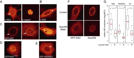
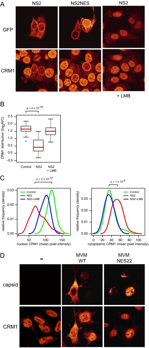
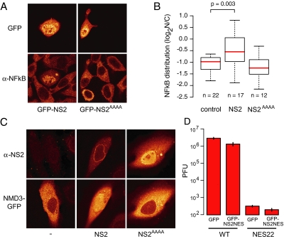
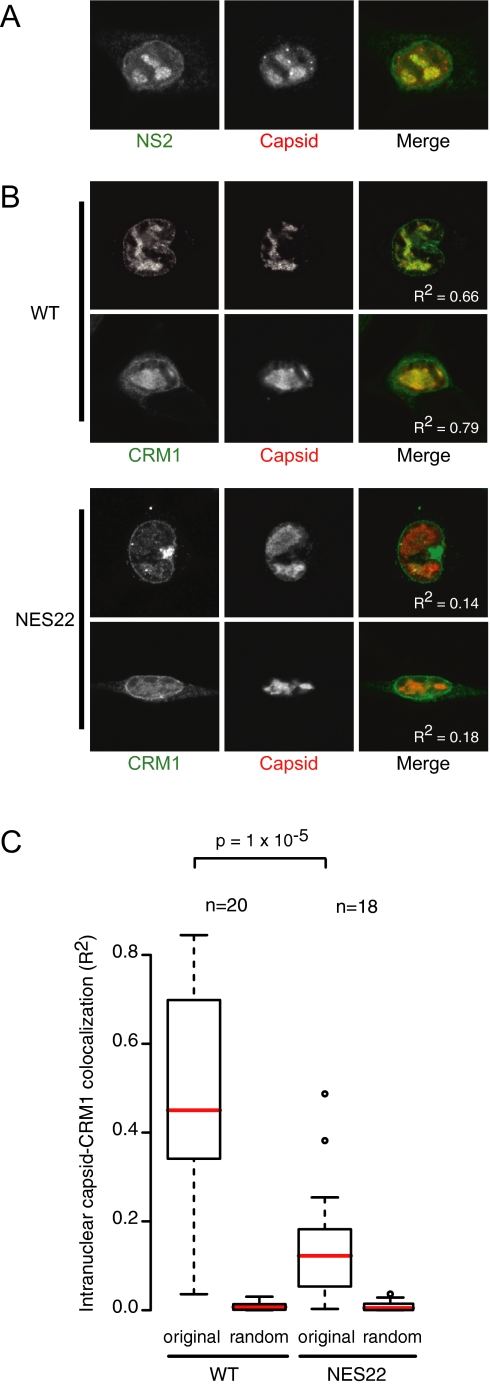
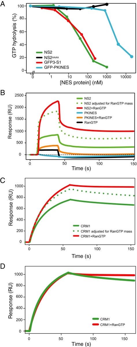
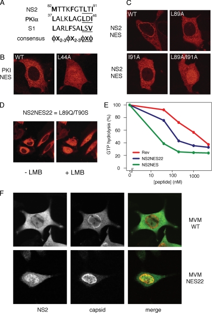
CRM1 exports proteins that carry a short leucine-rich peptide signal, the nuclear export signal (NES), from the nucleus. Regular NESs must have low affinity for CRM1 to function optimally. We previously generated artificial NESs with higher affinities for CRM1, termed supraphysiological NESs. Here we identify a supraphysiological NES in an endogenous protein, the NS2 protein of parvovirus Minute Virus of Mice (MVM). NS2 interacts with CRM1 without the requirement of RanGTP, whereas addition of RanGTP renders the complex highly stable. Mutation of a single hydrophobic residue that inactivates regular NESs lowers the affinity of the NS2 NES for CRM1 from supraphysiological to regular. Mutant MVM harboring this regular NES is compromised in viral nuclear export and productivity. In virus-infected mouse fibroblasts we observe colocalization of NS2, CRM1 and mature virions, which is dependent on the supraphysiological NS2 NES. We conclude that supraphysiological NESs exist in nature and that the supraphysiological NS2 NES has a critical role in active nuclear export of mature MVM particles before cell lysis.


Figures

Figure 1.
Figure 1.
NS2 localizes to the NE via Nup358. (A) A9 cells transfected with MVM expression plasmid pdBMVp for 24 h and stained with anti-NS2 and anti-capsid antibodies. (B) HeLa cells expressing untagged NS2 were stained after 24 h with anti-NS2 antibodies. MCF7 cells were transfected for 24 h with (C) NS2-GFP, (D) NS2AAAA–GFP, or (E) NS2-NES-GFP. White arrows indicate NE localization. (F and G) MCF7 cells were transfected with pSuper-Nup358 and Nup358 levels were determined after 72 h using Nup358 antibodies. (F) An example of GFP-NS2 localization in control cells or cells depleted of Nup358. (G) Boxplots showing relative amounts of NE-located GFP-tagged protein in control cells or Nup358 knockdown cells were determined by dividing the NE pixel intensity by the cytoplasmic pixel intensity, measured at three sites of the NE. Medians are indicated in red. p values were calculated using Mann-Whitney tests.
Figure 2.
Figure 2.
NS2 relocalizes CRM1 from the nucleus to the cytoplasm (A) MCF7 cells expressing NS2-GFP (left and right) or NS2-NES-GFP (middle). When indicated, cells were treated with LMB for 3 h. Cells were stained with anti-CRM1 antibodies. (B) Boxplot summarizing quantifications (n = 36 each) of CRM1 subcellular distribution exemplified in A. Medians are indicated in red. (C) Distribution of nuclear (left) and cytoplasmic (right) CRM1 intensities, showing both an NS2-dependent nuclear decrease and a cytoplasmic increase, which is LMB-sensitive. Densities were calculated using a bandwidth of 8. p values according to Mann-Whitney tests (D) A9 cells mock-infected (−) or infected with MVMp (wt) or MVMp-NES22 (NES22) for 48 h and stained for both capsid and CRM1.
Figure 3.
Figure 3.
NS2 interferes with the CRM1 export pathway. (A) NS2 inhibits the cytoplasmic localization of NFκB. L929 cells were transfected with GFP-NS2 or GFP-NS2AAAA and stained for NFκB. (B) Boxplots summarizing quantifications of NFκB subcellular distribution shown in (A). p value from Mann-Whitney test. (C) NS2 inhibits export of Nmd3. HeLa cells expressing Nmd3-GFP alone (right) Nmd3-GFP and untagged NS2 (middle), or Nmd3-GFP and untagged NS2AAAA (right). NS2 was detected with NS2 antibodies. (D) Exogenously expressed supraNES cannot rescue the defect in viral propagation of MVMNES22. A9 cells (n = 105) were transfected with either wild-type pdBMVp or pdBMV-NES22 (NES22) and cotransfected with either GFP or GFP-NS2-NES. After 48 h the extracellular virus pool was titered by a plaque assay using NB324K cells. Y-axis shows the plaque forming units (PFU). Infection with MVM-NES22 yielded microplaques. Error bars, SEs from independent experiments.
Figure 4.
Figure 4.
Intranuclear colocalization of CRM1 with mature virions is dependent on the NS2 supraNES. A9 cells transfected with pdBMVp (WT) or pdBMV-NES22 (NES22) for 48 h. (A) NS2 and capsid colocalize in SAABs. (B) CRM1 and capsid colocalize in SAABs in a supraNES-dependent manner. Colocalization values (R2) are displayed in the merged images. (C) Boxplots summarizing quantification of intranuclear colocalization of CRM1 with capsid. Pixel correlations (R) were calculated in the intranuclear region (excluding membrane invaginations) of CRM1 with original (original) or means of 25 randomized (random) capsid images using ImageJ. Correlations were squared to provide a linear approximation to colocalization. p values according to Mann-Whitney test.
Figure 5.
Figure 5.
NS2 binds stably to CRM1 in a RanGTP-independent manner. (A) Full-length NS2 protein binds CRM1 with supraphysiological affinities similar to the supraNES S1. The affinity for CRM1 of NS2, NS2AAAA, the regular PKI-NES, and the supraNES S1 was measured using the CRM1 GTPase assay. Complexes formed with Ran[γ-GT32P], purified recombinant NES-bearing proteins (x-axis) and CRM1 were subjected to RanGAP, and hydrolyzed 32P was measured (y-axis). 100% hydrolysis was recorded when CRM1-Ran[γ-GT32P] samples without cargo were subjected to RanGAP-stimulated hydrolysis. Full-length NS2 (green) and GFP3-S1 (red) were used as reference for supraphysiological affinity; GFP-PKI-NES (blue) was used as control for regular affinity, and full-length NS2AAAA protein (black) was used as negative control. (B–D) Surface plasmon resonance (SPR) analysis of the NS2/CRM1 interaction. (B) NS2 (green) or PKI NES (blue), alone or in combination with RanGTP (NS2 plotted in red and PKI NES plotted in orange) were streamed over chip-immobilized CRM1 for 40s. Resonance response (in RU) was measured to 150 s. The dotted line denotes the theoretical response of an NS2/RanGTP complex based on the NS2 response adjusted for the additional mass of Ran. RanGTP (black) alone displays minor affinity for CRM1. (C) CRM1 (green) or CRM1+RanGTP (red) was streamed over chip-immobilized NS2 for 60 s. Resonance response was measured to 150 s. The dotted line denotes the theoretical response of a CRM1/RanGTP complex based on the CRM1 response adjusted for the additional mass of Ran. (D) Superimposed CRM1 and CRM1+RanGTP response curves indicate a greater stability of NS2/CRM1/RanGTP (red) over NS2/CRM1 (green).
Figure 6.
Figure 6.
The NS2 supraNES is functionally required for viral nuclear export and infectivity. (A) Comparison of NES sequences of NS2, PKIα, and S1. Hydrophobic residues are marked bold. The core regions of the NESs are underlined. φ: L, I, F, M, or V; X: any other residue. (B and C) MCF7 cells were transfected with GFP-tagged NESs for 24 h. (B) Export of the PKIα-NES is inhibited upon mutation of one hydrophobic residue. (C) A single mutation in the NS2 supraNES downgrades the supraNES to a regular NES. To inactivate the NES activity an additional hydrophobic residues needs to be mutated. (D) NES22 behaves like a regular NES. Cells were transfected with GFP-NES22 and imaged using live confocal microscopy. + LMB: cells were treated with LMB for 10 min. (E) NES22 retains CRM1 binding activity NES. Affinity of NES22 was tested in GAP assay as described in Figure 2A. Synthetic peptides were used. NES22 (blue), supraNES (green), and RevNES (red). (F) NES22 viruses fail to be exported and the NS2-NES22 protein is retained in the nucleoplasm. Mouse fibroblasts expressing the WT or NES22 genome for 42 h were stained with anti-NS2 and anti-capsid antibodies.

Similar articles

Cited by

References

    1. Askjaer P., et al. RanGTP-regulated interactions of CRM1 with nucleoporins and a shuttling DEAD-box helicase. Mol. Cell. Biol. 1999;19:6276–6285. - PMC - PubMed
    1. Becskei A., Mattaj I. W. The strategy for coupling the RanGTP gradient to nuclear protein export. Proc. Natl. Acad. Sci USA. 2003;100:1717–1722. - PMC - PubMed
    1. Bernad R., Van Der Velde H., Fornerod M., Pickersgill H. Nup358/RanBP2 attaches to the nuclear pore complex via association with Nup88 and Nup214/CAN and plays a supporting role in CRM1-mediated nuclear protein export. Mol. Cell. Biol. 2004;24:2373–2384. - PMC - PubMed
    1. Bischoff F. R., Gorlich D. RanBP1 is crucial for the release of RanGTP from importin beta-related nuclear transport factors. FEBS Lett. 1997;419:249–254. - PubMed
    1. Bodendorf U., Cziepluch C., Jauniaux J. C., Rommelaere J., Salome N. Nuclear export factor CRM1 interacts with nonstructural proteins NS2 from parvovirus minute virus of mice. J. Virol. 1999;73:7769–7779. - PMC - PubMed

Publication types

MeSH terms