. 2008 Jun;19(6):2553-65.
doi: 10.1091/mbc.e07-09-0862. Epub 2008 Apr 2.

RAB-11 permissively regulates spindle alignment by modulating metaphase microtubule dynamics in Caenorhabditis elegans early embryos

Affiliations

RAB-11 permissively regulates spindle alignment by modulating metaphase microtubule dynamics in Caenorhabditis elegans early embryos

Haining Zhang et al. Mol Biol Cell. 2008 Jun.

Abstract

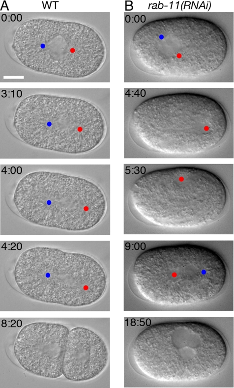
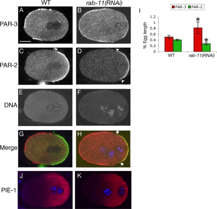
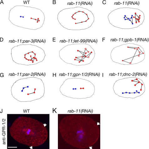
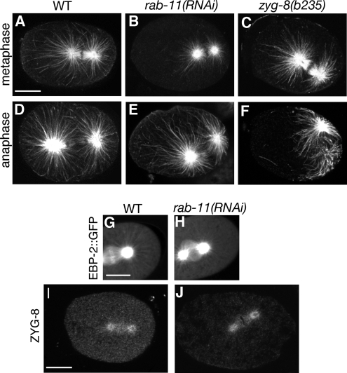
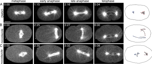
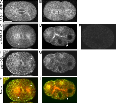
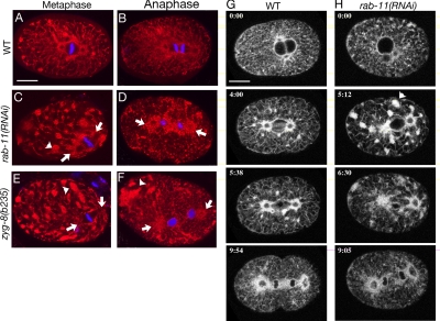
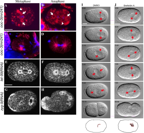
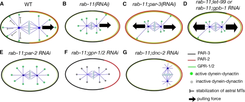
Alignment of the mitotic spindle along a preformed axis of polarity is crucial for generating cell diversity in many organisms, yet little is known about the role of the endomembrane system in this process. RAB-11 is a small GTPase enriched in recycling endosomes. When we depleted RAB-11 by RNAi in Caenorhabditis elegans, the spindle of the one-cell embryo failed to align along the axis of polarity in metaphase and underwent violent movements in anaphase. The distance between astral microtubules ends and the anterior cortex was significantly increased in rab-11(RNAi) embryos specifically during metaphase, possibly accounting for the observed spindle alignment defects. Additionally, we found that normal ER morphology requires functional RAB-11, particularly during metaphase. We hypothesize that RAB-11, in conjunction with the ER, acts to regulate cell cycle-specific changes in astral microtubule length to ensure proper spindle alignment in Caenorhabditis elegans early embryos.


Figures

Figure 1.
Figure 1.
rab-11(RNAi) embryos undergo violent spindle movements during one-cell anaphase. Time-lapse differential interference contrast (DIC) microscopy recordings of the first cell division of WT embryo (A, Video 1), and rab-11(RNAi) embryo (B, Video 2) after 44 h of feeding from prometaphase (t = 0) to the end of the first division. Blue dots, anterior centrosomes; red dots, posterior centrosomes. In the rab-11(RNAi) embryo, the P0 spindle did not align along the anterior-posterior axis of polarity and the posteriorly positioned centrosome (labeled with red dot) ended up in an anterior location at the end of anaphase due to the violent spindle movements. The anterior positioned centrosome was out of focus at some time points. Cytokinesis of the first division failed to complete in the rab-11(RNAi) embryo. Scale bar, 10 μm.
Figure 2.
Figure 2.
Polarity defects in rab-11(RNAi) embryos. WT (A, C, E, and G) and rab-11(RNAi) (B, D, F, and H) one-cell embryos stained with anti-PAR-3 (A and B), anti-PAR-2 (C and D) antibodies and Topro3 (E and F) to reveal DNA. Arrowheads mark the boundary of the PAR-2 domain. In the merged images (G and H), PAR-3 is red, PAR-2 is green, and DNA is blue. Arrows indicate the boundary of the overlapping region of PAR-2 and -3 in rab-11(RNAi) embryos. Note the extra pronucleus indicating the polar body was not extruded in the rab-11(RNAi) embryo. (I) Quantification of the egg length of PAR-2 and -3 domains before pronuclear centration in WT and rab-11(RNAi) embryos. n = 6 embryos for each measurement. Error bars, SD. Statistically significant difference between WT and rab-11(RNAi) embryos; *p < 0.05; Student's t test, two-tailed unequal variance. For PAR-2, p = 0.00016; PAR-3, p = 0.011. (J and K) Immunofluorescence staining of PIE-1 (red) in WT (J) and rab-11(RNAi) (K) embryos during pronuclear migration. DNA (blue) is labeled with Topro3. Scale bar, 10 μm.
Figure 3.
Figure 3.
Gα/GPR-1/2 activity may not be up-regulated in rab-11(RNAi) embryos. Schematic representations of the movements of one or both centrosomes in WT (A, Video 1), rab-11(RNAi) (B and C, Video 2), rab-11; par-3(RNAi) (D, Video 3, top), rab-11; let-99(RNAi) (E, Video 4, top), rab-11; gpb-1(RNAi) (F, Video 4, bottom), rab-11; par-2(RNAi) (G, Video 3, bottom), rab-11; gpr-1/2(RNAi) (H, Video 5, top), and rab-11; dnc-2(RNAi) embryo (I, Video 5, bottom). Each drawing is from a single representative embryo. These trajectories describe the path of centrosome movement from prometaphase to the end of anaphase (dots). The dots are not at the same time intervals. For rab-11; let-99(RNAi) and rab-11; gpb-1(RNAi) embryos (E and F), because the violent movements of the centrosomes begin before that observed in rab-11(RNAi), the traces of movements start from pronuclear centration (asterisks). In embryos whose spindles underwent violent movements, only the posterior centrosomes are shown as the anterior centrosomes were frequently out of the plane of focus. In the rab-11; par-2(RNAi) (G) and rab-11; dnc-2(RNAi) (I) embryos, the P0 spindles first set up perpendicular to the longitudinal axis. In rab-11; dnc-2(RNAi) embryo, the spindle failed to centrate due to disruption of DNC-2 activity. During anaphase, the spindles elongated and flipped to the longitudinal axis due to the constraints of the eggshell. (J and K) Immunofluorescence staining of GPR-1/2 (red) in WT (J) and rab-11(RNAi) (K) embryos during one-cell metaphase. DNA (blue) is labeled with Topro3. Arrows delineate regions of GPR-1/2 enrichment at posterior cortex. In the rab-11(RNAi) embryo, the P0 spindle did not rotate and the chromosomes did not align properly along the metaphase plate. Immunolabeled rab-11(RNAi) embryos are larger because they are pressure sensitive and flattened more than WT embryos during fixation. Scale bar, 10 μm.
Figure 4.
Figure 4.
rab-11(RNAi) and zyg-8(b235) embryos exhibit different MT length defects during one-cell division. (A–C) Metaphase MTs stained with anti-α-tubulin antibody in WT (A), rab-11(RNAi) (B), and zyg-8(b235) (C) embryos. (D--F) Anaphase MTs stained with anti-α-tubulin antibody in WT (D), rab-11(RNAi) (E), and zyg-8(b235) (F) embryos. (G and H) Projections of 100-frame EBP-2::GFP movies (89 s in length) in WT (G, Video 6, left) and rab-11(RNAi) (H, Video 6, right) embryos during metaphase (see Materials and Methods). Very few MTs were seen to grow out to the cortex in the rab-11(RNAi) embryos (H). Notice that EBP-2::GFP accumulation at the kinetochore–MT interface was also reduced in rab-11(RNAi) embryos. The significance of this defect is not known. (I and J) Immunofluorescence staining of ZYG-8 in WT (I) and rab-11(RNAi) (J) embryos. ZYG-8 localization to the spindle and centrosomes was normal in the rab-11(RNAi) embryos, although the cytoplasmic staining seemed to be reduced in rab-11(RNAi) embryo. Scale bar, 10 μm.
Figure 5.
Figure 5.
Nocodazole treatment can phenocopy the violent spindle movements in rab-11(RNAi) embryos. Multiphoton time series of embryos expressing β-tubulin::GFP show the following developmental stages: metaphase, early anaphase, late anaphase, and telophase. (A) β-tubulin::GFP embryos treated with the same dilution of DMSO (Video 7). (B and C) β-tubulin::GFP embryos treated with 50 μg/ml nocodazole during pronuclear migration or centration (Videos 8 and 9). Note that the metaphase MTs were shortened by nocodazole, but they resumed elongation during anaphase. Centrosomal positions that best describe the trace of the movements from metaphase to telophase are shown in the schematic drawings (blue dots, anterior centrosomes; red dots, posterior centrosomes; dots are not at the same time intervals). Scale bar, 10 μm.
Figure 6.
Figure 6.
RAB-11 colocalizes extensively with the ER. Metaphase (A) and anaphase (B) localization of RAB-11::GFP. One-cell anaphase embryo labeled with anti-RAB-11 (C), anti-HDEL (F), and merged (H). Four-cell embryo with ABa and ABp at anaphase, P2 and EMS at metaphase labeled with anti-RAB-11 (D), anti-HDEL (G), and merged (I). Arrow points to a structure unique to anti-RAB-11 labeling, and arrowhead to a structure unique to anti-HDEL labeling. In rab-11(RNAi) embryo (E), the anti-RAB-11 labeling is greatly reduced. Scale bar, 10 μm.
Figure 7.
Figure 7.
Metaphase ER morphology is disrupted in rab-11(RNAi) embryos. Immunofluorescence staining of the ER (anti-HDEL, red) and Topro3 staining of DNA (blue) in metaphase and anaphase WT embryos (A and B), rab-11(RNAi) embryo (C and D), and zyg-8(b235) mutant embryos (E and F). Arrows mark the positions of the mitotic spindle poles. Arrowhead shows one of the ER clumps. (G and H) Multiphoton time series of embryos expressing SP12::GFP show the following developmental stages: prometaphase (t = 0), metaphase, early anaphase, and late anaphase. Arrowhead shows one of the ER clumps in metaphase. Scale bar, 10 μm.
Figure 8.
Figure 8.
Microtubule and ER morphologies for gene disruptions and BFA treatment listed in Table 2. (A and B) Immunofluorescence staining of the ER (anti-HDEL, red) and Topro3 staining of DNA (blue) in ooc-3(mn241) mutant embryos during metaphase (A) and anaphase (B). Although previous work indicated that that the ER structure was less affected in ooc-5(it145) mutant embryos than in ooc-3(mn241) embryos (Basham and Rose, 2001), we observed a similar defect of ER morphology in these embryos (data not shown). Arrows mark the position of the mitotic spindle poles. Arrowheads show one of the ER clumps. (C and D) Immunofluorescence staining of the microtubules (red) and Topro3 staining of DNA (blue) in ooc-3(mn241) mutant embryos during one-cell metaphase (C) and anaphase (D). ooc-5(it145) mutant embryos showed similar phenotype (data not shown). ooc-3(mn241) and ooc-5(it145) mutant embryos are often smaller than the WT embryos (Basham and Rose, 1999). Scale bar, 10 μm. (E and F) SP12::GFP embryos treated with let-99 RNAi showed that ER morphology was not affected during either metaphase (E) or anaphase (F). (G and H) SP12::GFP embryos treated with zyg-9 RNAi showed that ER morphology was similar to that observed with nocodazole or tba-2(RNAi) treatment (Poteryaev et al., 2005). The accumulation of ER structure at the anterior during anaphase may be due to the exaggerated cytoplasmic flow resulting from short microtubules. (I and J) BFA treatment caused nuclear-centrosomal complex rocking during pronuclear centration. Nomarski images show the centrosome movements during pronuclear centration and the final stage of the first division (the last time point). (I, Video 10, top) WT meiosis II embryos treated with same dilution of DMSO as control (n = 15). (J, Video 10, bottom) WT meiosis II embryos treated with 150 μg/ml BFA (n = 14). Arrows mark the positions of the two centrosomes. Schematic representations of the movements of the posterior centrosome during pronuclear centration are drawn. Scale bar, 10 μm.
Figure 9.
Figure 9.
Model for the violent spindle movements in rab-11(RNAi) embryos. In the WT embryo (A), the posterior pulling force at metaphase is balanced by the tethering of astral MTs at the anterior cortex (Labbe et al., 2004). In the rab-11(RNAi) embryo (B), the metaphase MTs are short and no longer stabilized at the anterior, subjecting the spindle only to the pulling force from the posterior. The longer MT represents the few that do manage to reach to the posterior cortex where more abundant GPR-1/2 localizes, so that dynein-dynactin (shown in green dots) can exert pulling forces on the spindle. In the rab-11; par-3(RNAi) embryo (C), GPR-1/2 level is evenly elevated around the cortex (Colombo et al., 2003; Gotta et al., 2003). The spindle is then under equal pulling forces from both poles, and it undergoes violent movements in the center of the embryo. In the rab-11; let-99(RNAi) or rab-11; gpb-1(RNAi) embryo (D), the GPR-1/2 activity may be further up-regulated due to the disruption of these antagonizing regulators of G (Tsou et al., 2003), which results in even stronger pulling forces. Furthermore, because the embryo is still polarized, unlike in the rab-11; par-3(RNAi) embryo, the pulling forces may be unbalanced (not shown in the diagram) and thus lead to more violent and unpredictable spindle movements. The pulling force from the cortex is suppressed in the rab-11; par-2(RNAi) embryo (E) due to reduced GPR-1/2 activity (Colombo et al., 2003; Gotta et al., 2003). In the rab-11; gpr-1/2(RNAi) embryo (F), both GPR-1/2 and dynein-dynactin activities are reduced (Grill et al., 2003), accounting for the absence of pulling force from the posterior cortex. Inactivating the motor dynein-dynactin in rab-11; dnc-2(RNAi) embryo (G) also suppresses the pulling force from the cortex even though the GPR-1/2 activity may be normal.

Similar articles

Cited by

References

    1. Basham S. E., Rose L. S. Mutations in ooc-5 and ooc-3 disrupt oocyte formation and the reestablishment of asymmetric PAR protein localization in two-cell Caenorhabditis elegans embryos. Dev. Biol. 1999;215:253–263. - PubMed
    1. Basham S. E., Rose L. S. The Caenorhabditis elegans polarity gene ooc-5 encodes a Torsin-related protein of the AAA ATPase superfamily. Development. 2001;128:4645–4656. - PubMed
    1. Brenner S. The genetics of Caenorhabditis elegans. Genetics. 1974;77:71–94. - PMC - PubMed
    1. Boyd L., Guo S., Levitan D., Stinchcomb D. T., Kemphues K. J. PAR-2 is asymmetrically distributed and promotes association of P granules and PAR-1 with the cortex in C. elegans embryos. Development. 1996;122:3075–3084. - PubMed
    1. Capes-Davis A., Tolhurst O., Dunn J. M., Jeffrey P. L. Expression of doublecortin (DCX) and doublecortin-like kinase (DCLK) within the developing chick brain. Dev. Dyn. 2005;232:457–467. - PubMed

Publication types

MeSH terms

Substances

LinkOut - more resources