. 2008 Jun;19(6):2488-99.
doi: 10.1091/mbc.e07-08-0737. Epub 2008 Apr 2.

Cds1 controls the release of Cdc14-like phosphatase Flp1 from the nucleolus to drive full activation of the checkpoint response to replication stress in fission yeast

Affiliations

Cds1 controls the release of Cdc14-like phosphatase Flp1 from the nucleolus to drive full activation of the checkpoint response to replication stress in fission yeast

Helena Díaz-Cuervo et al. Mol Biol Cell. 2008 Jun.

Abstract

The Cdc14p-like phosphatase Flp1p (also known as Clp1p) is regulated by cell cycle-dependent changes in its subcellular localization. Flp1p is restricted to the nucleolus and spindle pole body until prophase, when it is dispersed throughout the nucleus, mitotic spindle, and medial ring. Once released, Flp1p antagonizes Cdc2p/cyclin activity by reverting Cdc2p-phosphorylation sites on Cdc25p. On replication stress, ataxia-telangiectasia mutated/ATM/Rad3-related kinase Rad3p activates Cds1p, which phosphorylates key proteins ensuring the stability of stalled DNA replication forks. Here, we show that replication stress induces changes in the subcellular localization of Flp1p in a checkpoint-dependent manner. Active Cds1p checkpoint kinase is required to release Flp1p into the nucleus. Consistently, a Flp1p mutant (flp1-9A) lacking all potential Cds1p phosphorylation sites fails to relocate in response to replication blocks and, similarly to cells lacking flp1 (Deltaflp1), presents defects in checkpoint response to replication stress. Deltaflp1 cells accumulate reduced levels of a less active Cds1p kinase in hydroxyurea (HU), indicating that nuclear Flp1p regulates Cds1p full activation. Consistently, Deltaflp1 and flp1-9A have an increased percentage of Rad22p-recombination foci during HU treatment. Together, our data show that by releasing Flp1p into the nucleus Cds1p checkpoint kinase modulates its own full activation during replication stress.


Figures

Figure 1.
Figure 1.
Flp1p-GFP accumulates in the nucleus during HU induced replication stress. (A) Asynchronous cultures of exponential growing flp1-GFP gar2-RFP cells were treated for 2 h with the ribonucleotide reductase inhibitory drug HU (12 mM). Localization of nucleolar protein Gar2p-RFP and nuclear Hoechst staining are included as reference controls. (B) Flp1p-GFP locates to the nucleolus and SPB in untreated interphase cells. (C) A 3.5× nuclei magnification of untreated interphase (top) and 12 mM HU-treated cells (bottom). Note that no physical nucleolar reorganization can be observed as a result of HU treatment. Bar, 4 μm.
Figure 2.
Figure 2.
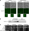
Functional interaction between Rad24p and Flp1p in response to blocks in DNA replication. (A) Flp1p does not change its subcellular localization in response to replicative stress in Δrad24 cells. Live imaging of Flp1p-GFP in a strain deleted for rad24 after 3 h of 12 mM HU treatment. Flp1p remains nucleolar, whereas in control cells the staining is nuclear, as described previously. Bars, 10 μm. A 3× magnification of nuclei is shown in the bottom panel. Bars, 1 μm. (B) Rad24p interacts physically in vivo with Flp1p in checkpoint response to HU. GST-Rad24p was purified from untreated cells and at different times of 12 mM HU treatment. Blots were incubated with α-Ha antibody to detect Flp1pHa. Purified GST was used as a control. In lane 1, 10 μg of whole cell protein extract and in lane 2, 1/200 of the input protein in the pull-down assay shown for reference. No Rad24p–Flp1p interaction is detected in untreated cells, whereas interaction is detected at all treatment points analyzed. (C) Sensitivity assay of Δrad24 and Δrad24 Δflp1 cells to chronic HU exposure at 25°C. Δrad24 is partially sensitive to HU treatment. Δrad24 Δflp1 presents a more sensitive phenotype than each simple mutant.
Figure 3.
Figure 3.
Nuclear accumulation of Flp1p depends on checkpoint kinase Cds1p (A) Flp1p-GFP localization and Hoechst costaining in untreated and 12 mM HU treated flp1-GFP cells. (B) Flp1p-GFP localizes normally in both Δcds1 and Δchk1 untreated cells. (C) Flp1p-GFP localization in 12 mM HU-treated cells lacking either Δcds1 or Δchk1. Whereas Δchk1 cells localize Flp1p-GFP to the nucleus in response to replication stress like wild-type cells, the phosphatase remains nucleolar in cells deleted for cds1. Nuclei magnified 3× are shown. Bar, 4 μm.
Figure 4.
Figure 4.
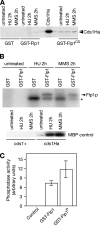
Cds1p interacts with Flp1p in vivo. (A) Flp1p and Cds1p interact physically in vivo. Pull-down assay showing physical interaction between Cds1p and Flp1p or catalytic inactive Flp1p (Flp1pCS). GST-Flp1p and GST-Flp1pCS were purified both from untreated and checkpoint activated cells, blots were incubated with α-Ha antibody to detect Cds1pHa. Stronger interaction was detected with the catalytic inactive Flp1p. (B) Checkpoint activated Cds1p phosphorylates Flp1p in vitro. Kinase assay of Cds1p by using Flp1p as substrate. Cds1pHa was immunoprecipitated from untreated cells and checkpoint activated cells, respectively, and assayed using purified GST-Flp1p and GSTp (control) as substrates. Activity of checkpoint activated Cds1pHa and of an untagged strain as a control were assayed using MBP as substrate (bottom). Flp1p is phosphorylated by the checkpoint-activated kinase. Phosphorylated Flp1p is marked by an arrow. The band marked by a star corresponds to a protein that coimmunoprecipitates with active Cds1p. (C) Cds1p-mediated Flp1p phosphorylation enhances the phosphatase activity of Flp1p in vitro. Quantification of phosphatase assay using DiFMUP as a substrate. Unphosphorylated and Cds1p phosphorylated GST-Flp1p were assayed in vitro for their ability to dephosphorylate the fluorescent substrate DiFMUP. Active immunoprecipitated Cds1p kinase was used as a negative control.
Figure 5.
Figure 5.
Characterization of the flp1-9A mutant. (A) Schematic representation of Flp1p showing Serine 468 phosphorylation site identified by mass spectrometry (bold) and the RXXS putative phosphorylation sites. (B) Phosphorylation assay of activated Cds1p using GST-Flp1p and RXXS sites mutants as substrates. Mutation of the complete set of sites was performed because phosphorylation of Flp1p is prevented in the flp1-9A mutant. (C) A 10-fold dilution plate assay showing the sensitivity of three different clones of flp1-9A-GFP to HU. It can be observed that the behavior is similar to that of Δflp1, presenting only a decrease on colony size compared with wild-type cells. (D) Mutation of the nine RXXS sites present in Flp1p does not alter the subcellular localization of the protein during the different stages of an unperturbed cell cycle. In vivo Flp1p-9A-GFP staining shows interphase and mitotic cells undergoing normal changes in the subcellular localization of Flp1p-9A as described before for wild-type Flp1p (Cueille et al., 2001). Dot lines represent the cells contour. Bar, 4 μm. (E) Flp1p-9A-GFP is not released to the nucleus in checkpoint response to HU treatment. Bars, 4 μm. Nuclei magnified 3× are shown in inner panels. Note that flp1-9A-GFP cells elongate indicating that they are responding to the checkpoint arrest.
Figure 6.
Figure 6.
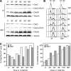
Kinetics of checkpoint-dependent phosphorylation of Cdc2p in Tyrosine 15 and Cdc25p protein accumulation are not significantly altered in Δflp1 cells in response to HU-induced replication arrest. (A) Western blot analysis of Cdc25p, Cdc2p, and Y15 phosphorylation of Cdc2p levels during 12 mM HU treatment in asynchronous cultures of wild-type and Δflp1 strains. (B) FACS analysis showing checkpoint induced S phase arrest of asynchronous wild-type and Δflp1 cells during 12 mM HU treatment. (C) Normalization of quantified Cdc2p Y15P and Cdc25p levels are shown in bar diagrams. Note that Y15P and Cdc25p levels increase during treatment in both strains after similar kinetics.
Figure 7.
Figure 7.
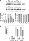
Cds1p protein levels and kinase activity in response to replication stress are altered in Δflp1 cells. (A) Western blot analysis of Cds1pHa and Chk1pHa protein levels in wild-type and Δflp1 cells treated with 20 mM HU. Samples were quantified and normalized to loading controls. Quantification is shown in bar diagrams. Whereas Δflp1 cells accumulate less Cds1p in checkpoint response to HU, Chk1p levels are similar in both strains. (B) Cells deleted for flp1 present decreased in vivo Cds1p activity in checkpoint response. Kinase assay of Cds1p using MBP as substrate. Active Cds1pHa was immunoprecipitated from wild-type and Δflp1 strains, both from HU and MMS treated cells. Activity was normalized to immunoprecipitated Cds1pHa in each sample and represented in a bar diagram. Cds1p-associated kinase activity in wild-type cells, both for HU and MMS treated cells, was arbitrarily normalized to 100%. Error bars are shown for three independent experiments.
Figure 8.
Figure 8.
Hydroxyurea induces Rad22p recombination foci in flp1 mutants. (A) Quantification of nuclei containing single or multiple Rad22p-YFP foci in flp1 mutant strains in asynchronous exponentially growing cells (untreated) and after 2 h of 12 mM HU treatment. Control counts in checkpoint proficient wild-type cells and checkpoint mutant Δcds1 are shown. Results are representative of at least two independent experiments. Standard deviations are as follows: untreated cells, wt % no foci 1.16, % one focus 1.64, % multi foci 0.48, Δflp1 % no foci 1.05, % one focus 0.80, % multi foci 0.24, flp1-9A % no foci 3.02, % one focus 3.40, % multi foci 0.37, Δcds1 % no foci 3.51, % one focus 1.09, % multi foci 2.41; HU treated cells, wt % no foci 1.74, % one focus 1.47, % multi foci 0.27, Δflp1 % no foci 6.32, % one focus 5.16, % multi foci 1.15, flp1-9A % no foci 0.75, % one focus 1.25, % multi foci 0.5, Δcds1 % no foci 0.36, % one focus 1.34, % multi foci 0.97. (B) Live imaging of Rad22p-YFP foci (bright spots) in flp1 mutants and control strains after 2 h of 12 mM HU treatment. Bar, 4 μm. Although recombination foci are rarely seen in a wild-type strain, single and multiple Rad22p-YFP foci are detected in both flp1 mutants nuclei. This effect is however weaker than in checkpoint mutant Δcds1.
Figure 9.
Figure 9.
Quantification of checkpoint defects of Δflp1 mutants in synchronized cultures. (A) cdc10-129, cdc10-129 Δflp1, and cdc10-129 Δrad3 strains were synchronized in G1 (4.5 h at 36.5°C), and then they were released from the G1 block (at 25°C) in the presence of 12 mM HU. The percentages of binucleated and cut cells were estimated by counting cell samples stained with DAPI every indicated time point and plotted. At least 200 cells were counted for every strain for each time point and the experiment was repeated twice. Labels: c, cdc10-129 control; Δf, cdc10-129 Δflp1, and Δ3, cdc10-129 Δrad3 mutants. (B) Deletion of flp1+ and rad3+ causes cdc10-129 cells to accumulate binucleated and cut cells upon HU treatment. Nuclear staining (DAPI) of cdc10-129, cdc10-129 Δflp1, and cdc10-129 Δrad3 cells synchronized in G1 and released in the presence of 12 mM HU (120 min of treatment). Note the presence of binucleated and cut cells (indicated by arrows). Bar, 10 μm. (C) A 10-fold dilution plate assay showing the different sensitivity of cdc10-129, cdc10-129 Δflp1, and cdc10-129 Δrad3 strains to chronic exposure to 5 mM HU (at 32°C).

Similar articles

Cited by

References

    1. Baber-Furnari B. A., Rhind N., Boddy M. N., Shanahan P., Lopez-Girona A., Russell P. Regulation of mitotic inhibitor Mik1 helps to enforce the DNA damage checkpoint. Mol. Biol. Cell. 2000;11:1–11. - PMC - PubMed
    1. Bailis J. M., Luche D. D., Hunter T., Forsburg S. L. Minichromosome maintenance proteins interact with checkpoint and recombination proteins to promote S-phase genome stability. Mol. Cell. Biol. 2008;28:1724–1738. - PMC - PubMed
    1. Boddy M. N., Furnari B., Mondesert O., Russell P. Replication checkpoint enforced by kinases Cds1 and Chk1. Science. 1998;280:909–912. - PubMed
    1. Boddy M. N., Russell P. DNA replication checkpoint. Curr. Biol. 2001;11:R953–R956. - PubMed
    1. Brondello J. M., Boddy M. N., Furnari B., Russell P. Basis for the checkpoint signal specificity that regulates Chk1 and Cds1 protein kinases. Mol. Cell. Biol. 1999;19:4262–4269. - PMC - PubMed

Publication types

MeSH terms