

Power Doppler imaging in acute renal vein occlusion and recanalization: a canine model
- PMID: 18385559
- PMCID: PMC2627218
- DOI: 10.3348/kjr.2008.9.2.128
Power Doppler imaging in acute renal vein occlusion and recanalization: a canine model
Abstract
Objective: To evaluate the dynamic changes of the power Doppler (PD) in acute renal vein occlusion and recanalization in a canine model.
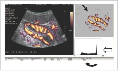
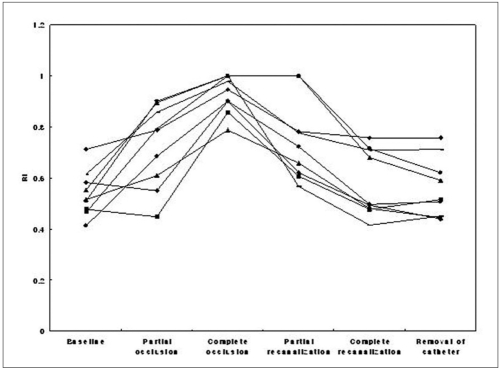
Materials and methods: We performed a PD of the kidney during graded renal vein occlusion and recanalization induced by balloon inflation and deflation in nine dogs. The PD images were transferred to a personal computer, and the PD signals were quantified.
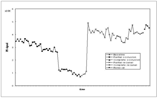
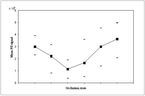
Results: We observed the temporal change of the PD signal during renal vein occlusion and recanalization, with a decrease in the PD signal during occlusion and an increase during recanalization. The mean PD signal decreased gradually as the renal vein was occluded, and conversely increased gradually with sequential relief of occlusion. The sequential change of the mean value of the PD signal was statistically significant.
Conclusion: The PD can detect a change in renal blood flow during acute renal vein occlusion and recanalization in a canine model. The PD may be used as a helpful tool for the early detection of acute renal vein thrombosis and the monitoring of renal perfusion.
Figures

Similar articles
-
Renal vascular perfusion index in a canine model.Ultrasound Med Biol. 2009 Jan;35(1):36-43. doi: 10.1016/j.ultrasmedbio.2008.06.010. Epub 2008 Sep 21. Ultrasound Med Biol. 2009. PMID: 18805627
-
The unsuitability of implantable Doppler probes for the early detection of renal vascular complications - a porcine model for prevention of renal transplant loss.PLoS One. 2017 May 25;12(5):e0178301. doi: 10.1371/journal.pone.0178301. eCollection 2017. PLoS One. 2017. PMID: 28542429 Free PMC article.
-
Quantitative estimation of renal blood flow by power Doppler ultrasonography in renovascular hypertensive dogs.Kidney Int. 2005 Dec;68(6):2781-6. doi: 10.1111/j.1523-1755.2005.00749.x. Kidney Int. 2005. PMID: 16316353
-
Nontraumatic vascular emergencies: imaging and intervention in acute venous occlusion.Eur Radiol. 2002 Nov;12(11):2627-43. doi: 10.1007/s00330-002-1615-8. Epub 2002 Aug 22. Eur Radiol. 2002. PMID: 12386751 Review.
-
[Color-Doppler semiology in transplanted kidney].Radiol Med. 1993 May;85(5 Suppl 1):68-74. Radiol Med. 1993. PMID: 8332816 Review. Italian.
Cited by
-
Evaluation of Multimode Color Doppler Flow Imaging in the Diagnosis of Solid Renal Tumor.Contrast Media Mol Imaging. 2021 Apr 1;2021:6656877. doi: 10.1155/2021/6656877. eCollection 2021. Contrast Media Mol Imaging. 2021. PMID: 33880110 Free PMC article.
-
Color Doppler ultrasound diagnosis of intrarenal vein thrombosis: A rare case report and literature review.Medicine (Baltimore). 2018 Mar;97(13):e0284. doi: 10.1097/MD.0000000000010284. Medicine (Baltimore). 2018. PMID: 29595692 Free PMC article.
References
-
- Hibbert J, Howlett DC, Greenwood KL, MacDonald LM, Saunders AJ. The ultrasound appearances of neonatal renal vein thrombosis. Br J Radiol. 1997;70:1191–1194. - PubMed
-
- Llach F, Papper S, Massry SG. The clinical spectrum of renal vein thrombosis: acute and chronic. Am J Med. 1980;69:819–827. - PubMed
-
- Laplante S, Patriquin HB, Robitaille P, Filiatrault D, Grignon A, Decarie JC. Renal vein thrombosis in children: evidence of early flow recovery with Doppler US. Radiology. 1993;189:37–42. - PubMed
-
- Paling MR, Wakefield JA, Watson LR. Sonography of experimental acute renal vein occlusion. J Clin Ultrasound. 1985;13:647–653. - PubMed
-
- Rosenfield AT, Zeman RK, Cronan JJ, Taylor KJ. Ultrasound in experimental and clinical renal vein thrombosis. Radiology. 1980;137:735–741. - PubMed
Publication types
MeSH terms
LinkOut - more resources
Full Text Sources
