. 2008 Sep;128(9):2190-7.
doi: 10.1038/jid.2008.81. Epub 2008 Apr 3.

Activation of keratinocyte protein kinase C zeta in psoriasis plaques

Affiliations

Activation of keratinocyte protein kinase C zeta in psoriasis plaques

Yuming Zhao et al. J Invest Dermatol. 2008 Sep.

Abstract

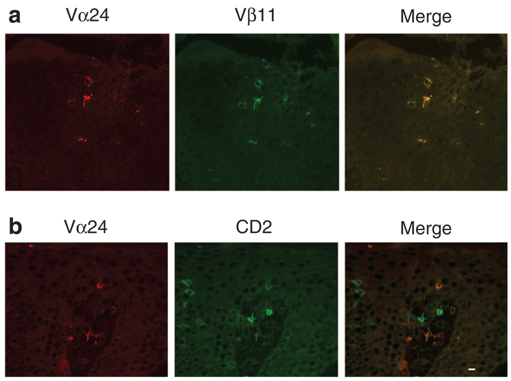
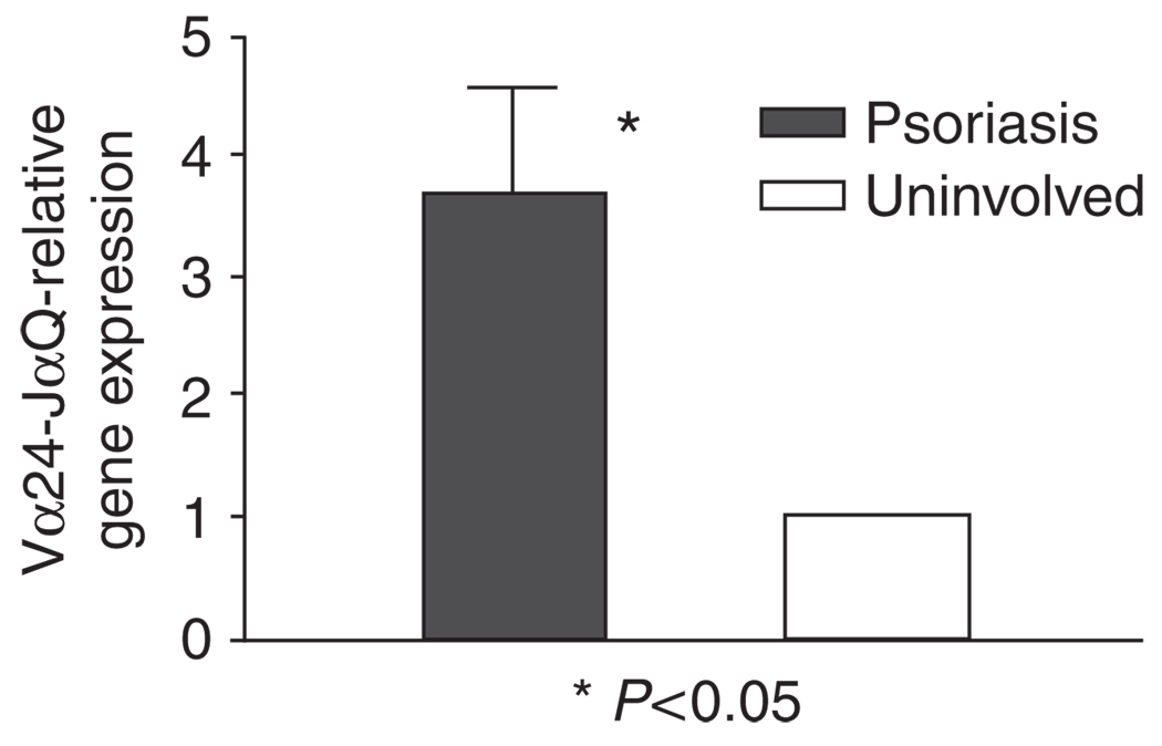
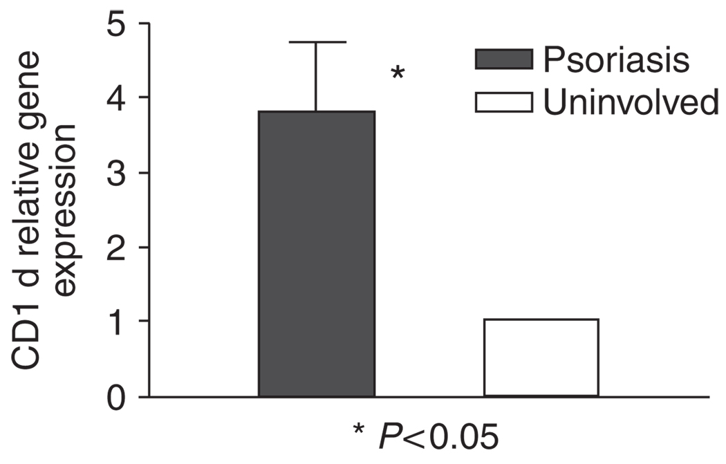
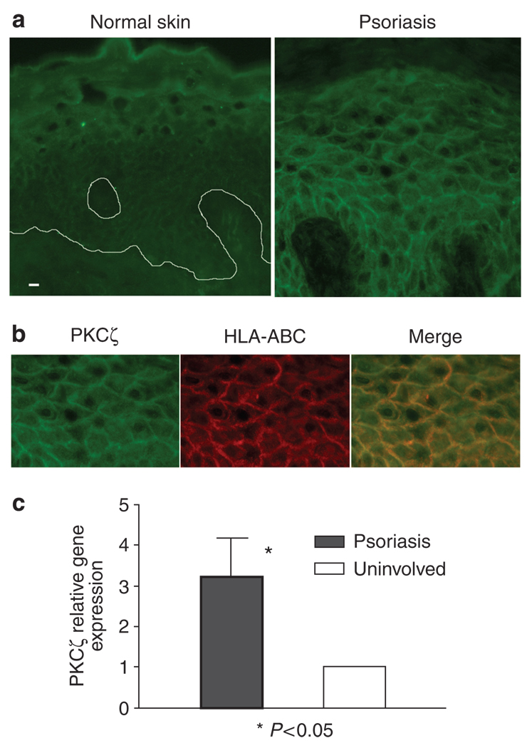
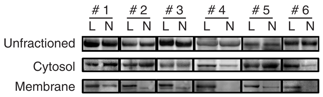
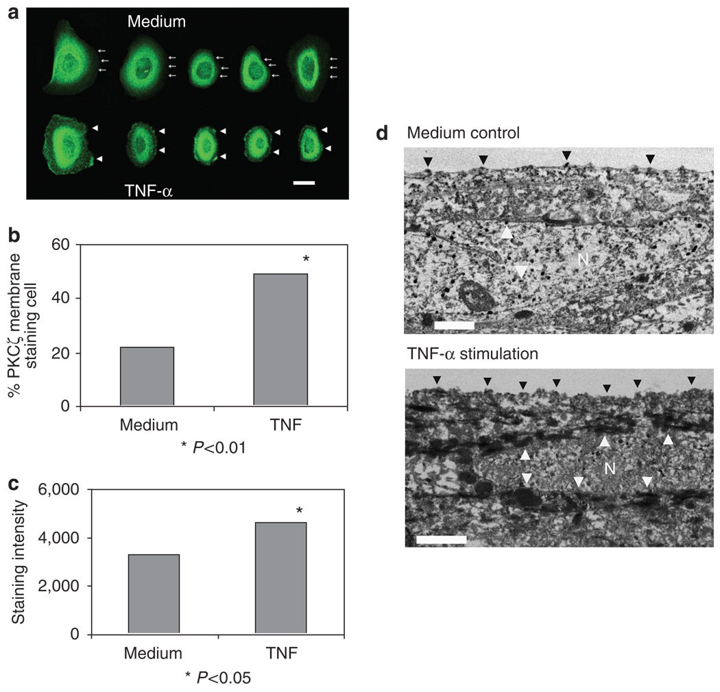
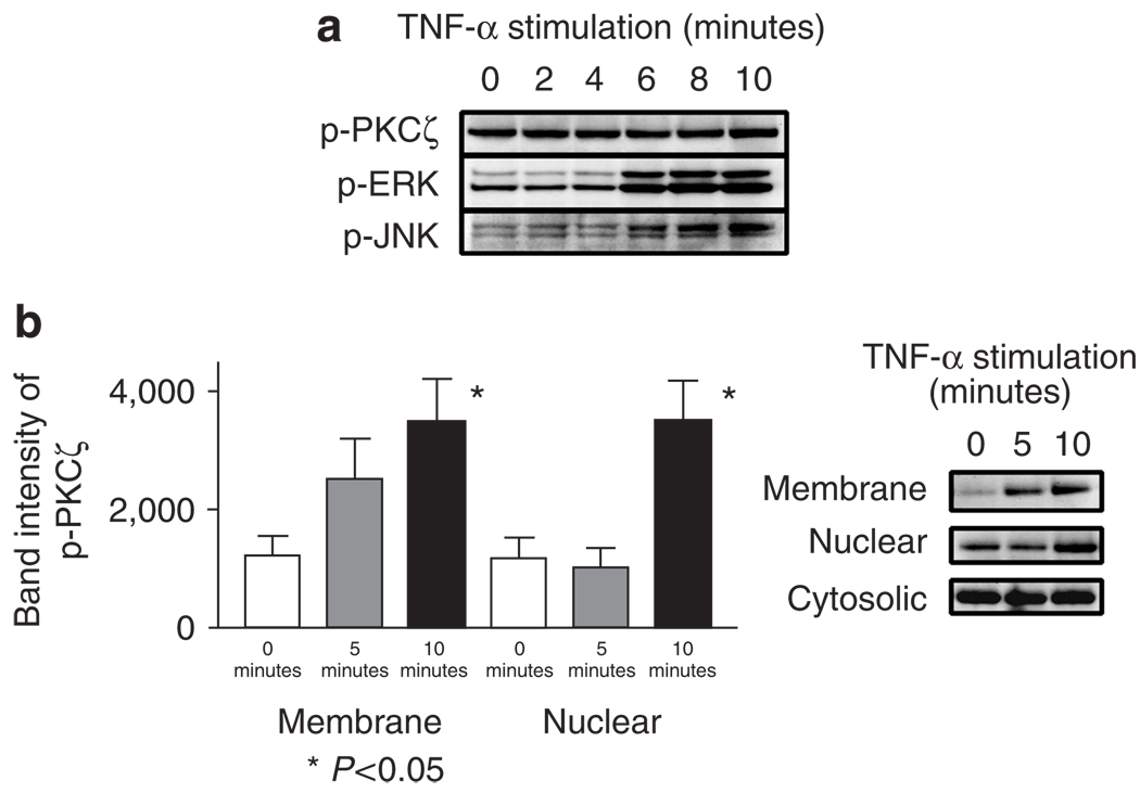
PKCzeta (protein kinase C-zeta), a member of protein kinase C family, plays an important role in cell proliferation, differentiation, and apoptosis. It acts as a downstream molecule for TNF-alpha (tumor necrosis factor) signal transduction and also regulates the expression of CD1d, an HLA-class I-like molecule. The interaction of CD1d with natural killer T (NKT) cells has been shown to be important in their Th1 cytokine production in psoriasis. In this study, we examined PKCzeta in psoriasis in order to define its role in the pathogenesis of the disease. We found that T-cell receptor (TCR) V alpha24+ V beta11+ NKT cells and CD1d molecules within psoriatic skin were increased. Moreover, there was an associated increase in PKCzeta mRNA and protein expression with membrane translocation in psoriasis lesions compared to uninvolved skin. Furthermore, cultured keratinocytes exhibited increased PKCzeta activity and membrane translocation upon stimulation by TNF-alpha, a cytokine known to play an important role in the pathogenesis of psoriasis. These results implied that PKCzeta is an important transduction molecule downstream of TNF-alpha signaling and is associated with increased expression of CD1d that may enhance CD1d-NKT cell interactions in psoriasis lesions. This makes PKCzeta a tempting target for possible pharmacological intervention in modifying the downstream effects of TNF-alpha in psoriasis.


Conflict of interest statement

CONFLICT OF INTEREST

The authors state no conflict of interest.

Figures

Figure 1
Figure 1. NKT cells increased in psoriasis
(a) Increase of Vα24 + Vβ11 + double positive NKT cells in the epidermis of a psoriatic plaque. All Vα24-positive cells in psoriasis were also Vβ11-positive. The isotype controls were negative and Vα24 and Vβ11 single labeling showed no isotype cross-reactivity (not shown). (b) CD2 + Vα24 + double positive NKT cells (merge), as well as Va24 (red) or CD2 (green) single positive cells, in the psoriatic epidermis and dermal papilla. Bar = 10 µm.
Figure 2
Figure 2. Infiltrating lymphocytes in psoriatic plaques express increased invariant Vα24-JαQ transcripts
Relative Vα24-JαQ gene expression was measured using real-time PCR to analyze gene expression from six psoriasis (lesional and paired uninvolved skin). There is significant increase in relative Vα24-JαQ gene expression in psoriasis lesions (P < 0.05, Wilcoxon signed-rank test). Values are mean ± SD, error bar = 1 SD.
Figure 3
Figure 3. CD1d gene expression is increased in psoriatic plaques
Increased CD1d mRNA in psoriatic plaques was shown compared with uninvolved skin in the six patients by real-time PCR (P < 0.05, Wilcoxon rank test). Values are mean ± SD, error bar=1 SD.
Figure 4
Figure 4. Increased membrane expression of PKCζ by psoriatic plaques
(a) Immunofluorescence of PKCζ in psoriasis and normal control skin showed increased cytoplasmic and membrane staining for PKCζ in psoriasis. Bar = 10 µm. (b) Double labeling with anti-HLA-ABC antibody showed membrane localization of PKCζ in psoriasis lesion. (c) When mRNA from six pairs of both lesional and uninvolved skin specimens was examined by real-time PCR, PKCζ gene expression was increased in all psoriasis samples compared with uninvolved skin (P < 0.05, Wilcoxon rank test). Values are mean ± SD, error bar = 1 SD.
Figure 5
Figure 5. Increased phosphorylated PKCζ in membrane fractions in psoriasis plaques
Whereas no consistent difference was demonstrated in phosphorylated PKCζ in the whole lysates or in the cytosolic fractions of psoriasis and their uninvolved skin (n = 6), the membrane fractions of psoriasis lesions (L) showed significant increase in phospho-PKCζ compared with their uninvolved counterparts (N). P < 0.01, Student’s t-test. Values are mean ± SD, error bar = 1 SD.
Figure 6
Figure 6. Increased PKCζ and membrane translocation in KCs stimulated with TNF
(a) Confocal images: KCs cultured on coverslips stimulated with TNF-α showed prominent membrane staining for PKCζ (arrowheads) than the unstimulated cells (arrows at the dim cell boundaries). Bar = 10 µm. The picture is representative of three separate experiments. (b) The cells with membrane staining were increased (48.9%) significantly in TNF-α-stimulated cells compared with the medium alone (22.1%; P < 0.01 by Fisher’s exact test). One hundred cells were counted for each condition. (c) The fluorescence staining intensity of TNF-α stimulated cells increased significantly than those in medium alone. The intensities were measured by image analysis system of the fluorescence micrographs. P < 0.05, Student’s t-test. (d) Electron microscopic images of HaCaT on culture inserts stimulated with or without TNF-α, 100 ng ml−1, for 10 minutes. Cell stimulated with TNF-α showed stronger staining (arrowheads) for PKCζ in the plasma membrane as well as in the nuclear and perinuclear areas apart from cytoplasmic staining, compared with unstimulated control cells. Note: N denotes nucleus. Bar = 1 µm.
Figure 7
Figure 7. Increased phosphorylated PKCζ in membrane fractions of KCs stimulated with TNF
(a) KCs were stimulated with 100 ng ml−1 of TNF-α for 0, 2, 4, 6, 8, and 10 minutes. While no significant change in the quantity of phospho-PKCζ was observed, pERK and pJNK were increased 6 minutes after TNF-α stimulation. (b) Increased phospho-PKCζ in the membrane and nuclear, but not in the cytosolic fractions, of primary KCs stimulated with TNF-α 100 ng ml−1 for 5 and 10 minutes (Student’s t-test). These blots are representations of three separate experiments. Values are mean ± SD, error bar = 1 SD.

Similar articles

Cited by

References

    1. Berra E, Díaz-Meco MT, Lozano J, Frutos S, Municio MM, Sánchez P, et al. Evidence for a role of MEK and MAPK during signal transduction by protein kinase C zeta. EMBO J. 1995;14:6157–6163. - PMC - PubMed
    1. Bonish B, Jullien D, Dutronc Y, Huang BB, Modlin R, Spada FM, et al. Overexpression of CD1d by keratinocytes in psoriasis and CD1d-dependent IFN-gamma production by NK-T cells. J Immunol. 2000;165:4076–4085. - PubMed
    1. Bos JD. Psoriasis, innate immunity, and gene pools. J Am Acad Dermatol. 2007;56:468–471. - PubMed
    1. Cameron AL, Kirby B, Fei W, Griffiths CE. Natural killer and natural killer-T cells in psoriasis. Arch Dermatol Res. 2002;294:363–369. - PubMed
    1. Cohen EE, Lingen MW, Zhu B, Zhu H, Straza MW, Pierce C, et al. Protein kinase C zeta mediates epidermal growth factor-induced growth of head and neck tumor cells by regulating mitogen-activated protein kinase. Cancer Res. 2006;66:6296–6303. - PubMed

Publication types

MeSH terms