Review
. 2008 Jul;130(1):21-54.
doi: 10.1007/s00418-008-0420-0. Epub 2008 Apr 3.

The desmosome and pemphigus

Affiliations
Review

The desmosome and pemphigus

Jens Waschke. Histochem Cell Biol. 2008 Jul.

Abstract

Desmosomes are patch-like intercellular adhering junctions ("maculae adherentes"), which, in concert with the related adherens junctions, provide the mechanical strength to intercellular adhesion. Therefore, it is not surprising that desmosomes are abundant in tissues subjected to significant mechanical stress such as stratified epithelia and myocardium. Desmosomal adhesion is based on the Ca(2+)-dependent, homo- and heterophilic transinteraction of cadherin-type adhesion molecules. Desmosomal cadherins are anchored to the intermediate filament cytoskeleton by adaptor proteins of the armadillo and plakin families. Desmosomes are dynamic structures subjected to regulation and are therefore targets of signalling pathways, which control their molecular composition and adhesive properties. Moreover, evidence is emerging that desmosomal components themselves take part in outside-in signalling under physiologic and pathologic conditions. Disturbed desmosomal adhesion contributes to the pathogenesis of a number of diseases such as pemphigus, which is caused by autoantibodies against desmosomal cadherins. Beside pemphigus, desmosome-associated diseases are caused by other mechanisms such as genetic defects or bacterial toxins. Because most of these diseases affect the skin, desmosomes are interesting not only for cell biologists who are inspired by their complex structure and molecular composition, but also for clinical physicians who are confronted with patients suffering from severe blistering skin diseases such as pemphigus. To develop disease-specific therapeutic approaches, more insights into the molecular composition and regulation of desmosomes are required.


Figures
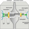
Fig. 1
Fig. 1
Molecular model of the desmosome. The desmosomal cadherins desmoglein and desmocollin undergo homophilic and heterophilic binding via interaction with the amino-terminal extracellular (EC) 1 domain of partner molecules on the same (cis) as well as on the neighbouring cell (trans). The cytoplasmic domains are largely embedded in the outer dense plaque (ODP) where they are associated with plakoglobin and plakophilin. In the inner dense plaque (IDP), desmoplakin links these adaptor molecules to the intermediate filament cytoskeleton
Fig. 2
Fig. 2
Ultrastructure of the desmosome. The electron micrograph of a keratinocyte desmosome shows the desmosomal plaque with inserting cytokeratin intermediate filaments as well as some fuzzy material within the extracellular space likely reflecting the extracellular domains of desmosomal cadherins
Fig. 3
Fig. 3
Ultrastructure of the area composita of a myocardial intercalated disc. The electron micrograph shows an intercalated disc containing a gap junction (GJ) in its longitudinal section as well as an adhering junction with an extensive electron-dense plaque in the section perpendicular to the cellular axis. Note that insertion of actin filaments, which is typical for adherens junctions, is present in some parts of the junction (asterisk) but not in others (hash key). Based on the recent finding that all parts of these adhering junctions contain the same set of desmosomal components, they are now defined as area composita
Fig. 4
Fig. 4
Expression patterns of desmosomal components in the epidermis. The schematic drawing of the epidermis (left) indicates the basal (BL), spinous (SL), granular (GL) and corneal (CL) layer of the epidermis. On the right, the expression patterns of desmosomal components in the specific epidermal layers are illustrated. For instance, Dsg 1 and Pkp 1 are most prominent in the superficial layers, whereas expression of Dsg 3 and Dsc 3 is strongest in the deep epidermis. Dsg desmoglein, Dsc desmocollin, Pkp plakophilin, PG plakoglobin, DP desmoplakin
Fig. 5
Fig. 5
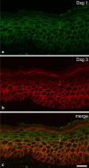
Immunostaining of Dsg 1 and Dsg 3 in human epidermis. Intact human epidermis was immunostained using monoclonal antibodies against Dsg 1 (a) and Dsg 3 (b). A merge of both panels is shown in c. Dsg 1 is most abundant in the superficial epidermis but is also present in the basal layer. Dsg 3 is expressed in the basal layer as well as throughout the spinous layer indicating that in human epidermis the expression patterns of these two proteins broadly overlap. Scale bar is 20 μm
Fig. 6
Fig. 6
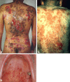
Clinical phenotype of Pemphigus vulgaris and Pemphigus foliaceus. Patients suffering from the mucocutaneous form of pemphigus vulgaris (PV) usually have flaccid blisters and erosions on the trunk (a) accompanied by mucosal ulcerations in the mouth (b). In contrast, Pemphigus foliaceus patients are characterized by crusted epidermal erosions (c) whereas involvement of mucous membranes is absent
Fig. 7
Fig. 7
Typical histology of epidermal lesions from pemphigus patients. Hematoxylin eosin-stained paraffin sections from PV (a) and PF (b) patients showed suprabasal epidermal cleavage in the PV and superficial granular blistering in PF. Scale bar is 50 μm
Fig. 8
Fig. 8
The two principal mechanisms underlying pemphigus skin blistering. Two principal mechanisms have been proposed by which autoantibodies specific for Dsg 1 and Dsg 3 could impair desmosomal adhesion. First, antibodies could directly interfere with desmoglein transinteraction (a). Second, antibody binding has been shown to trigger intracellular signalling pathways, which indirectly results in loss of desmoglein-mediated binding (b)
Fig. 9
Fig. 9
The desmoglein compensation hypothesis. Based on the different autoantibody profiles in PV and PF together with the findings that Dsg 3 is present in the deep epidermis only whereas Dsg 1 is primarily expressed in the superficial epidermis, the desmoglein compensation hypothesis has been proposed to explain the epidermal cleavage planes typical for PV and PF. According to this model, blistering in PF affects the superficial epidermis because Dsg 3 is present in the deep epidermis to compensate for the autoantibody-induced loss of Dsg 1 binding. In PV, epidermal involvement would occur only when autoantibodies against both Dsg 1 and Dsg 3 are present because Dsg 1 is found in all epidermal layers and could compensate for loss of Dsg 3 binding when antibodies to Dsg 3 are solely present
Fig. 10
Fig. 10
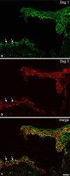
Immunostaining of Dsg 1 and Dsg 3 in PV lesional epidermis. Epidermis from a patient with mucocutaneous PV was stained for Dsg 1 (a) and Dsg 3 (b). A merge of both panels is shown in c. Both Dsg 1 and Dsg 3 are expressed in the basal layer underneath the blister as well as in keratinocytes in the blister roof. However, Dsg 3 staining appears to be fragmented throughout the epidermis whereas Dsg 1 staining is more continuous. Note that in the level of the cleavage plane the apical membrane of basal cells shows strong immunostaining for Dsg 1 and Dsg 3 (arrows). Therefore, based on the desmoglein compensation hypothesis the expression patterns of Dsg 1 and Dsg 3 cannot explain why the cleavage plane is located suprabasally in PV but not in other epidermal layers. Scale bar is 20 μm
Fig. 11
Fig. 11
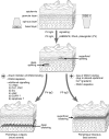
The mechanisms involved in pemphigus acantholysis. Accumulating evidence indicates that PV-IgG and PF-IgG initiate keratinocyte dissociation via intracellular signalling pathways including p38 MAPK, Rho A and plakoglobin (PV only). In addition, other mechanisms such as direct inhibition of Dsg 3 binding and Dsg 3 depletion from desmosomes as well as other signalling events seem to contribute to PV pathogenesis, whereas their role for acantholysis in PF is unclear. These mechanisms may account for the more severe clinical phenotype of PV compared to PF. PLC phospholipase C, PKC protein kinase C, cdk 2 cyclin-dependent kinase 2, EGFR epidermal growth factor receptor

Similar articles

Cited by

References

    1. {'text': '', 'ref_index': 1, 'ids': [{'type': 'PMC', 'value': 'PMC41522', 'is_inner': False, 'url': 'https://pmc.ncbi.nlm.nih.gov/articles/PMC41522/'}, {'type': 'PubMed', 'value': '7604000', 'is_inner': True, 'url': 'https://pubmed.ncbi.nlm.nih.gov/7604000/'}]}
    2. Aberle H, Bierkamp C, Torchard D, Serova O, Wagner T, Natt E, Wirsching J, Heidkamper C, Montagna M, Lynch HT et al (1995) The human plakoglobin gene localizes on chromosome 17q21 and is subjected to loss of heterozygosity in breast and ovarian cancers. Proc Natl Acad Sci USA 92:6384–6388 - PMC - PubMed
    1. {'text': '', 'ref_index': 1, 'ids': [{'type': 'PubMed', 'value': '17065638', 'is_inner': True, 'url': 'https://pubmed.ncbi.nlm.nih.gov/17065638/'}]}
    2. Ahmed AR, Spigelman Z, Cavacini LA, Posner MR (2006) Treatment of pemphigus vulgaris with rituximab and intravenous immune globulin. New Engl J Med 355:1772–1779 - PubMed
    1. {'text': '', 'ref_index': 1, 'ids': [{'type': 'PubMed', 'value': '17965884', 'is_inner': True, 'url': 'https://pubmed.ncbi.nlm.nih.gov/17965884/'}]}
    2. Akat K, Bleck CK, Lee YM, Haselmann-Weiss U, Kartenbeck J (2008) Characterization of a novel type of adherens junction in meningiomas and the derived cell line HBL-52. Cell Tissue Res 331:401–412 - PubMed
    1. {'text': '', 'ref_index': 1, 'ids': [{'type': 'PMC', 'value': 'PMC32271', 'is_inner': False, 'url': 'https://pmc.ncbi.nlm.nih.gov/articles/PMC32271/'}, {'type': 'PubMed', 'value': '11294891', 'is_inner': True, 'url': 'https://pubmed.ncbi.nlm.nih.gov/11294891/'}]}
    2. Akhtar N, Hotchin NA (2001) RAC1 regulates adherens junctions through endocytosis of E-cadherin. Molec Biol Cell 12:847–862 - PMC - PubMed
    1. {'text': '', 'ref_index': 1, 'ids': [{'type': 'PubMed', 'value': '18064004', 'is_inner': True, 'url': 'https://pubmed.ncbi.nlm.nih.gov/18064004/'}]}
    2. Al-Amoudi A, Diez DC, Betts MJ, Frangakis AS (2007) The molecular architecture of cadherins in native epidermal desmosomes. Nature 450:832–837 - PubMed

Publication types