Review
. 2008 Jun;19(3):245-51.
doi: 10.1016/j.semcdb.2008.02.003. Epub 2008 Feb 23.

Role of adenomatous polyposis coli (APC) and microtubules in directional cell migration and neuronal polarization

Affiliations
Review

Role of adenomatous polyposis coli (APC) and microtubules in directional cell migration and neuronal polarization

Angela I M Barth et al. Semin Cell Dev Biol. 2008 Jun.

Abstract

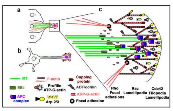
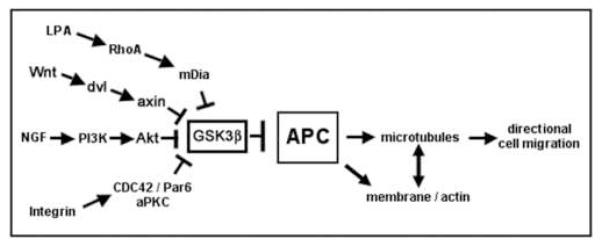
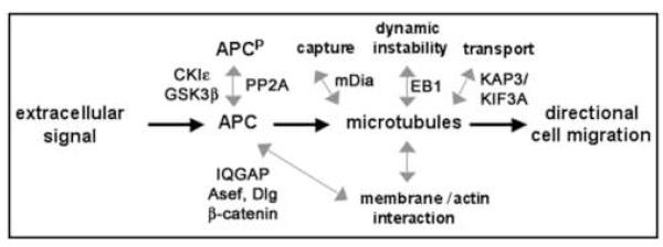
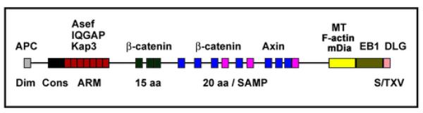
In response to extracellular signals during embryonic development, cells undergo directional movements to specific sites and establish proper connections to other cells to form organs and tissues. Cell extension and migration in the direction of extracellular cues is mediated by the actin and microtubule cytoskeletons, and recent results have shed new light on how these pathways are activated by neurotrophins, Wnt or extracellular matrix. These signals lead to modifications of microtubule-associated proteins (MAPs) and point to glycogen synthase kinase (GSK) 3beta as a key regulator of microtubule function during directional migration. This review will summarize these results and then focus on the role of microtubule-binding protein adenomatous polyposis coli (APC) in neuronal polarization and directed migration, and on its regulation by GSK3beta.


Figures

Fig 1
Fig 1. Organization of cytoskeleton and signaling proteins in directional cell migration and extension
(A) Epithelial or astrocyte migrating into a wound area; (B) neuronal cell extending axon and dendrites; (C) Cytoskeletal organization and signaling proteins at the leading edge: The actin (G- & F-actin) cytoskeleton (red) provides a membrane protrusive force by actin polymerization at the leading edge. The microtubule (MT) cytoskeleton (green) delivers membrane components via the secretory pathway and provides a protrusive force by microtubule polymerization (pioneering) and bundling. The microtubule-organizing center (MTOC, green dots in A and B) orients towards the direction of extension, and microtubules orient along the axis of extension, being laterally guided by cortical actin bundles attached to focal adhesions. Growth factor signals and integrin-matrix adhesion at the leading edge stimulate small GTPases Cdc42, Rac and Rho. Cdc42 and Rac promote actin polymerization by activating the WASP- and WAVE-Arp2/3 actin-nucleation complexes in filopodia and lamellipodia. In epithelial cells, Rho promotes directed migration by inducing actin/myosin contractility in the rear and contraction forces at actin bundle-focal adhesion connections in the front. In neuronal cells, an actin arc separates the central microtubule rich area of the growth cone from peripheral actin rich lamellipodia and filopodia and Rho activity promotes growth cone repulsion. Small GTPases also activate the leading edge-enriched MAP APC. The mechanism of this activation is poorly understood but activated APC promotes directed cell extension probably by interacting with the microtubule plus-end binding protein EB1 and regulating microtubule dynamics at the leading edge. APC can interact with actin either directly via its actin binding domain or indirectly via binding to the CDC42/Rac effector and actin binding protein IQGAP.
Fig. 2
Fig. 2
Signaling pathways that induce activation of APC in cell migration.
Fig. 3
Fig. 3. APC as a common target of signals that regulate cell motility
grey double arrows indicate potential changes in APC phosphorylation and interaction with binding partners that need further investigation.
Fig. 4
Fig. 4. APC protein-protein interaction domains
Grey: dimerization domain; Black: conserved APC domain. Red: armadillo repeat domain that binds Asef, IQGAP and Kap3. The N-terminal 1121 amino acids also contain a binding site for the B56 regulatory subunit of PP2A. Dark green and dark purple: 3 repeats of 15 amino acids (15aa) and 7 repeats of 20 amino acids (20 aa), respectively, which bind β~catenin. Light purple: 3 SAMP domains in between the 20 aa repeats which bind axin. Yellow: microtubule/F-actin/mDia-binding region between amino acids 2219 and 2580. Olive: C-terminal region that binds to the plus-end microtubule binding protein EB1. Pink: C-terminal S/TXV motif binds to PDZ domains of Dlg, PSD-95.

References

    1. Thiery JP. Mechanisms of cell migration in the vertebrate embryo. Cell Differ. 1984;15:1–15. - PubMed
    1. Hynes RO, Lander AD. Contact and adhesive specificities in the associations, migrations, and targeting of cells and axons. Cell. 1992;68:303–22. - PubMed
    1. Ridley AJ, Schwartz MA, Burridge K, Firtel RA, Ginsberg MH, Borisy G, et al. Cell migration: integrating signals from front to back. Science. 2003;302:1704–9. - PubMed
    1. Gordon MD, Nusse R. Wnt signaling: multiple pathways, multiple receptors, and multiple transcription factors. J Biol Chem. 2006;281:22429–33. - PubMed
    1. Lykissas MG, Batistatou AK, Charalabopoulos KA, Beris AE. The role of neurotrophins in axonal growth, guidance, and regeneration. Curr Neurovasc Res. 2007;4:143–51. - PubMed

Publication types

Substances

LinkOut - more resources