

Donor age and corneal endothelial cell loss 5 years after successful corneal transplantation. Specular microscopy ancillary study results
- PMID: 18387408
- PMCID: PMC2959119
- DOI: 10.1016/j.ophtha.2008.01.004
Donor age and corneal endothelial cell loss 5 years after successful corneal transplantation. Specular microscopy ancillary study results
Abstract
Objective: To determine whether endothelial cell loss 5 years after successful corneal transplantation is related to the age of the donor.
Design: Multicenter, prospective, double-masked clinical trial.
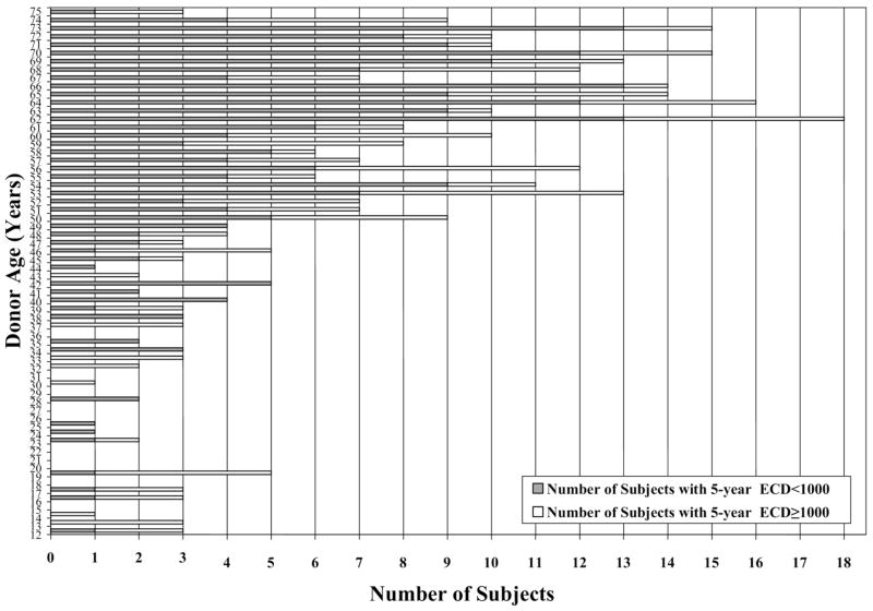
Participants: Three hundred forty-seven subjects participating in the Cornea Donor Study who had not experienced graft failure 5 years after corneal transplantation for a moderate-risk condition (principally Fuchs' dystrophy or pseudophakic corneal edema).
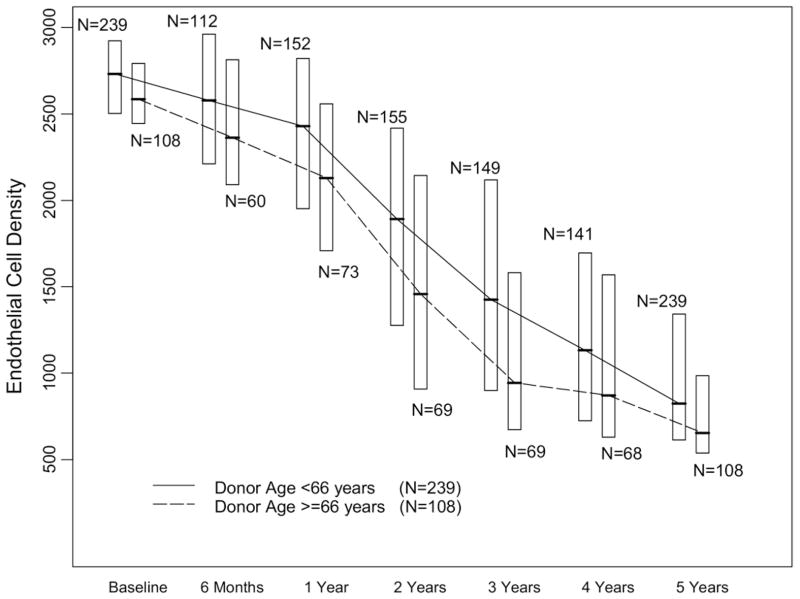
Testing: Specular microscopic images of donor corneas obtained before surgery and postoperatively at 6 months, 12 months, and then annually through 5 years were submitted to a central reading center to measure endothelial cell density (ECD).
Main outcome measure: Endothelial cell density at 5 years.
Results: At 5 years, there was a substantial decrease in ECD from baseline for all donor ages. Subjects who received a cornea from a donor 12 to 65 years old experienced a median cell loss of 69% in the study eye, resulting in a 5-year median ECD of 824 cells/mm(2) (interquartile range, 613-1342), whereas subjects who received a cornea from a donor 66 to 75 years old experienced a cell loss of 75%, resulting in a median 5-year ECD of 654 cells/mm(2) (interquartile range, 538-986) (P [adjusted for baseline ECD] = 0.04). Statistically, there was a weak negative association between ECD and donor age analyzed as a continuous variable (r [adjusted for baseline ECD] = -0.19; 95% confidence interval, -0.29 to -0.08).
Conclusions: Endothelial cell loss is substantial in the 5 years after corneal transplantation. There is a slight association between cell loss and donor age. This finding emphasizes the importance of longer-term follow-up of this cohort to determine if this relationship affects graft survival.
Figures


References
-
- Eye Bank Association of America. Medical Standards. Washington: EBAA; 2006. pp. 1–36.
-
- Abbott RL, Fine M, Guillet E. Long-term changes in corneal endothelium following penetrating keratoplasty: a specular microscopic study. Ophthalmology. 1983;90:676–85. - PubMed
-
- Culbertson WW, Abbott RL, Forster RK. Endothelial cell loss in penetrating keratoplasty. Ophthalmology. 1982;89:600–4. - PubMed
-
- Zacks CM, Abbott RL, Fine M. Long-term changes in corneal endothelium after keratoplasty: a follow-up study. Cornea. 1990;9:92–7. - PubMed
-
- Bertelmann E, Pleyer U, Rieck P. Risk factors for endothelial cell loss post-keratoplasty. Acta Ophthalmol Scand. 2006;84:766 –70. - PubMed
Publication types
MeSH terms
Grants and funding
LinkOut - more resources
Full Text Sources
Medical
