. 2008 Jun 13;283(24):16928-39.
doi: 10.1074/jbc.M801454200. Epub 2008 Apr 3.

PATE gene clusters code for multiple, secreted TFP/Ly-6/uPAR proteins that are expressed in reproductive and neuron-rich tissues and possess neuromodulatory activity

Affiliations

PATE gene clusters code for multiple, secreted TFP/Ly-6/uPAR proteins that are expressed in reproductive and neuron-rich tissues and possess neuromodulatory activity

Fiana Levitin et al. J Biol Chem. .

Abstract

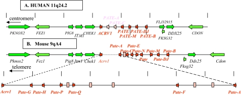
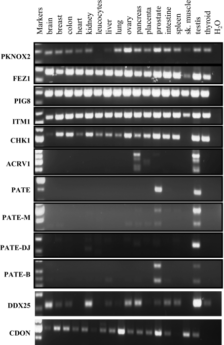
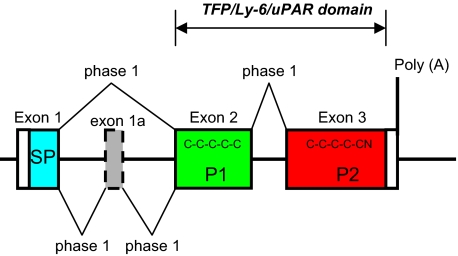
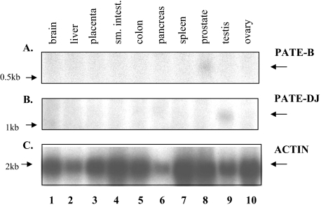
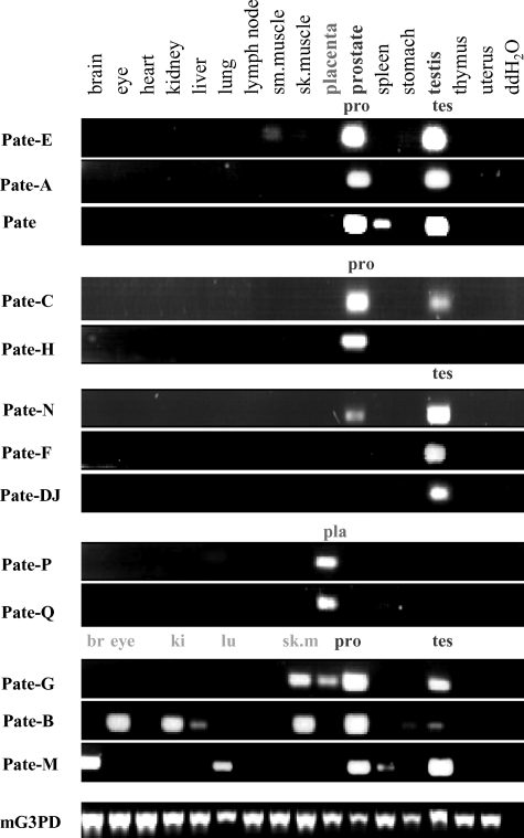
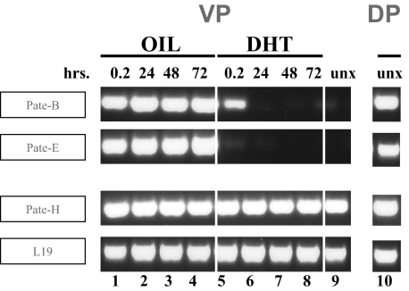
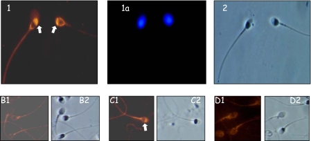
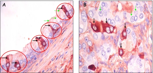
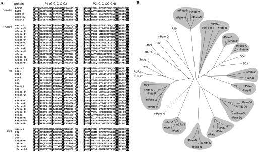
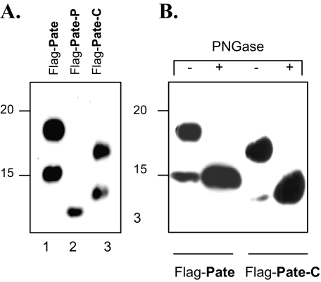
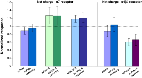
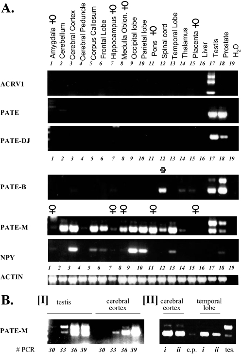
We report here syntenic loci in humans and mice incorporating gene clusters coding for secreted proteins each comprising 10 cysteine residues. These conform to three-fingered protein/Ly-6/urokinase-type plasminogen activator receptor (uPAR) domains that shape three-fingered proteins (TFPs). The founding gene is PATE, expressed primarily in prostate and less in testis. We have identified additional human PATE-like genes (PATE-M, PATE-DJ, and PATE-B) that co-localize with the PATE locus, code for novel secreted PATE-like proteins, and show selective expression in prostate and/or testis. Anti-PATE-B-specific antibodies demonstrated the presence of PATE-B in the region of the sperm acrosome and at high levels on malignant prostatic epithelial cells. The syntenic mouse Pate-like locus encompasses 14 active genes coding for secreted proteins, which are all, except for Pate-P and Pate-Q, expressed primarily in prostate and/or testis. Pate-P and Pate-Q are expressed solely in placental tissue. Castration up-regulates prostate expression of mouse Pate-B and Pate-E, whereas testosterone ablates this induced expression. The sequence similarity between TFP/Ly-6/uPAR proteins that modulate activity of nicotinic acetylcholine receptors and the PATE (Pate)-like proteins stimulated us to see whether these proteins possess analogous activity. Pharmacological studies showed significant modulation of the nicotinic acetylcholines by the PATE-B, Pate-C, and Pate-P proteins. In concert with these findings, certain PATE (Pate)-like genes were extensively expressed in neuron-rich tissues. Taken together, our findings indicate that in addition to participation of the PATE (Pate)-like genes in functions related to fertility and reproduction, some of them likely act as important modulators of neural transmission.


Figures

FIGURE 1.
FIGURE 1.
Arrangement of genes within the locus comprising the human PATE-like genes and the corresponding syntenic murine genomic locus. A, known human genes lie within a genomic segment on chromosome 11q24 initiating at the centromeric side with PKNOX2 at nucleotide 124,726,419 (numbering as in the Genome Browser May 2004 release), and terminating at the telomeric side with CDON at nucleotide 125,438,397. Arrows indicate direction of transcription. The genes coding for proteins containing the distinctive 10-cysteine motif are shown in red, and inactive pseudogenes are stippled. Accession numbers are as follows: ACRV1, NM_001612; PATE, NM_138294; PATE-M, NM_212555; PATE-B, AK123042. Accession number for PATE-DJ is EU703625. B, syntenic murine (chromosome 9qA4) genomic locus. Note the 0.8-Mbp insertion in the mouse genome between Acrv1 and Pate-A. Accession numbers are as follows: Acrv1, NM_007391; Pate-H, BY721155; Pate-Q, BQ032923; Pate-F, BY721010; Pate-A, AK020329; Pate-C, NM_026593; Pate-E:, AV379335; Pate, AK033745; Pate-M, BY721028; Pate-B (svs7), NM_020264. Accession numbers for Pate-G, Pate-P, Pate-N, and Pate-DJ are EU703627, EU703629, EU703628, and EU703626, respectively.
FIGURE 2.
FIGURE 2.
RT-PCR expression analysis of human PATE-like genes and flanking genes. RT-PCR analysis of the human PATE-like genes and flanking genes was performed with cDNAs obtained from the indicated human tissues. Forward and reverse primers were chosen such that they always spanned an intron, and the observed RT-PCR product at all times corresponded to the size expected of a spliced mRNA. For the ACRV1, PATE, PATE-M, PATE-DJ, and PATE-B genes, the forward and reverse primers were located in the first and third exons (coding for the signal peptide and cysteines 6-10, respectively). PCR was performed for 35 cycles. Note that PIG8 (p53 induced gene 8) is ubiquitously expressed in all tissues, serving as a convenient internal control for cDNA integrity.
FIGURE 3.
FIGURE 3.
Exon structure of PATE (Pate)-like genes. The canonical exon structure of the PATE (Pate)-like genes is presented. Optional extra exons are represented as dotted boxes. The coding region is in color: blue, signal peptide (SP); green, pattern 1 (P1) containing C1 to C5; red, pattern 2 (P2) containing C6 to C10N.
FIGURE 4.
FIGURE 4.
Northern blot analyses of PATE-B and PATE/DJ expression. PATE-B, PATE/DJ, and actin cDNAs were radioactively (32P) labeled and each used to sequentially probe, under stringent wash conditions, a Northern blot (Clontech) of total RNA derived from the indicated human tissues. The blot was stripped between sequential probing. Expression of PATE-B and PATE/DJ mRNAs is clearly visible in prostatic and testicular tissues (lanes 8 and 9, respectively); no signal was seen in any other tissues examined.
FIGURE 5.
FIGURE 5.
RT-PCR expression analyses of mouse Pate-like genes. RT-PCR analyses of the mouse Pate-like genes were performed with cDNAs (Clontech) obtained from different mouse tissues as indicated. Forward and reverse primers were chosen such that they always spanned an intron, and all RT-PCR products corresponded to the sizes expected of spliced mRNA. Results presented here used forward and reverse primers located in the second and third exons (coding for cysteines 1-5 and cysteines 6-10, respectively); similar results were obtained when the analysis was repeated with forward and reverse primers located in the first and third exons (data not shown). PCR was performed for 35 cycles, and the PCR products were analyzed as described under “Experimental Procedures.” The ubiquitously expressed mouse glyceraldehyde-3-phosphate dehydrogenase (mG3PDH) served as a control for cDNA integrity.
FIGURE 6.
FIGURE 6.
Effect of castration and subsequent DHT administration on Pate-like gene expression in the ventral and dorsal lobes of the mouse prostate. Mice were castrated and 14 days later injected (subcutaneously) at time 0, 24, and 48 h either with oil or with DHT dissolved in the oil. Mice were sacrificed 12 min and 24, 48, and 72 h, respectively, and the ventral and dorsal prostate lobes were isolated, followed by RNA isolation and cDNA preparation. RT-PCR analyses were performed using forward and reverse primers located in the first and third exons. PCR was performed for 35 cycles, and the PCR products were analyzed as described under “Experimental Procedures.” The ubiquitously expressed mouse L19 gene served as a control for cDNA integrity. unx, uncastrated.
FIGURE 7.
FIGURE 7.
Localization of the PATE-B protein to the acrosomal region of human sperm. Panels 1, 1a, and 2, human sperm was incubated with anti-PATE-B antisera followed by fluorescently labeled anti-rabbit antibodies. Specific staining was observed in the region of the sperm acrosome (panel 1). Staining with 4′,6-diamidino-2-phenylindole demonstrated the sperm nucleus (panel 1a), and Nomarski optics showed the whole sperm (panel 2). Panels B-D, human sperm similarly investigated with preimmune serum (panel B1) or with immune anti-PATE-B antisera plus a nonrelevant protein (panel C1) or competing soluble PATE-B protein (panel D1) attested to the specificity of staining, indicated by the white arrow. Nomarski optics demonstrated the sperm cells (panels B2, C2, and D2).
FIGURE 8.
FIGURE 8.
Human PATE-B protein expression in discrete prostatic apical epithelial cells and in malignant epithelial cells. A, tissue sections of paraffin-embedded prostate tissue containing both normal and malignant prostate cells were reacted with anti-PATE-B rabbit polyclonal antisera, and antibody binding was detected with polymer-horseradish peroxidase-conjugated anti-rabbit antibodies followed by 3-amino-9-ethylcarbazole chromagen. Staining is seen in isolatedcells (blackarrows) suggesting that a specific subpopulation of the ductal epithelial cells express PATE-B; other cells in this duct (green arrows indicating selected cells) are barely stained indicating very low PATE-B expression. B, malignant prostate cells from a different region but contained in the section as in A. Discrete malignant cells are very intensely stained (black arrows) indicating high PATE-B expression, whereas other malignant cells (green arrows indicating selected cells) show very little staining.
FIGURE 9.
FIGURE 9.
Multiple sequence alignment of C10N motif and a phylogenetic tree of PATE (Pate)-like proteins from human, mouse, rat, and dog. A, the amino acid sequences of the human, mouse, rat, and dog PATE (Pate)-like proteins are presented. The first (P1) and second (P2) blocks show sequences extending from C1 to C5 and C6 to C10N, respectively. The C10N motifs are highlighted. Where possible Pate-like nomenclature has been used for the interspecies orthologs. Otherwise arbitrary designations, such as R05, were used for illustration. Expression of all human and mouse PATE (Pate)-like genes presented have been demonstrated experimentally by RT-PCR analyses. B, same multiple sequence alignment was used to build a phylogenetic tree. The tree is a Neighbor-joining consensus tree based on 1000 replicates. Apparent orthologous groups are marked with a gray background.
FIGURE 10.
FIGURE 10.
Expression of Pate, Pate-P, and Pate-C proteins, N-glycosylation of Pate and Pate-C in HEK293 (human kidney) cells. A, HEK293 cells were transfected with constructs encoding the proteins that comprised the Pate, Pate-P, or Pate-C sequences, tagged at their N termini with a FLAG epitope, and the secreted Pate-like proteins purified as described under “Experimental Procedures.” The purified proteins were analyzed by 11% SDS-PAGE, Western-blotted, and probed with anti-FLAG antibodies (A and B). B, Western blot analysis of FLAG-tagged Pate-like proteins treated with PNGase F. Proteins were treated as described in A, with the difference that the proteins were treated with PNGase F to remove N-linked sugars (lanes indicated by +).
FIGURE 11.
FIGURE 11.
Modulation of the activity of nAChRs by PATE (Pate)-like proteins. The indicated nAChRs were expressed in Xenopus oocytes as described under “Experimental Procedures.” After obtaining initial control responses to 8-s applications of ACh, cells were washed for 4 min with either control Ringer or Ringer's plus the PATE-like proteins (60 or 200 nm) and then challenged with ACh or ACh plus the PATE-like proteins, respectively. Responses to ACh plus the PATE-like proteins were normalized to the net charge responses to ACh alone for each oocyte. ACh concentrations were 30 μm for α4β2-expressing cells and 60 μm for α7-expressing cells. To test for recovery, oocytes were then washed for an additional 4 min in the absence of acetylcholine and the PATE-like proteins, and then acetylcholine (30 or 60 μm) was applied for 8 s and the net charge calculated to give the recovery values.
FIGURE 12.
FIGURE 12.
Expression of human PATE-like genes in different regions of the brain. A, upper panel, cDNAs prepared from different individual regions of the brain as indicated, were subjected to RT-PCR analyses. Expression analysis of the human PATE-like genes was performed with forward and reverse primers that always spanned an intron and the observed RT-PCR product at all times corresponded to the size expected of a spliced mRNA. For the ACRV1, PATE, PATE-M, PATE-DJ, and PATE-B genes, the forward and reverse primers were located in the first and third exons (coding for the signal peptide and cysteines 6-10, respectively). In addition, cDNAs derived from testes and prostate on the one hand and liver and placenta on the other hand were also analyzed for expression of the PATE-like genes and served as positive and negative controls, respectively. PCR was performed for 35 cycles. Control for cDNA integrity was provided by the actin primers, as indicated, and expression analysis of the brain neuropeptide NPY served as a positive control for expression of a brain neuropeptide in specific regions of the brain. B, panel I, to assess the relative PATE-M expression levels in testis and cerebral cortex, a semiquantitative RT-PCR analysis was performed wherein samples were taken following 30, 33, 36, and 39 PCR cycles and subjected to agarose gel analysis. The fact that there are two differently sized PATE-M isoforms, each possessing a distinct duplex melting temperature, precluded real time PCR analysis for this gene. Panel II, to assess the generality of PATE-M gene expression in the brain, cDNA samples deriving from cerebral cortex and temporal lobe isolated from different individuals (i and ii) were subjected to PATE-M RT-PCR analysis. cDNAs prepared from testis (tes) and cerebral peduncle (c.p.) served as positive and negative controls, respectively.

References

    1. Bera, T. K., Maitra, R., Iavarone, C., Salvatore, G., Kumar, V., Vincent, J. J., Sathyanarayana, B. K., Duray, P., Lee, B. K., and Pastan, I. (2002) Proc. Natl. Acad. Sci. U. S. A. 99 3058-3063 - PMC - PubMed
    1. Fry, B. G., Wuster, W., Kini, R. M., Brusic, V., Khan, A., Venkataraman, D., and Rooney, A. P. (2003) J. Mol. Evol. 57 110-129 - PubMed
    1. Ploug, M., and Ellis, V. (1994) FEBS Lett. 349 163-168 - PubMed
    1. Ploug, M. (2003) Curr. Pharm. Des. 9 1499-1528 - PubMed
    1. Tsetlin, V. I., and Hucho, F. (2004) FEBS Lett. 557 9-13 - PubMed

Publication types