

Case Reports
doi: 10.3174/ajnr.A1071.
Epub 2008 Apr 3.
Endovascular treatment of giant serpentine aneurysms
Affiliations
- PMID: 18388210
- PMCID: PMC8119165
- DOI: 10.3174/ajnr.A1071
Item in Clipboard
Case Reports
Endovascular treatment of giant serpentine aneurysms
AJNR Am J Neuroradiol.
2008 Aug.
Abstract
Giant serpentine aneurysms are fusiform partially thrombosed aneurysms with a separate outflow tract to normal distal cerebral vessels. Three patients with giant serpentine aneurysms of the anterior and middle cerebral arteries were treated with endovascular occlusion of the aneurysmal lumen with coils or glue after balloon test occlusion of the involved vessel. In all 3 patients, leptomeningeal collateral circulation was sufficient to prevent distal ischemia.
Figures
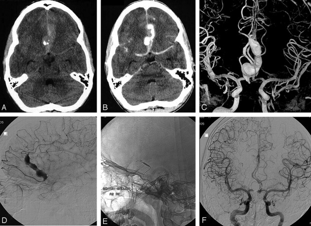
A 16-year-old boy presented with acute subarachnoid hemorrhage from a giant serpentine anterior cerebral artery aneurysm. A and B, Native (A) and contrast-enhanced (B) CT scans show subarachnoid hemorrhage from a calcified partially thrombosed fusiform aneurysm in the frontal interhemispheric fissure. C, Bilateral internal carotid artery 3D angiography reveals a duplicated right A2 segment with the aneurysm located on the lateral branch. D, Selective angiography of the aneurysm-bearing segment demonstrates branches arising both proximal and distal from the fusiform dilated segment. E, Microballoon for test occlusion. F, Complete exclusion from the circulation after coil occlusion of the proximal part of the lumen. No infarction developed.
References
-
- Day AL, Gaposchkin CG, Yu CJ, et al. Spontaneous fusiform middle cerebral artery aneurysms: characteristics and a proposed mechanism of formation. J Neurosurg 2003;99:228–40 - PubMed
-
- Vishteh AG, Spetzler RF. Evolution of a dolichoectatic aneurysm into a giant serpentine aneurysm during long-term follow-up: case illustration. J Neurosurg 1999;91:346. - PubMed
-
- Anson JA, Lawton MT, Spetzler RF. Characteristics and surgical treatment of dolichoectatic and fusiform aneurysms. J Neurosurg 1996;84:185–93 - PubMed
-
- Anson JA. Treatment strategies for intracranial fusiform aneurysms. Neurosurg Clin N Am 1998;9:743. - PubMed
-
- Coley SC, Hodgson TJ, Jakubowski J. Coil embolization of giant serpentine aneurysms: report of two cases arising from the posterior cerebral artery. Br J Neurosurg 2002;16:43–47 - PubMed
Publication types
MeSH terms
LinkOut - more resources
Full Text Sources
Medical