

Case Reports
doi: 10.3174/ajnr.A1069.
Epub 2008 Apr 3.
MR imaging-based decision in thrombolytic therapy for stroke on awakening: report of 2 cases
Affiliations
- PMID: 18388211
- PMCID: PMC8119137
- DOI: 10.3174/ajnr.A1069
Item in Clipboard
Case Reports
MR imaging-based decision in thrombolytic therapy for stroke on awakening: report of 2 cases
AJNR Am J Neuroradiol.
2008 Aug.
Abstract
Patients with stroke on awakening are denied the potential benefit of thrombolysis on the grounds that the onset time is unknown. Relying on clinical and MR imaging to indicate the most appropriate treatment could be more rational. We report 2 cases of stroke with unknown onset time. In both cases, anamnesis and MR imaging indicated that we might still be within 6 hours from stroke onset, with salvageable tissue. Arterial recanalization was successfully performed in both cases.
Figures
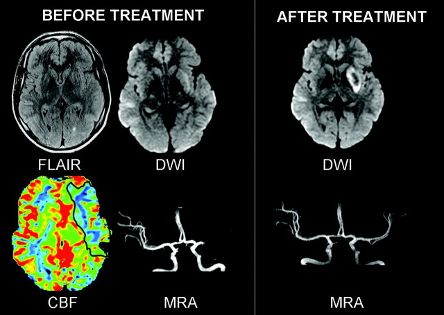
Case 1. Before treatment, FLAIR was normal and DWI showed a mildly hyperintense left putamen. Perfusion abnormalities (CBF decrease) existed in the superficial MCA territory, which was normal on DWI (PWI-DWI mismatch). MRA showed a left proximal MCA occlusion. After IA thrombolysis, the flow was restored in the left MCA. A deep MCA infarct with petechiae and a small cortical infarct in the left inferior frontal gyrus (not illustrated) were seen on 24-hour DWI follow-up.
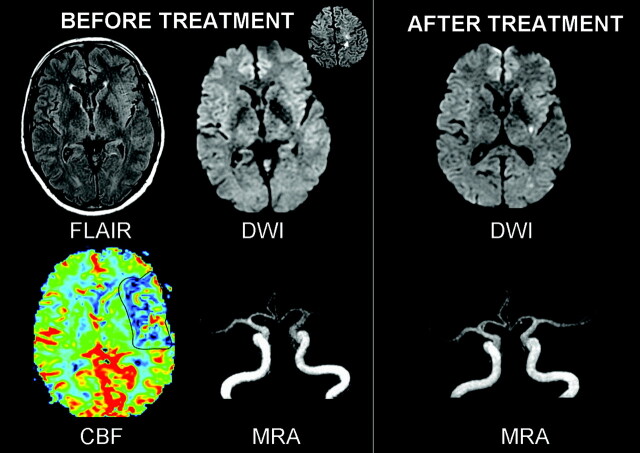
Case 2. Before treatment, FLAIR was normal while DWI showed punctuate hyperintensities in the superficial MCA territory (inset) and a left MCA occlusion. Perfusion abnormalities existed in the superficial MCA territory, consistent with a large PWI/DWI mismatch. 24-hour MR imaging follow-up showed an additional lesion in the left putamen and MCA recanalisation after IA thrombolysis.
Similar articles
-
Comparison of MRI-based thrombolysis for patients with middle cerebral artery occlusion<or=3 h and 3-6 h.Neurol India. 2009 Jul-Aug;57(4):426-33. doi: 10.4103/0028-3886.55615. Neurol India. 2009. PMID: 19770543 Clinical Trial.
-
Prognostic significance of blood pressure variability after thrombolysis in acute stroke.Neurology. 2008 Aug 19;71(8):552-8. doi: 10.1212/01.wnl.0000318294.36223.69. Epub 2008 Jun 11. Neurology. 2008. PMID: 18550860
-
Systemic thrombolysis with recombinant tissue plasminogen activator and tirofiban in acute middle cerebral artery occlusion.Stroke. 2004 Mar;35(3):705-9. doi: 10.1161/01.STR.0000117094.41638.EE. Epub 2004 Jan 29. Stroke. 2004. PMID: 14752128 Clinical Trial.
-
Magnetic resonance imaging criteria for thrombolysis in acute cerebral infarct.Stroke. 2005 Feb;36(2):388-97. doi: 10.1161/01.STR.0000152268.47919.be. Epub 2004 Dec 23. Stroke. 2005. PMID: 15618445 Review.
-
Thrombolysis in acute ischaemic stroke.Postgrad Med J. 2001 Mar;77(905):166-71. doi: 10.1136/pmj.77.905.166. Postgrad Med J. 2001. PMID: 11222823 Free PMC article. Review.
Cited by
-
Wake-up stroke and CT perfusion: effectiveness and safety of reperfusion therapy.Neurol Sci. 2018 Oct;39(10):1705-1712. doi: 10.1007/s10072-018-3486-z. Epub 2018 Jul 10. Neurol Sci. 2018. PMID: 29987433
-
Wake-up stroke and stroke of unknown onset: a critical review.Front Neurol. 2014 Aug 12;5:153. doi: 10.3389/fneur.2014.00153. eCollection 2014. Front Neurol. 2014. PMID: 25161646 Free PMC article. Review.
-
What to do With Wake-Up Stroke.Neurohospitalist. 2015 Jul;5(3):161-72. doi: 10.1177/1941874415576204. Neurohospitalist. 2015. PMID: 26288674 Free PMC article. Review.
-
Wake-up stroke: clinical characteristics, imaging findings, and treatment option - an update.Front Neurol. 2014 Mar 26;5:35. doi: 10.3389/fneur.2014.00035. eCollection 2014. Front Neurol. 2014. PMID: 24723908 Free PMC article. Review.
-
Intravenous thrombolysis in unwitnessed stroke onset: MR WITNESS trial results.Ann Neurol. 2018 May;83(5):980-993. doi: 10.1002/ana.25235. Epub 2018 Apr 27. Ann Neurol. 2018. PMID: 29689135 Free PMC article. Clinical Trial.
References
-
- Serena J, Davalos A, Segura T, et al. Stroke on awakening: looking for a more rational management. Cerebrovasc Dis 2003;16:128–33 - PubMed
-
- Fink JN, Kumar S, Horkan C, et al. The stroke patient who woke up: clinical and radiological features, including diffusion and perfusion MRI. Stroke 2002;33:988–93 - PubMed
-
- Schellinger PD, Thomalla G, Fiehler J, et al. MRI-based and CT-based thrombolytic therapy in acute stroke within and beyond established time windows: an analysis of 1210 patients. Stroke 2007;38:2640–45 - PubMed
-
- Schellinger PD, Fiebach JB, Hacke W. Imaging-based decision making in thrombolytic therapy for ischemic stroke: present status. Stroke 2003;34:575–83 - PubMed
Publication types
MeSH terms
Substances
LinkOut - more resources
Full Text Sources
Medical