

Performance evaluation of radiologists with artificial neural network for differential diagnosis of intra-axial cerebral tumors on MR images
- PMID: 18388216
- PMCID: PMC8118852
- DOI: 10.3174/ajnr.A1037
Performance evaluation of radiologists with artificial neural network for differential diagnosis of intra-axial cerebral tumors on MR images
Abstract
Background and purpose: Previous studies have suggested that use of an artificial neural network (ANN) system is beneficial for radiological diagnosis. Our purposes in this study were to construct an ANN for the differential diagnosis of intra-axial cerebral tumors on MR images and to evaluate the effect of ANN outputs on radiologists' diagnostic performance.
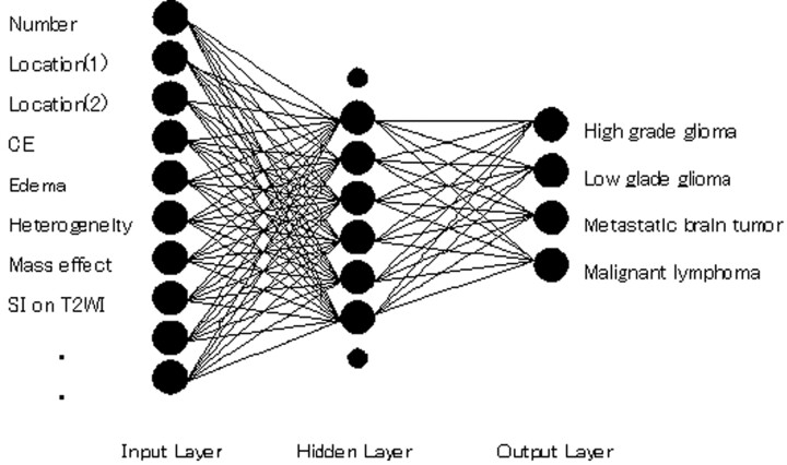
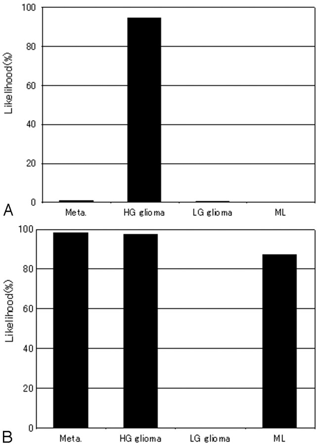
Materials and methods: We collected MR images of 126 patients with intra-axial cerebral tumors (58 high-grade gliomas, 37 low-grade gliomas, 19 metastatic tumors, and 12 malignant lymphomas). We constructed a single 3-layer feed-forward ANN with a Levenberg-Marquardt algorithm. The ANN was designed to differentiate among 4 categories of tumors (high-grade gliomas, low-grade gliomas, metastases, and malignant lymphomas) with use of 2 clinical parameters and 13 radiologic findings in MR images. Subjective ratings for the 13 radiologic findings were provided independently by 2 attending radiologists. All 126 cases were used for training and testing of the ANN based on a leave-one-out-by-case method. In the observer test, MR images were viewed by 9 radiologists, first without and then with ANN outputs. Each radiologist's performance was evaluated through a receiver operating characteristic (ROC) analysis on a continuous rating scale.
Results: The averaged area under the ROC curve for ANN alone was 0.949. The diagnostic performance of the 9 radiologists increased from 0.899 to 0.946 (P < .001) when they used ANN outputs.
Conclusions: The ANN can provide useful output as a second opinion to improve radiologists' diagnostic performance in the differential diagnosis of intra-axial cerebral tumors seen on MR imaging.
Figures


References
-
- Abdolmaleki P, Mihara F, Masuda K, et al. Neural networks analysis of astrocytic gliomas from MRI appearances. Cancer Lett 1997;118:69–78 - PubMed
-
- Kobayashi T, Xu XW, MacMahon H, et al. Effect of a computer-aided diagnosis scheme on radiologists' performance in detection of lung nodules on radiographs. Radiology 1996;199:843–48 - PubMed
-
- Ashizawa K, MacMahon H, Ishida T, et al. Effect of an artificial neural network on radiologists' performance in the differential diagnosis of interstitial lung disease using chest radiographs. AJR Am J Roentgenol 1999;172:1311–15 - PubMed
-
- Nakamura K, Yoshida H, Engelmann R, et al. Computerized analysis of the likelihood of malignancy in solitary pulmonary nodules with use of artificial neural networks. Radiology 2000;214:823–30 - PubMed
-
- Matsuki Y, Nakamura K, Watanabe H, et al. Usefulness of an artificial neural network for differentiating benign from malignant pulmonary nodules on high-resolution CT: evaluation with receiver operating characteristic analysis. AJR Am J Roentgenol 2002;178:657–63 - PubMed
Publication types
MeSH terms
LinkOut - more resources
Full Text Sources
Medical