Metabolic assessment of gliomas using 11C-methionine, [18F] fluorodeoxyglucose, and 11C-choline positron-emission tomography
- PMID: 18388218
- PMCID: PMC8118839
- DOI: 10.3174/ajnr.A1008
Metabolic assessment of gliomas using 11C-methionine, [18F] fluorodeoxyglucose, and 11C-choline positron-emission tomography
Abstract
Background and purpose: Positron-emission tomography (PET) is a useful tool in oncology. The aim of this study was to assess the metabolic activity of gliomas using (11)C-methionine (MET), [(18)F] fluorodeoxyglucose (FDG), and (11)C-choline (CHO) PET and to explore the correlation between the metabolic activity and histopathologic features.
Materials and methods: PET examinations were performed for 95 primary gliomas (37 grade II, 37 grade III, and 21 grade IV). We measured the tumor/normal brain uptake ratio (T/N ratio) on each PET and investigated the correlations among the tracer uptake, tumor grade, tumor type, and tumor proliferation activity. In addition, we compared the ease of visual evaluation for tumor detection.
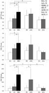
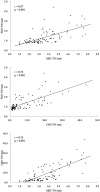
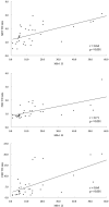
Results: All 3 of the tracers showed positive correlations with astrocytic tumor (AT) grades (II/IV and III/IV). The MET T/N ratio of oligodendroglial tumors (OTs) was significantly higher than that of ATs of the same grade. The CHO T/N ratio showed a significant positive correlation with histopathologic grade in OTs. Tumor grade and type influenced MET uptake only. MET T/N ratios of more than 2.0 were seen in 87% of all of the gliomas. All of the tracers showed significantly positive correlations with Mib-1 labeling index in ATs but not in OTs and oligoastrocytic tumors.
Conclusion: MET PET appears to be useful in evaluating grade, type, and proliferative activity of ATs. CHO PET may be useful in evaluating the potential malignancy of OTs. In terms of visual evaluation of tumor localization, MET PET is superior to FDG and CHO PET in all of the gliomas, due to its straightforward detection of "hot lesions".
Figures


Comment in
-
FDG, MET or CHO? The quest for the optimal PET tracer for glioma imaging continues.Nat Clin Pract Neurol. 2008 Sep;4(9):470-1. doi: 10.1038/ncpneuro0863. Epub 2008 Jul 15. Nat Clin Pract Neurol. 2008. PMID: 18628750
-
Technetium Tc99m tetrofosmin single-photon emission CT for the assessment of glioma proliferation.AJNR Am J Neuroradiol. 2008 Nov;29(10):e96. doi: 10.3174/ajnr.A1183. AJNR Am J Neuroradiol. 2008. PMID: 19008320 Free PMC article. No abstract available.
References
-
- Delbeke D, Meyerowitz C, Lapidus RL, et al. Optimal cutoff levels of F-18 fluorodeoxyglucose uptake in the differentiation of low-grade from high-grade brain tumors with PET. Radiology 1995;195:47–52 - PubMed
-
- Derlon JM, Bourdet C, Bustany P, et al. [11c]l-Methionine uptake in gliomas. Neurosurgery 1989;25:720–28 - PubMed
-
- Herholz K, Holzer T, Bauer B, et al. 11c-Methionine pet for differential diagnosis of low-grade gliomas. Neurology 1998;50:1316–22 - PubMed
-
- Kaschten B, Stevenaert A, Sadzot B, et al. Preoperative evaluation of 54 gliomas by PET with fluorine-18-fluorodeoxyglucose and/or carbon-11-methionine. J Nucl Med 1998;39:778–85 - PubMed
-
- De Witte O, Goldberg I, Wikler D, et al. Positron emission tomography with injection of methionine as a prognostic factor in glioma. J Neurosurg 2001;95:746–50 - PubMed
MeSH terms
Substances
LinkOut - more resources
Full Text Sources
Medical
Miscellaneous
