. 2008 Apr 15;121(Pt 8):1243-51.
doi: 10.1242/jcs.017517.

Recycling of IRAP from the plasma membrane back to the insulin-responsive compartment requires the Q-SNARE syntaxin 6 but not the GGA clathrin adaptors

Affiliations

Recycling of IRAP from the plasma membrane back to the insulin-responsive compartment requires the Q-SNARE syntaxin 6 but not the GGA clathrin adaptors

Robert T Watson et al. J Cell Sci. .

Erratum in

  • J Cell Sci. 2008 Jul 1;121(Pt 13):2275. Hou, June C [added]

Abstract

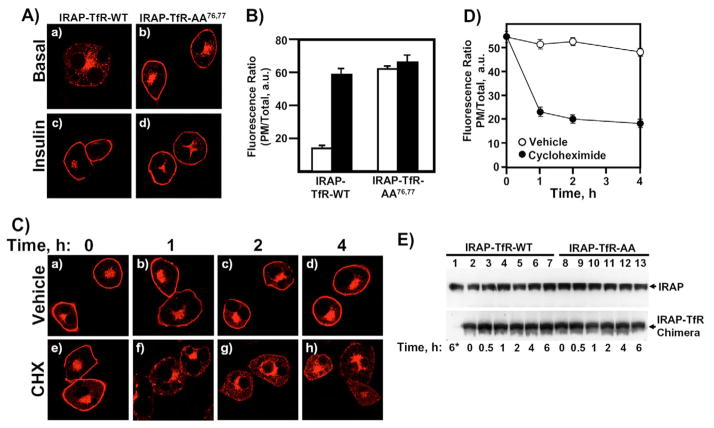
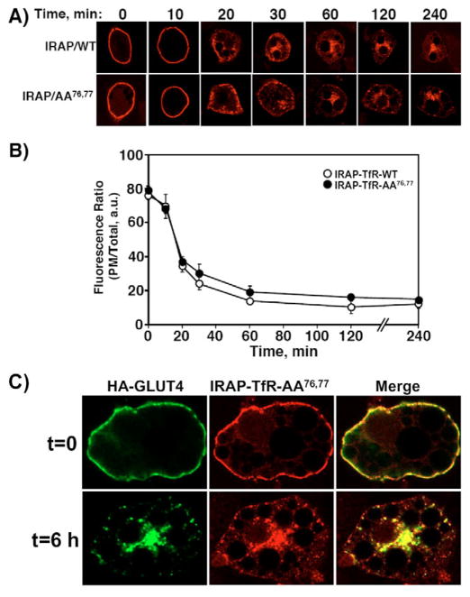
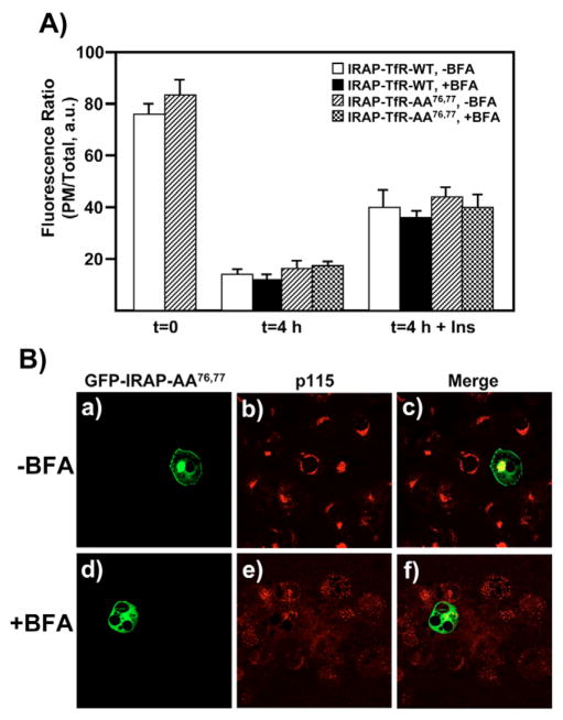
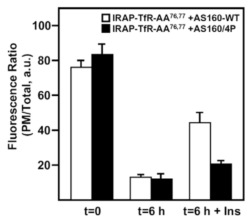
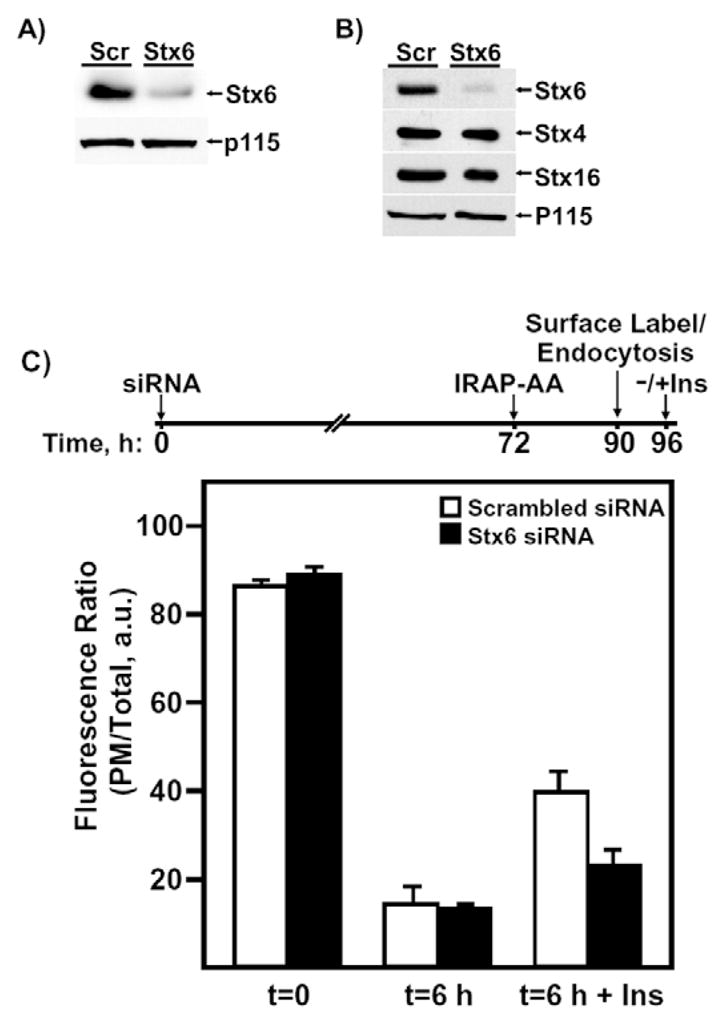
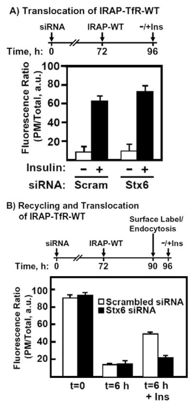
Insulin recruits two transmembrane proteins, GLUT4 and IRAP, to the plasma membrane of muscle cells and adipocytes. The subcellular trafficking and localization of GLUT4, and to a lesser extent IRAP, have been intensely studied, yet the molecular mechanisms responsible for their insulin-responsive compartmentalization remain unknown. Herein we have investigated the endocytosis and recycling of IRAP from the cell surface back to the insulin-responsive compartment (IRC). Our results show that a key dileucine motif at position 76,77 (LL76,77), although required for the initial biosynthetic entry of IRAP into the IRC, is dispensable for entry into the IRC via the endosomal system. Indeed, we found that an AA76,77 mutant of IRAP is fully capable of undergoing endocytosis and is correctly routed back to the IRC. To verify that the AA76,77 mutant enters the bona fide IRC, we show that the internalized IRAP-AA76,77 construct is sequestered in an IRC that is insensitive to brefeldin A yet sensitive to a dominant-interfering mutant of AS160 (AS160-4P). In addition, we show that the GGA clathrin adaptors are not required for the re-entry of IRAP from the cell surface back into the IRC, whereas the Q-SNARE syntaxin 6 is required for this process.


Figures

Fig. 1
Fig. 1
Newly synthesized IRAP-TfR-AA76,77 defaults to the plasma membrane, where it accumulates under steady-state conditions. (A) Differentiated 3T3L1 adipocytes were electroporated with 50 μg of IRAP-TfR-WT (panels a and c) or IRAP-TfR-AA76,77 (panels b and d) and, 16 hours later, cells were serum-starved and treated without (panels a and b) or with (panels c and d) 100 nM insulin for 20 minutes. The cells were then fixed, permeabilized and labeled with an anti-TfR antibody followed by Texas-Red-conjugated secondary antibody, and were then visualized with a Zeiss LSM510 scanning laser confocal microscope. (B) The data in A were quantified as the ratio of surface fluorescence:total fluorescence, as described in the Materials and Methods. White bars, basal cells; black bars, insulin-stimulated cells. (C) Differentiated 3T3L1 adipocytes were electroporated with 50 μg of IRAP-TfR-AA76,77 and, 16 hours later, cells were incubated with vehicle alone (panels a–d) or vehicle plus 10 μg/ml cycloheximide (CHX) (panels e–h) for 0, 1, 2 or 4 hours. The cells were then processed for confocal microscopy as indicated in A. (D) The data presented in C were quantified by calculating the ratio of plasma membrane:total fluorescence, as described under Materials and Methods. Results are from three independent experiments, with fifteen cells per experiment (mean ± s.e.m.). (E) Fully differentiated 3T3L1 adipocytes were transfected with 50 μg IRAP-TfR-WT or 50 μg IRAP-TfR-AA76,77 cDNA. After an overnight recovery period, cells were incubated for 0, 0.5, 1, 2, 4 or 6 hours in 10 μg/ml CHX. In lane 1, cells were plated immediately in CHX after electroporation and were then incubated for 6 hours. At the indicated time points, cell lysates were prepared and subjected to western blotting. An anti-IRAP antibody was used to detect both endogenous IRAP (upper panel) and the expressed IRAP-TfR chimera (lower panel). a.u., arbitrary units.
Fig. 2
Fig. 2
IRAP-TfR-AA76,77 displays a normal rate and extent of plasma membrane endocytosis. Differentiated 3T3L1 adipocytes were transfected with 50 μg of IRAP-TfR-WT or IRAP-TfR-AA76,77 and, 16 hours later, stimulated with 100 nM insulin (this is only necessary to induce TfR-IRAP-WT translocation but was done for both constructs for internal consistency). The cells were then cooled to 4°C and the cell surface was labeled with the anti-TfR antibody. The cells were washed and warmed to 37°C for various times, as indicated. The cells were then fixed, permeabilized and labeled with Texas-Red-conjugated anti-TfR antibody. (A) Representative cell images are shown. (B) The percentage of cell-surface signal was calculated using the Zeiss LSM 510 software package (mean ± s.e.m. of three independent experiments). (C) Fully differentiated 3T3L1 adipocytes were co-electroporated with 50 μg of both HA-GLUT4 and IRAP-TfR-AA76,77, and were allowed to recover overnight. Cells were then stimulated with 100 nM insulin for 20 minutes, and then surface-labeled on ice with a mouse monoclonal anti-TfR antibody and a polyclonal rabbit HA antibody for 60 minutes. After extensive washing with ice-cold PBS, cells were either fixed in paraformaldehyde (t=0) or were warmed to 37°C for 6 hours (t=6 h), fixed and labeled with Texas-Red-conjugated donkey anti-mouse and Alexa-Fluor-488-conjugated donkey anti-rabbit secondary antibodies. Images were collected on a Zeiss LSM 510 META confocal microscope. a.u., arbitrary units.
Fig. 3
Fig. 3
IRAP-TfR-WT and IRAP-TfR-AA76,77 both recycle from the cell surface back to an IRC that is BFA-insensitive. (A) Fully differentiated 3T3L1 adipocytes were transfected with 50 μg of either IRAP-TfR-WT or IRAP-TfR-AA76,77 and, 16 hours later, cells were stimulated with insulin and surface-labeled with the anti-TfR antibody on ice for 60 minutes. Cells were then washed with PBS and incubated at 37°C for 4 hours. For the BFA treatment, cells were incubated with BFA (5 μg/ml) during the last 30 minutes of the washout period and also during the second round of insulin treatment. Control cells were incubated with vehicle alone. Following the second round of treatment without or with insulin (100 nM, 30 minutes), cells were fixed, permeabilized and labeled with Texas-Red-conjugated secondary antibody, as described under Materials and Methods. The ratio of plasma membrane fluorescence:total fluorescence was determined using the Zeiss LSM software package (mean ± s.e.m. of three independent experiments). (B) Control experiments demonstrating the efficacy of 5 μg/ml BFA. Cells were transfected with 50 μg of GFP-IRAP-AA76,77 reporter construct and immediately plated in media either without (panels a-c) or with (panels d–f) 5 μg/ml BFA. After a 3-hour recovery period, cells were fixed and labeled with an anti-p115 monoclonal antibody, followed by Texas Red secondary antibody. a.u., arbitrary units.
Fig. 4
Fig. 4
A dominant-interfering mutant form of AS160 (AS160-4P) inhibits the insulin-stimulated exit of IRAP-TfR-AA76,77 from the IRC following endocytosis from the cell surface. Fully differentiated 3T3L1 adipocytes were electroporated with 50 μg of IRAP-TfR-AA76,77 and either AS160-WT (white bars), or AS160-4P (black bars). Following a 16-hour recovery period, cells were surface-labeled with the anti-TfR antibody on ice for 60 minutes. Cells were then warmed to 37°C for 6 hours to allow endocytosis to occur, then treated without or with 100 nM insulin for 30 minutes, fixed, permeabilized and labeled with Texas-Red-conjugated secondary antibody as described under Materials and Methods. The ratio of plasma membrane fluorescence to total fluorescence was determined using the Zeiss LSM software package (mean ± s.e.m. of three independent experiments). a.u., arbitrary units.
Fig. 5
Fig. 5
GGA clathrin adaptors function specifically during the biosynthetic entry of IRAP into the IRC. (A) Fully differentiated 3T3L1 adipocytes were transfected with 50 μg of IRAP-TfR-WT or IRAP-TfR-AA76,77 and, 16 hours later, were stimulated with 100 nM insulin. Following surface labeling with the anti-TfR antibody, endocytosis time courses were performed as described in Fig. 1 and in the Materials and Methods. (B) Fully differentiated 3T3L1 adipocytes were electroporated with 50 μg of IRAP-TfR-AA76,77 and 200 μg of either GGA-WT or VHS-GAT. After an overnight recovery, cells were surface-labeled with the anti-TfR antibody as described in Fig. 3 and in the Materials and Methods. Following a 6-hour washout period at 37°C, cells were treated without or with 100 nM insulin for 30 minutes, fixed, permeabilized and labeled with Texas-Red-conjugated secondary antibody as described under Materials and Methods. The ratio of plasma membrane fluorescence to total fluorescence was determined using the Zeiss LSM software package (mean ± s.e.m. of three independent experiments). a.u., arbitrary units.
Fig. 6
Fig. 6
Stx6 functions during the re-entry of IRAP into the IRC following endocytosis from the cell surface. (A) Western blot showing the extent of Stx6 knockdown by siRNA (5 nmol) 72 hours after transfection. (B) Western blot showing the extent and specificity of Stx6 knockdown by siRNA (5 nmol) 96 hours after transfection. Lane 1, scrambled-siRNA-transfected cells; lane 2, Stx6-siRNA-transfected cells. For the 96-hour time point, the same membrane was stripped and re-probed with anti-Stx4, -Stx16 and -p115 antibodies. p115 is a Golgi-tethering factor and is used here as a protein loading control. (C) Cells were transfected with either 5 nmol of scrambled or Stx6 siRNA and, 72 hours later, were transfected with IRAP-TfR-WT reporter. Following an 18-hours recovery period, cells were stimulated with insulin and surface-labeled on ice with the anti-TfR monoclonal antibody. Following a 6-hour washout period at 37°C, cells were re-stimulated with or without insulin, fixed, labeled with Texas-Red-secondary antibody and processed for confocal microscopy (mean ± s.e.m. of three independent experiments). A timeline for the transfections is shown above the bar graph for clarity. a.u., arbitrary units.
Fig. 7
Fig. 7
Stx6 function is not required for the insulin-stimulated exit of IRAP from the IRC. (A) Fully differentiated 3T3L1 adipocytes were transfected with 5 nmol of scrambled or Stx6 siRNA and, 72 hours later, were transfected with 50 μg EGFP-IRAP-TfR-WT reporter. Following a 24-hour recovery period, cells were treated with or without insulin, fixed and labeled with Texas Red secondary antibody. The ratio of plasma membrane to total fluorescence was determined using Zeiss LSM software. (B) Cells were transfected with either 5 nmol of scrambled or Stx6 siRNA and, 72 hours later, were transfected with IRAP-TfR-WT reporter. Following an 18-hour recovery period, cells were stimulated with insulin and surface-labeled on ice with anti-TfR monoclonal antibody. Following a 6-hour washout at 37°C, cells were re-stimulated with or without insulin, fixed, labeled with Texas Red secondary antibody and processed for confocal microscopy (mean ± s.e.m. of three independent experiments). A timeline for the transfections is shown above the bar graphs for clarity. Scram, scrambled siRNA. a.u., arbitrary units.

Similar articles

Cited by

References

    1. Abel ED, Graveleau C, Betuing S, Pham M, Reay PA, Kandror V, Kupriyanova T, Xu Z, Kandror KV. Regulation of insulin-responsive aminopeptidase expression and targeting in the insulin-responsive vesicle compartment of glucose transporter isoform 4-deficient cardiomyocytes. Mol Endocrinol. 2004;18:2491–2501. - PubMed
    1. Arias EB, Kim J, Funai K, Cartee GD. Prior exercise increases phosphorylation of Akt substrate of 160 kDa (AS160) in rat skeletal muscle. Am J Physiol Endocrinol Metab. 2007;292:E1191–E1200. - PubMed
    1. Bao S, Smith RM, Jarett L, Garvey WT. The effects of brefeldin A on the glucose transport system in rat adipocytes. Implications regarding the intracellular locus of insulin-sensitive Glut4. J Biol Chem. 1995;270:30199–30204. - PubMed
    1. Bock JB, Lin RC, Scheller RH. A new syntaxin family member implicated in targeting of intracellular transport vesicles. J Biol Chem. 1996;271:17961–17965. - PubMed
    1. Bock JB, Klumperman J, Davanger S, Scheller RH. Syntaxin 6 functions in trans-Golgi network vesicle trafficking. Mol Biol Cell. 1997;8:1261–1271. - PMC - PubMed