. 2008 May 9;102(9):1118-26.
doi: 10.1161/CIRCRESAHA.108.173948. Epub 2008 Apr 3.

IP3 constricts cerebral arteries via IP3 receptor-mediated TRPC3 channel activation and independently of sarcoplasmic reticulum Ca2+ release

Affiliations

IP3 constricts cerebral arteries via IP3 receptor-mediated TRPC3 channel activation and independently of sarcoplasmic reticulum Ca2+ release

Qi Xi et al. Circ Res. .

Erratum in

  • Circ Res. 2009 Jul 2;105(1):e1

Abstract

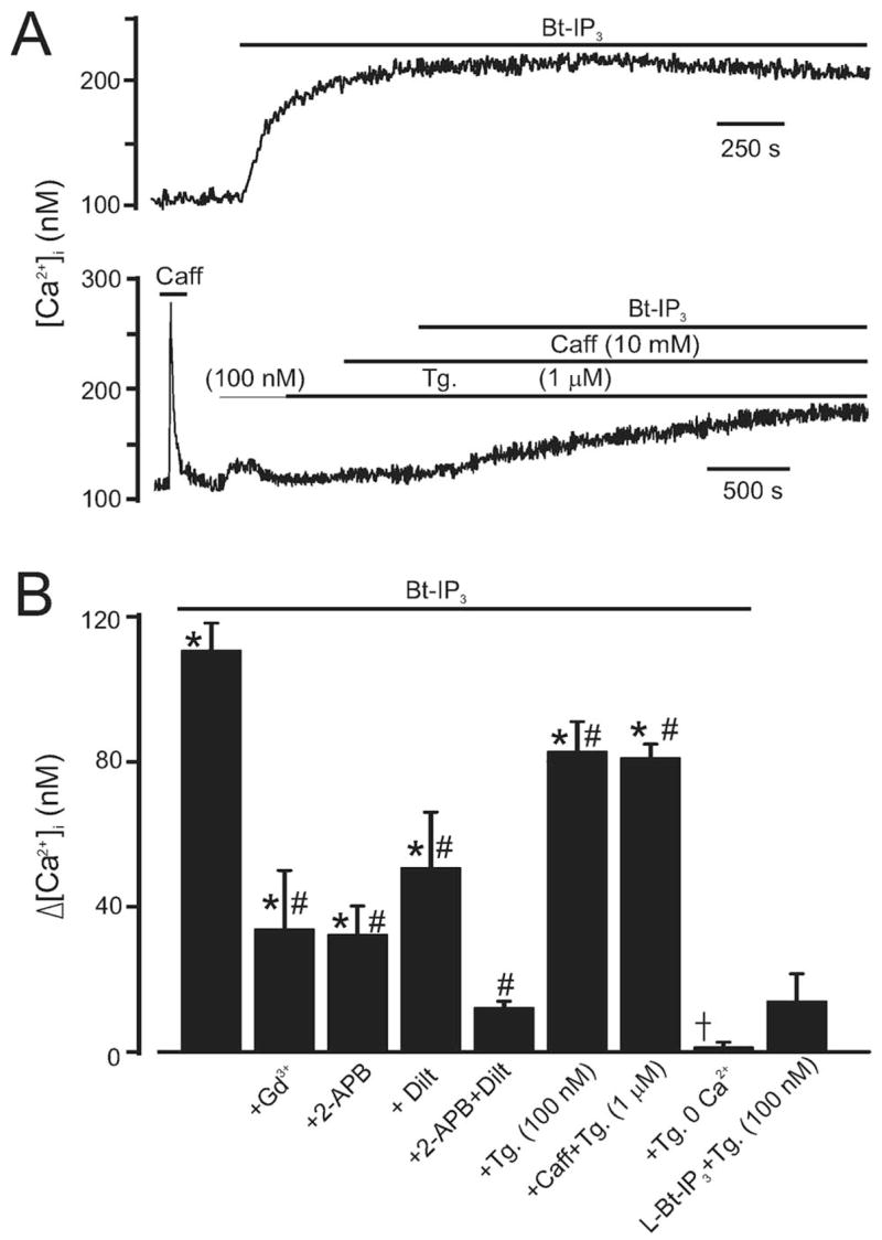
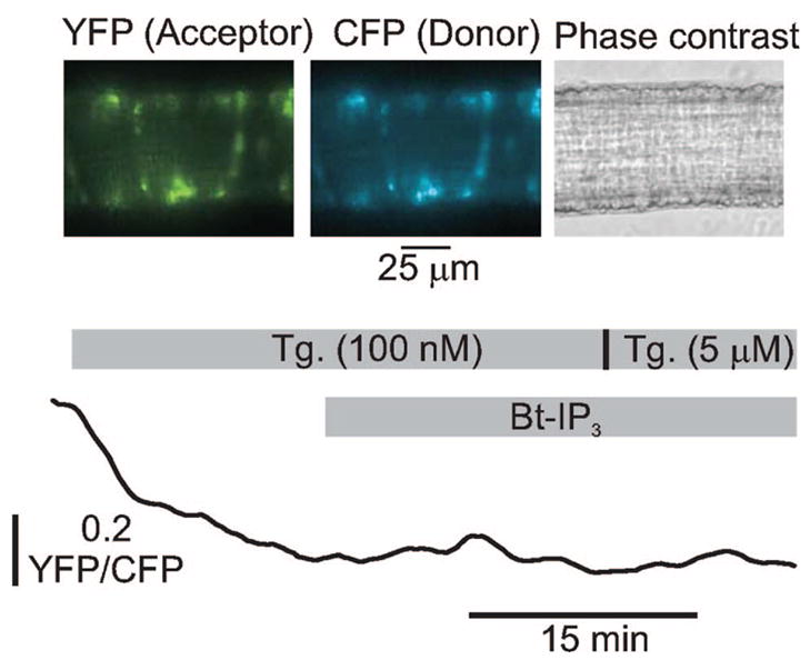
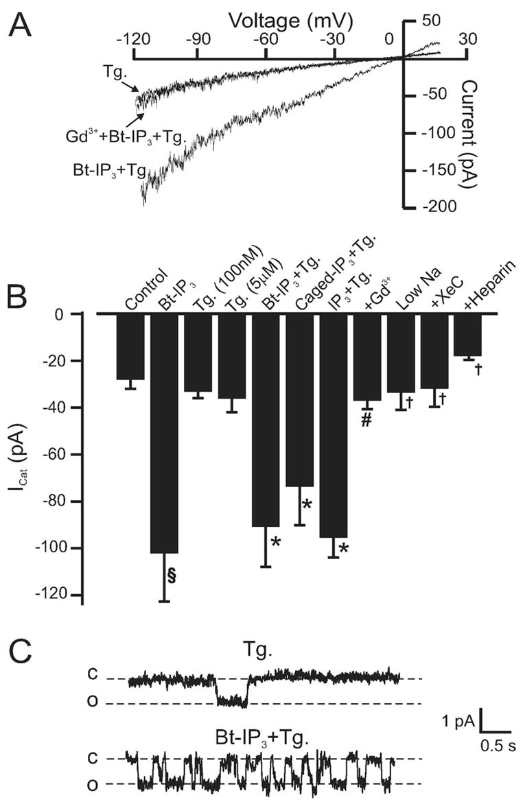
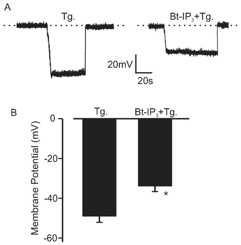
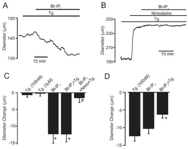
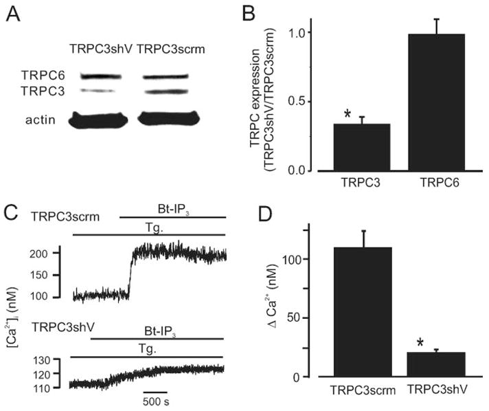
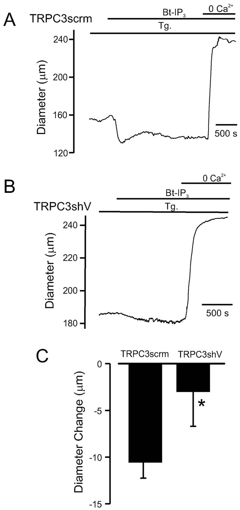
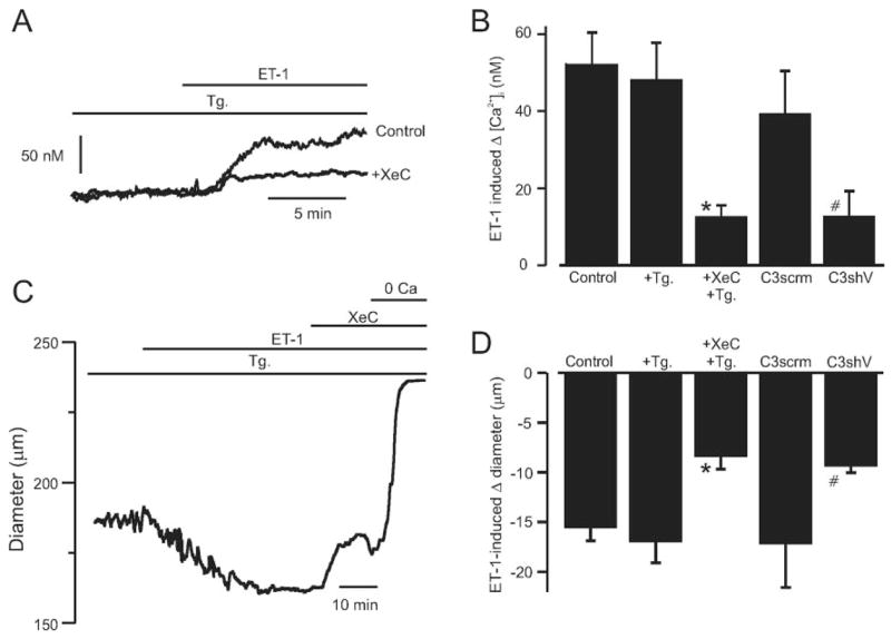
Vasoconstrictors that bind to phospholipase C-coupled receptors elevate inositol-1,4,5-trisphosphate (IP(3)). IP(3) is generally considered to elevate intracellular Ca(2+) concentration ([Ca(2+)](i)) in arterial myocytes and induce vasoconstriction via a single mechanism: by activating sarcoplasmic reticulum (SR)-localized IP(3) receptors, leading to intracellular Ca(2+) release. We show that IP(3) also stimulates vasoconstriction via a SR Ca(2+) release-independent mechanism. In isolated cerebral artery myocytes and arteries in which SR Ca(2+) was depleted to abolish Ca(2+) release (measured using D1ER, a fluorescence resonance energy transfer-based SR Ca(2+) indicator), IP(3) activated 15 pS sarcolemmal cation channels, generated a whole-cell cation current (I(Cat)) caused by Na(+) influx, induced membrane depolarization, elevated [Ca(2+)](i), and stimulated vasoconstriction. The IP(3)-induced I(Cat) and [Ca(2+)](i) elevation were attenuated by cation channel (Gd(3+), 2-APB) and IP(3) receptor (xestospongin C, heparin, 2-APB) blockers. TRPC3 (canonical transient receptor potential 3) channel knockdown with short hairpin RNA and diltiazem and nimodipine, voltage-dependent Ca(2+) channel blockers, reduced the SR Ca(2+) release-independent, IP(3)-induced [Ca(2+)](i) elevation and vasoconstriction. In pressurized arteries, SR Ca(2+) depletion did not alter IP(3)-induced constriction at 20 mm Hg but reduced IP(3)-induced constriction by approximately 39% at 60 mm Hg. [Ca(2+)](i) elevations and constrictions induced by endothelin-1, a phospholipase C-coupled receptor agonist, were both attenuated by TRPC3 knockdown and xestospongin C in SR Ca(2+)-depleted arteries. In summary, we describe a novel mechanism of IP(3)-induced vasoconstriction that does not occur as a result of SR Ca(2+) release but because of IP(3) receptor-dependent I(Cat) activation that requires TRPC3 channels. The resulting membrane depolarization activates voltage-dependent Ca(2+) channels, leading to a myocyte [Ca(2+)](i) elevation, and vasoconstriction.


Figures

Figure 1
Figure 1
Bt-IP3 elevates [Ca2+]i in arterial myocytes with intact and depleted SR Ca2+. A, Original traces illustrating [Ca2+]i regulation by Bt-IP3 (10 μmol/L) in myocytes with intact (top) and depleted (bottom) SR Ca2+. B, Mean data. Control [Ca2+]i was 114±7 nmol/L (n=53). *P<0.05 compared with control in the same cell, #P<0.05 compared with Bt-IP3, †P<0.05 compared with Bt-IP3+thapsigargin (Tg) (100 nmol/L). The experimental numbers for bars from left to right were 6, 5, 4, 5, 5, 7, 9, 5, and 4, respectively.
Figure 2
Figure 2
Thapsigargin (100 nmol/L to 5 μmol/L) depletes myocyte SR Ca2+. Images illustrate D1ER fluorescence in myocytes of a cerebral artery segment. Original trace is shown illustrating the time course of normalized D1ER yellow fluorescent protein (YFP)/cyan fluorescent protein (CFP) ratio change in the same artery in response to thapsigargin and Bt-IP3 (10 μmol/L). Data are representative of 6 separate experiments.
Figure 3
Figure 3
IP3 activates a Na+-permeant ICat as a result of IP3R activation in myocytes with intact and depleted SR Ca2+. A, Original recordings obtained using the perforated-patch configuration illustrating that Bt-IP3 (10 μmol/L) activates a Gd3+-sensitive (30 μmol/L) whole-cell ICat in a myocyte with depleted SR Ca2+. B, Mean data: control perforated-patch (p-p), n=9; Bt-IP3 (10 μmol/L) p-p, n=9; thapsigargin (Tg) (100 nmol/L; p-p [n=12] and conventional whole-cell [w-c] [n=9] were statistically similar and pooled, giving n=21; Tg [5 μmol/L] p-p, n=4; Bt-IP3+Tg [100 nmol/L] p-p, n=5; caged IP3 [2 μmol/L]+ Tg [100 nmol/L] w-c, n=14; IP3 [20 μmol/L]+ Tg [100 nmol/L] w-c, n=17; +Gd3+ [30 μmol/L] [n=5] represents Bt-IP3 [10 μmol/L, n=2] and caged IP3 [2 μmol/L, n=3], which were statistically similar and pooled; IP3 [20 μmol/L]+ Tg [100 nmol/L] with 20 mmol/L bath Na+ w-c, n=5; IP3 [20 μmol/L]+ Tg [100 nmol/L]+ XeC [20 μmol/L, via pipette], w-c, n=6; IP3 [20 μmol/L]+ Tg [100 nmol/L]+ heparin [1 mg/mL via pipette], w-c, n=7). Bt-IP3 shifted the reversal potential (Erev) from −9±1 to −5±0.4 mV. C, Bt-IP3 (10 μmol/L) activates single channel currents in a myocyte (cell-attached configuration) with depleted SR Ca2+ (Tg 100 nmol/L) at −60 mV. c indicates closed; o, open. §P<0.05 compared with control, *P<0.05 compared with Tg, #P<0.05 compared with Bt-IP3 and IP3, †P<0.05 compared with IP3.
Figure 4
Figure 4
IP3 depolarizes pressurized arteries with depleted SR Ca2+. A, Original traces illustrating membrane depolarization induced by Bt-IP3 (1 μmol/L) in the same SR Ca2+ depleted (100 nmol/L thapsigargin [Tg]; >15 minutes) pressurized (20 mm Hg) artery. Negative deflections from 0 voltage (dotted line) illustrate intracellular microelectrode impalement to measure membrane potential, followed by subsequent removal. B, Mean data (Tg, n=5; Bt-IP3+Tg, n=9). *P<0.05 compared with Tg.
Figure 5
Figure 5
Bt-IP3 constricts pressurized arteries with intact and depleted [Ca2+]SR. A, Bt-IP3 (10 nmol/L) constricts an artery at 20 mm Hg with depleted SR Ca2+ (thapsigargin [Tg],100 nmol/L; >15 minutes). B, At 20 mm Hg, nimodipine (1 μmol/L) caused a large vasodilation and attenuated Bt-IP3–induced vasoconstriction. C, Mean data illustrating Bt-IP3–induced vasoconstriction at 20 mm Hg in arteries with intact (n=8) and depleted (n=7) [Ca2+]SR and inhibition by nimodipine (n=5). D, Mean data illustrating vasoregulation at 60 mm Hg by thapsigargin (n=10), Bt-IP3 (n=7), and Bt-IP3+thapsigargin (n=7). Mean myogenic tone was (in %): 20 mm Hg, 24±2 (n=25); 60 mm Hg, 42.1±10.7 (n=7). *P<0.05 compared with respective control or thapsigargin, #P<0.05 compared with Bt-IP3+thapsigargin.
Figure 6
Figure 6
TRPC3 knockdown attenuates Bt-IP3–induced [Ca2+]i elevations in arterial myocytes with depleted SR Ca2+. A, Original Western blot data illustrating that TRPC3shV reduces TRPC3 protein but does not change TRPC6 in the same arteries. Lanes and bands shown were obtained from the same original Western blot that was reprobed with antibodies for TRPC3, TRPC6, and actin and combined for presentation. For clarity, an unrelated lane that was between the 2 illustrated lanes was not shown. B, Mean data (n=5). C, TRPC3 knockdown attenuates Bt-IP3–induced (10 μmol/L) [Ca2+]i elevations in SR Ca2+-depleted myocytes (thapsigargin [Tg], 100 nmol/L; >15 minutes). D, Mean data (TRPC3scrm, n=14; TRPC3shV, n=19). *P<0.05 when compared with TRPC3scrm.
Figure 7
Figure 7
TRPC3 knockdown reduces vasoconstriction induced by Bt-IP3. Original traces illustrating Bt-IP3–induced (10 nmol/L) diameter responses in pressurized (20 mm Hg) arteries treated with TRPC3scrm (A) or TRPC3shV (B). C, Mean Bt-IP3–induced vasoconstriction in arteries treated with TRPC3scrm (n=7) or TRPC3shV (n-6). *P<0.05 compared with TRPC3scrm.
Figure 8
Figure 8
ET-1–induced [Ca2+]i elevation and vasoconstriction requires TRPC3 channels and is attenuated by IP3R inhibition in SR Ca2+-depleted arteries. A, Original recordings illustrating ET-1–induced (300 pmol/L) [Ca2+]i elevations and inhibition by XeC (3 μmol/L) in SR Ca2+-depleted (thapsigargin [Tg], 100 nmol/L; >15 minutes) arteries. B, Mean data for ET-1–induced [Ca2+]i elevations in arteries in control (n=6), the presence of Tg (100 nmol/L; >15 minutes, n=16), and Tg (100 nmol/L)+ XeC (3 μmol/L, n=5) or in arteries treated with TRPC3scrm (n=4) or TRPC3shV (n=4). C, XeC (3 μmol/L) partially reverses an ET-1–induced (300 pmol/L) vasoconstriction in a pressurized (20 mm Hg) artery with depleted SR Ca2+. D, Mean data for ET-1–induced vasoconstriction in arteries in control (n=5), in the presence of Tg (100 nmol/L; >15 minutes, n=6), or Tg+XeC (n=7) or in arteries treated with TRPC3scrm (n=5) or TRPC3shV (n=4). *P<0.05 compared with ET-1+Tg, #P<0.05 compared with TRPC3scrm.

References

    1. Davis MJ, Hill MA. Signaling mechanisms underlying the vascular myogenic response. Physiol Rev. 1999;79:387–423. - PubMed
    1. Clapham DE, Julius D, Montell C, Schultz G. International Union of Pharmacology. XLIX. Nomenclature and structure-function relationships of transient receptor potential channels. Pharmacol Rev. 2005;57:427–450. - PubMed
    1. Jaggar JH. Intravascular pressure regulates local and global Ca2+ signaling in cerebral artery smooth muscle cells. Am J Physiol. 2001;281:C439–C448. - PubMed
    1. Palmer AE, Jin C, Reed JC, Tsien RY. Bcl-2-mediated alterations in endoplasmic reticulum Ca2+ analyzed with an improved genetically encoded fluorescent sensor. Proc Natl Acad Sci U S A. 2004;101:17404–17409. - PMC - PubMed
    1. Dixit M, Zhuang D, Ceacareanu B, Hassid A. Treatment with insulin uncovers the motogenic capacity of nitric oxide in aortic smooth muscle cells: dependence on Gab1 and Gab1-SHP2 association. Circ Res. 2003;93:e113–e123. - PubMed

Publication types

MeSH terms