. 2008 Jun;16(6):1089-97.
doi: 10.1038/mt.2008.56. Epub 2008 Mar 25.

Extended transgene expression from a nonintegrating adenoviral vector containing retroviral elements

Affiliations

Extended transgene expression from a nonintegrating adenoviral vector containing retroviral elements

Changyu Zheng et al. Mol Ther. 2008 Jun.

Abstract

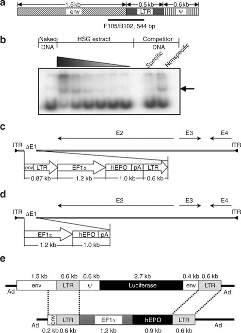
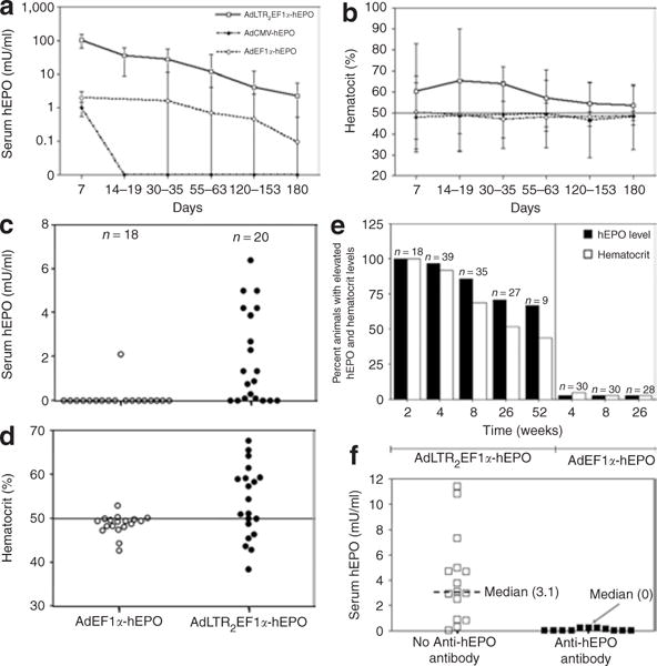
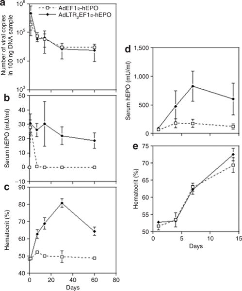
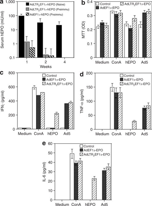
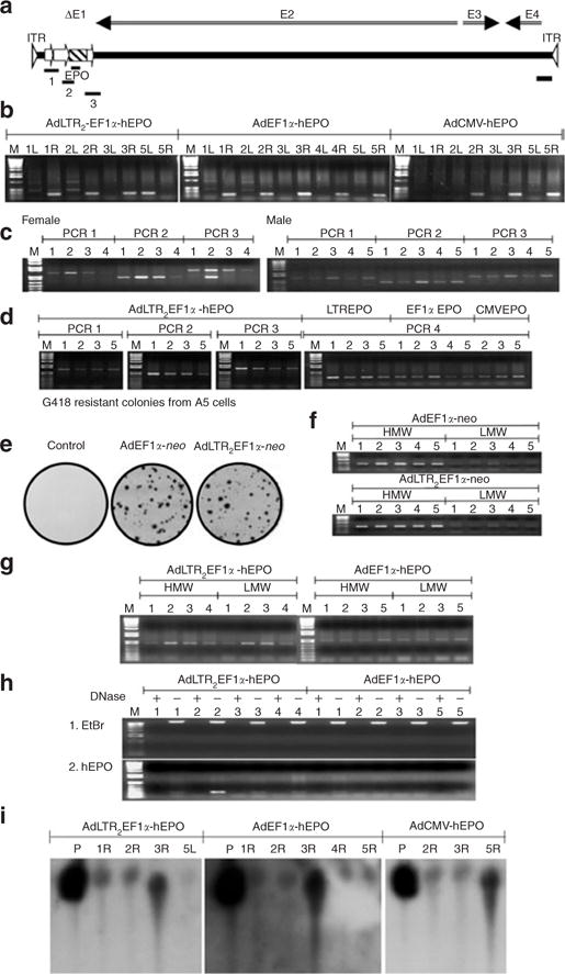
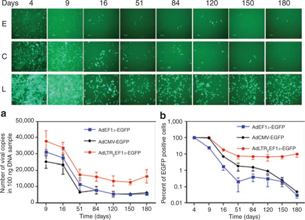
We studied the effects of specific retroviral elements in a first-generation serotype 5 adenoviral (Ad5) vector, AdLTR(2)EF1alpha-hEPO. This vector contains 858 base pair (bp) [251-bp envelope sequence plus 607-bp long-terminal repeat (LTR)] from Moloney murine leukemia virus (MoMLV), upstream of the human elongation factor-1alpha (EF1alpha) promoter and human erythropoietin (hEPO) cDNA, with the LTR sequence downstream of the polyadenylation signal. We compared expression of AdLTR(2)EF1alpha-hEPO with corresponding expressions of two conventional Ad5 vectors, AdEF1alpha-hEPO and AdCMV-hEPO, in vivo in submandibular glands in rats. Both the conventional vectors yielded low serum hEPO levels by day 7, and little change in hematocrits. In contrast, after receiving AdLTR(2)EF1alpha-hEPO, the rats showed elevated hEPO levels and hematocrits for 1-3 months. In vitro studies showed that the integration efficiencies of all the vectors were similar (approximately 10(-3)). Approximately 0.1% of the vector genomes were present 1 year after delivery in the case of each of the three vectors, primarily as intact linear double-strand DNA. The unique results seen with AdLTR(2)EF1alpha-hEPO are partly because of LTR enhancer activity. However, other cis-acting activity, which is not immunomodulatory but nevertheless influences promoter methylation, appears to be involved. A vector such as AdLTR(2)EF1alpha-hEPO may prove useful in clinical applications in which extended, but not "permanent," transgene expression is desirable.


Figures

Figure 1
Figure 1. Vector design
(a) Schematic representation of the 2.7 kilobase (kb) Moloney murine leukemia virus element used in AdLTR-luc, and used as a template to amplify 10 different DNA fragments for protein– DNA binding studies with HSG cell nuclear proteins. F105/B102 indicates the position of a 544-base pair (bp) fragment exhibiting the greatest interaction. (b) Autoradiograph of a representative gel shift assay. The interacting DNA–protein complex is denoted by the arrow, which shows that the F105/B102 544-bp fragment has strong and specific binding with increasing concentrations of the HSG cell nuclear protein extract. (c) Schematic depiction of AdLTR2EF1α-hEPO. (d) Schematic depiction of AdEF1α-hEPO. (e) Schematic comparison of the transgene cassettes in AdLTR-luc and AdLTR2EF1α-hEPO (reported herein). See text for additional details. Ad, adenovirus; EF1α, elongation factor-1α; env, envelope; hEPO, human erythropoietin; ITR, inverted terminal repeat; LTR, long-terminal repeat; pA, polyadenylation.
Figure 2
Figure 2. Effects of vector administration on human erythropoietin (hEPO), hematocrit, and antibody levels
(a) Time-course of hEPO expression after vector (109 particles) delivery to one submandibular gland in each rat. The data shown are the mean values ± SD for results from a total of 82 male rats. The animals received AdLTR2EF1α-hEPO (n = 37), AdEF1α-hEPO (n = 25), or AdCMV-hEPO (n = 5). None of the 15 untreated rats followed over this time-course exhibited any measurable hEPO in their sera (not shown). (b) Time-course of hematocrit levels (mean value ±SD) measured for the same rats as are shown in panel (a). The control rats exhibited hematocrits between 47 and 50 over this duration of time (not shown). (c) Serum hEPO levels measured within a single experiment 6 months after delivery of either AdEF1α-hEPO or AdLTR2EF1α-hEPO. (d) Hematocrit levels of rats in experiment shown in panel (c). (e) Summary of all in vivo experiments shown in this figure after delivery of 109 particles of either AdEF1α-hEPO or AdLTR2EF1α-hEPO to rats, at various time-points. Any measurable hEPO value was considered to be elevated, as were all hematocrit values >50. (f) Differences in serum hEPO levels 6 months after AdLTR2EF1α-hEPO administration, measured in rats with and without serum antibodies to hEPO. Ad, adenovirus; CMV, cytomegalovirus; EF1α, elongation factor-1α; LTR, long-terminal repeat.
Figure 3
Figure 3. Early time-course of glandular vector copy number, serum human erythropoietin (hEPO) expression, and hematocrit levels after vector delivery to submandibular glands and femoral veins in rats
(a) Time-course of vector copy numbers after vector (109 particles) delivery in rats to one submandibular gland in each animal. The animals received either AdLTR2EF1α-hEPO or AdEF1α-hEPO. The data shown are mean values ± SD for glands from three rats. The vector copy number was determined using quantitative PCR and primers for the hEPO cDNA (see Materials and Methods). The data were analyzed using a one-way analysis of variance, and no significant differences over this time-course were found between the two vectors. (b) Time-course of hEPO levels measured for the same rats as are shown in a. The data shown are mean values ± SD. (c) Time-course of hematocrit levels measured for the same rats as are shown in a. The data shown are mean values ± SD. (d) Time-course of hEPO levels after systemic vector (109 particles) delivery to 6 rats through the femoral vein. The animals (n = 3/group) received either AdLTR2EF1α-hEPO or AdEF1α-hEPO. The data shown are mean values ± SD. Note the difference in the y-axis scale from b. (e) Time-course of hematocrit levels measured for the same rats as are shown in d. The data shown are mean values ± SD. Ad, adenovirus; EF1α, elongation factor-1α; LTR, long-terminal repeat.
Figure 4
Figure 4. Immune reactivity following vector administration to salivary glands in rats
(a) The rats were either pretreated with a conventional Ad5 vector (AdCMV-luc) by intramuscular injection or not (naive) on day –21. On day 0 the rats were administered either AdLTR2EF1α-hEPO or AdEF1α-hEPO through their submandibular glands, as indicated. The data shown are the mean values ± SD (n = 5/group). (b–e) Spleens were removed 2 months after vector administration. Splenocytes were prepared and used for performing cellular immune response assays. (b) Cellular proliferation was measured using the MTT assay. (c,d) IFN-γ and TNF-α production, i.e., TH1 responses. (e) IL-6 production, i.e., a TH2 response. The data shown in b–e are the mean values ± SD of results from 2 rats/group, each assayed in triplicate. Medium: the negative control, cells incubated in culture medium only. Con A: the positive control, cells incubated in medium supplemented with concanavalin A (10 μg/ml). hEPO: results from cells incubated with 500 mU/ml recombinant hEPO protein. Ad5: results from cells incubated with AdCMV-luc at a multiplicity of infection (MOI) = 10 particles/cell. See text for additional details. Ad, adenovirus; CMV, cytomegalovirus; EF1α, elongation factor-1α; hEPO, human erythropoietin; IFN-γ, interferon-γ; IL-6, interleukin-6; LTR, long-terminal repeat; MTT, 3-(4,5-dimethylthiazol-2-yl)-2,5-diphenyltetrazolium bromide, TNF-α, tumor necrosis factor-α.
Figure 5
Figure 5. Assessment of vector DNA in situ
(a) Diagram of PCR products amplified with AdLTR2EF1α-hEPO. The PCR1 amplicon goes from the beginning of the upstream Moloney murine leukemia virus element to the middle of the EF1α promoter. The PCR2 amplicon goes from the EF1α promoter to the middle of the hEPO cDNA. The EPO amplicon is from the middle of the hEPO cDNA. The PCR3 amplicon goes from the hEPO cDNA into the Ad5 E2 region. The PCR4 amplicon is at the 3′-end of the vector in the Ad5 E4 region. (b) Detection of the EPO amplicon in targeted glands 6 months after vector delivery. Note that vectors were delivered to the right (R) submandibular glands of all animals except rat #5 in the AdLTR2EF1α-hEPO group, which received the vector in the left (L) gland. (c) Detection of the amplicons PCR 1, 2, and 3 in targeted glands 1 year after AdLTR2EF1α-hEPO delivery to female and male rats. (d) Detection of the amplicons PCR 1, 2, and 3 in targeted glands 6 months after AdLTR2EF1α-hEPO delivery, and PCR 4 after delivery of AdLTR2EF1α-hEPO (LTREPO), AdEF1α-hEPO (EF1αEPO), and AdCMV-hEPO (CMVEPO). (e) G418-resistant colonies obtained from A5 cells transduced with either AdEF1α-neo or AdLTR2EF1α-neo (neo, neomycin resistance gene). (f) PCR assays (using EF1αF4 and neoB3 primers) with high-molecular-weight (HMW) and low-molecular-weight (LMW) DNA extracts from G418-resistant colonies. (g) Assay for the PCR2 amplicon using HMW and LMW DNA extracts from female rats 1 year after administration of either AdEF1α-hEPO or AdLTR2EF1α-hEPO. (h) Plasmid-safe DNase assays to determine the general vector DNA structure (linear double strand or circular) in rats whose submandibular glands had been transduced with either AdLTR2EF1α-hEPO or AdEF1α-hEPO 1year earlier. Plus means presence of plasmid-safe DNase in the incubation mixture, Minus means without plasmid-safe DNase present. (i) Southern hybridization of undigested DNA samples from targeted glands 6 months after delivery of AdLTR2EF1α-hEPO, AdEF1α-hEPO, or AdCMV-hEPO. P refers to positive control samples from intact vectors spiked into glands from naive rats during genomic DNA extraction. Ad, adenovirus; CMV, cytomegalovirus; hEF1α, human elongation factor-1α; EtBr, ethidium bromide; hEPO, human erythropoietin; LTR, long-terminal repeat.
Figure 6
Figure 6. Time-course of vector transduction of A5 cells in vitro
The Ad5 vectors used were AdLTR2EF1α-EGFP, AdEF1α-EGFP, and AdCMV-EGFP. The top panel shows one of five representative fields at each time-point. E: AdEF1α-EGFP-transduced A5 cells. C: AdCMV-EGFP-transduced A5 cells. L: AdLTR2EF1α-EGFP-transduced A5 cells. (a) Time-course of viral vector copy numbers present. (b) Time-course of EGFP positive A5 cells. The data in b were normalized to the maximum EGFP expression seen, i.e., at day 4 (100%). The data shown in panels a and b are the mean values ± SD of results from all five fields. See text for additional details. Ad, adenovirus; CMV, cytomegalovirus; EF1α, elongation factor-1α; EGFP, enhanced-green fluorescent protein; LTR, long-terminal repeat.

Similar articles

Cited by

References

    1. Greber UF, Suomalainen M, Stidwill RP, Boucke K, Ebersold MW, Helenius A. The role of the nuclear pore complex in adenovirus DNA entry. EMBO J. 1997;16:5998–6007. - PMC - PubMed
    1. Greber UF, Webster P, Weber J, Helenius A. The role of the adenovirus protease on virus entry into cells. EMBO J. 1996;15:1766–1777. - PMC - PubMed
    1. Schaack J, Ho WY, Freimuth P, Shenk T. Adenovirus terminal protein mediates both nuclear matrix association and efficient transcription of adenovirus DNA. Genes Dev. 1990;4:1197–1208. - PubMed
    1. Harui A, Suzuki S, Kochanek S, Mitani K. Frequency and stability of chromosomal integration of adenovirus vectors. J Virol. 1999;73:6141–6146. - PMC - PubMed
    1. Yang Y, Li Q, Ertl HC, Wilson JM. Cellular and humoral immune responses to viral antigens create barriers to lung-directed gene therapy with recombinant adenoviruses. J Virol. 1995;69:2004–2015. - PMC - PubMed

Publication types

LinkOut - more resources