. 2008 May;16(5):845-53.
doi: 10.1038/mt.2008.39. Epub 2008 Mar 11.

Reduced pathology and improved behavioral performance in Alzheimer's disease mice vaccinated with HSV amplicons expressing amyloid-beta and interleukin-4

Affiliations

Reduced pathology and improved behavioral performance in Alzheimer's disease mice vaccinated with HSV amplicons expressing amyloid-beta and interleukin-4

Maria E Frazer et al. Mol Ther. 2008 May.

Abstract

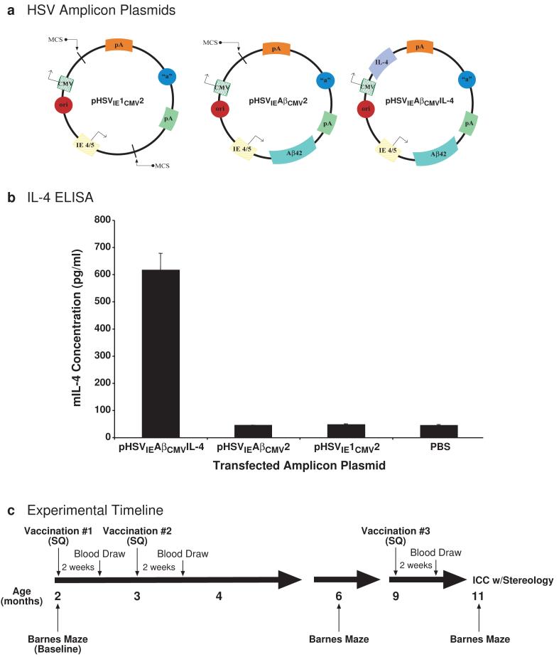
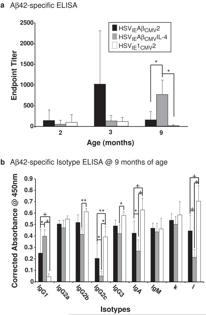
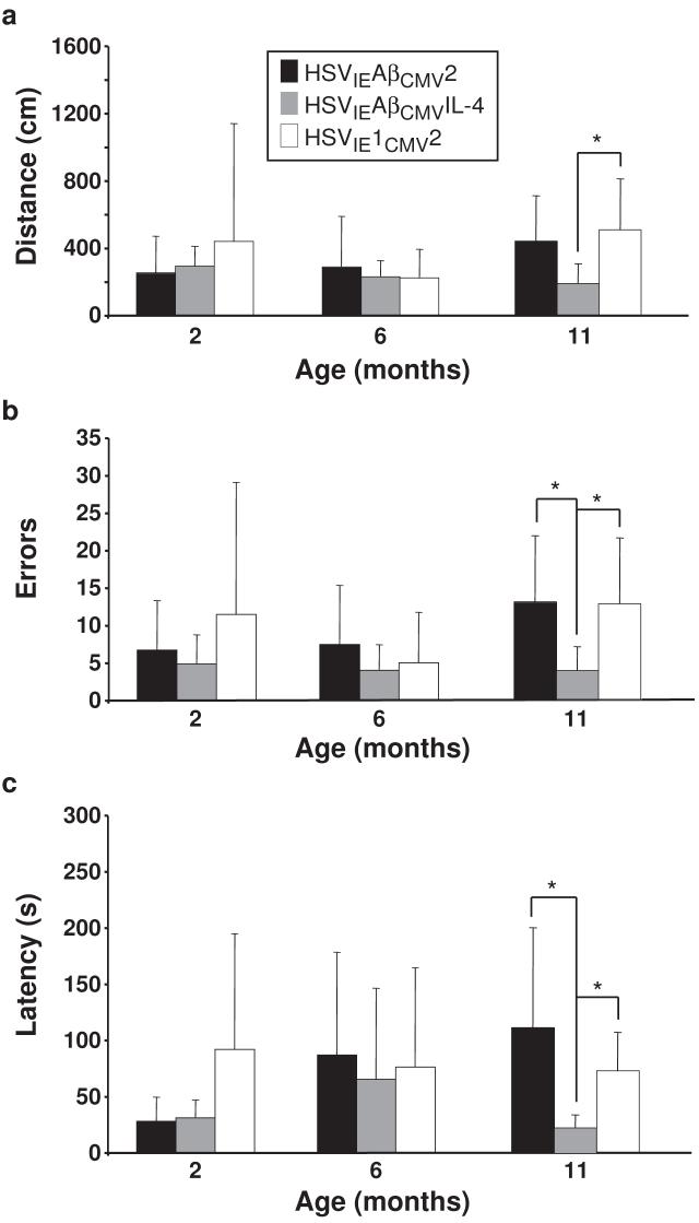
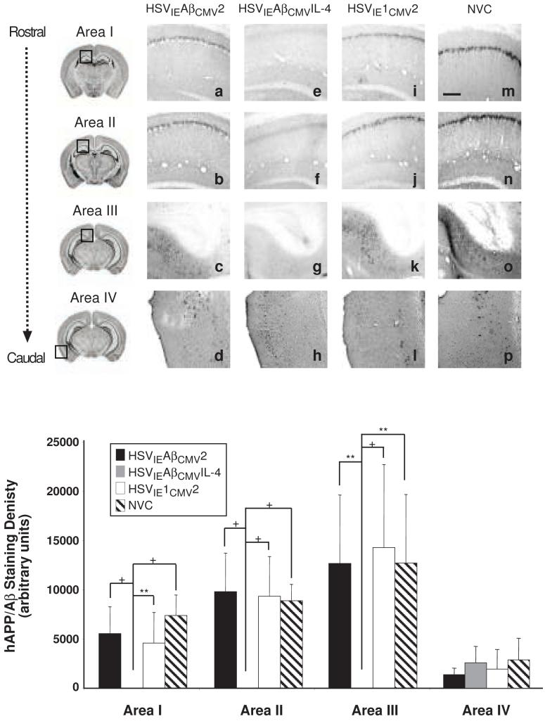
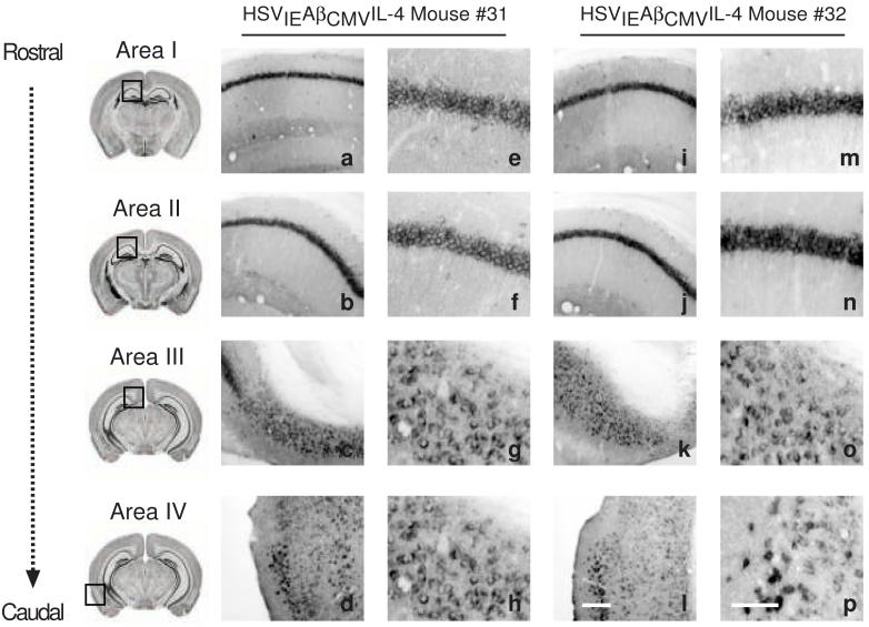
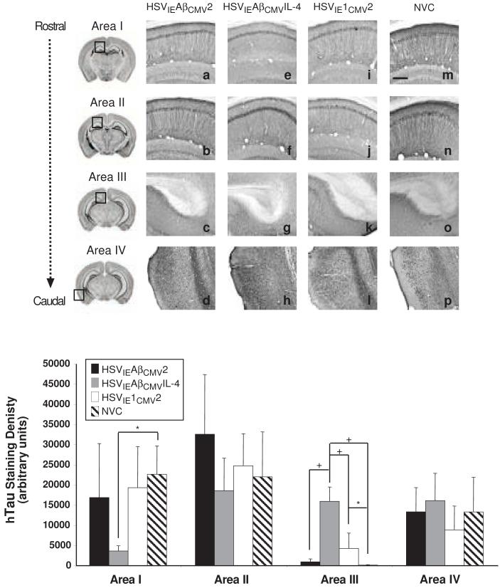
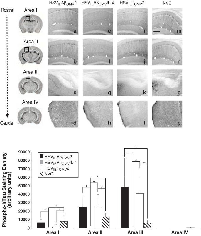
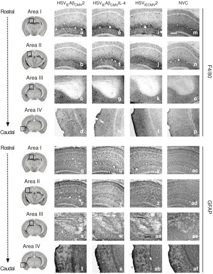
Immunotherapeutics designed to dissolve existing amyloid plaques or to interrupt amyloid-beta (Abeta) accumulation may be feasible for treatment and/or prevention of Alzheimer's disease (AD). "Shaping" the immune responses elicited against Abeta is requisite toward generating an efficacious and safe outcome; this can be achieved by minimizing the possibility of deleterious inflammatory reactions in the brain as observed in clinical testing of Abeta peptide/adjuvant-based modalities. Herpes simplex virus (HSV)-based amplicons can coexpress multiple antigens and/or immunomodulatory genes due to their large genetic size capacity, thereby facilitating antigen-specific immune response shaping. We have constructed an amplicon (HSV(IE)Abeta(CMV)IL-4) that co-delivers Abeta(1-42) with interleukin-4 (IL-4), a cytokine that promotes the generation of Th2-like T-cell responses, which are favored in the setting of AD immunotherapy. Triple-transgenic AD (3xTg-AD) mice, which progressively develop both amyloid and neurofibrillary tangle pathology, were vaccinated thrice with HSV(IE)Abeta(CMV)IL-4, or a set of control amplicon vectors. Increased Th2-related, Abeta-specific antibodies, improved learning and functioning of memory, and prevention of AD-related amyloid and tau pathological progression were observed significantly more in the HSV(IE)Abeta(CMV)IL-4 vaccinated mice as compared to the other experimental groups. Our study underscores the potential of Abeta immunotherapy for AD and highlights the potency of amplicons in facilitating the immune response modulation to a disease-relevant antigen.


Figures

Figure 1
Figure 1. Schematic representation of newly constructed amplicon vectors, in vitro confirmation of amplicon-mediated murine IL-4 expression, and study design
(a) Three kanamycin-resistant HSV amplicons plasmids were constructed: one that served as an empty vector control (pHSVIE1CMV2) with HSV origin of replication (ori) and HSV packaging signal (“a”), a second (pHSVIECMV2) that expressed the Aβ1-42 peptide derived from human amyloid precursor protein (APP) under the transcriptional control of the HSV immediate-early (IE) 4/5 gene promoter and SV40 polyadenylation signal (pA), and a third (pHSVIECMVIL-4) that expressed Aβ1-42 via the IE4/5 promoter/SV40 pA transcription unit and murine Interleukin-4 under the separate transcriptional control of the cytomegalovirus (CMV) immediate-early promoter and bovine growth hormone polyadenylation signal. All amplicons were packaged using a previously described helper virus-free method [23]. (b) Each amplicon plasmid was transiently transfected into baby hamster kidney (BHK) cells and culture supernatants were analyzed by ELISA to assess murine IL-4 expression (pg/ml) from the pHSVIECMVIL-4 amplicon (N=4 per experimental group). A phosphate-buffered saline (PBS) group served as a no-vector control condition. (c) A Barnes maze behavioral assessment was performed to determine baseline learning and memory functioning at 2 months of age. Each packaged vector (1 × 10⁶ transduction units) was delivered subcutaneously (SQ) to a randomized cohort of male 3xTg-AD mice [22] (N=6 per experimental group). Amplicons were administered to each animal thrice, and humoral assessments were performed 2 weeks after each vaccination. An intermediate Barnes maze assessment was performed at 6 months of age. Antibody isotype analysis was performed on sera obtained at the 9-month post-initial vaccination time point. Vaccinated mice were sacrificed at 11 months of age at which time endpoint behavioral, histological, and stereological analyses were performed.
Figure 2
Figure 2. Elicitation of distinctive humoral responses in HSV amplicon vector-vaccinated 3xTg-AD mice
(a) Helper virus-free HSVIECMV2 (black bars), HSVIECMVIL-4 (grey bars), and HSVIE1CMV2 (open bars) were delivered subcutaneously thrice to 3xTg-AD mice beginning at 2 months of age (1×10⁶ transduction units per vaccination). Serum was obtained from each vaccinated mouse according to the schema illustrated in Figure 1 and serum samples were analyzed for levels of antibodies binding specifically to the Aβ1-42 peptide in quadruplicate by ELISA. Levels of Aβ-specific antibodies arising from each vaccination were corrected using serum isolated from control mice, and are expressed as endpoint titers. (b) Isotypes of α-Aβ1-42 antibodies were determined by ELISA using sera obtained at the 9-month time point from vaccinated 3xTg-AD mice. Levels of Aβ-specific antibody isotypes arising from each vaccination were corrected using serum isolated from control mice, and are expressed as “Corrected Absorbance @ 450 nm”. Error bars represent standard deviation. “*” equals P<0.05, “**” equals P<0.01, and “+” equals P<0.001 as determined by two-way ANOVA with Bonferroni post-hoc analysis.
Figure 3
Figure 3. HSVIECMVIL-4 vaccinated 3xTg-AD mice exhibit improved performance in the Barnes maze learning and memory task
Helper virus-free HSVIECMV2 (black bars), HSVIECMVIL-4 (grey bars), and HSVIE1CMV2 (open bars) were delivered subcutaneously thrice to 3xTg-AD mice beginning at 2 months of age (1×10⁶ transduction units per vaccination). Mice were tested on the Barnes maze at 2, 6, and 11 months of age to assess the effects of amplicon vaccination on learning/memory functioning using established distance (a), errors (b), and latency (c) criteria. Error bars represent standard deviation. “*” equals P<0.05 and “**” equals P<0.01 as determined by one-way ANOVA with Bonferroni post-hoc analysis.
Figure 4
Figure 4. HSVIECMVIL-4 vaccinated 3xTg-AD mice are devoid of amyloid/Aβ burden
Helper virus-free HSVIECMV2 (a, b, c, d), HSVIECMVIL-4 (e, f, g, h), and HSVIE1CMV2 (i, j, k, l) were delivered subcutaneously thrice to 3xTg-AD mice beginning at 2 months of age (1×10⁶ transduction units per vaccination). Coronal mouse brain sections (30 μm) were prepared from vaccinated mice sacrificed at 11 months of age and were processed for 6E10 immunohistochemistry to assess the extent of human APP/Aβ burden. Age-matched, non-vaccinated control (NVC) 3xTg-AD mice (m, n, o, p) were sacrificed at 11 months of age and brains processed identically for comparison purposes. Images were obtained for four areas of the brain. Area I represents the CA1 hippocampal region at -1.28 mm from Bregma (a, e, i, m), area II represents the CA1 region at -2.12 mm from Bregma (b, f, j, n), area III represents the subiculum at -2.75 mm from Bregma (c, g, k, o), and area IV represents the entorhinal cortex at -2.50 to -3.80 mm from Bregma (d, h, l, p). The optical densities of immunopositive staining were quantified (q). Black bars represent HSVIECMV2, grey bars represent HSVIECMVIL-4, open bars represent HSVIE1CMV2, and hatched bars represent non-vaccinated control mice. The scale bar depicted in m represents 250 μm. Error bars indicate standard deviation. “*” equals P<0.05 and “**” equals P<0.01, and “+” equals P<0.001 as determined by one-way ANOVA with Bonferroni post-hoc analysis.
Figure 5
Figure 5. Immunohistochemical analysis of HSVIECMVIL-4 vaccinated 3xTg-AD mice using a human APP-specific antibody demonstrates that vaccination does not affect hAPPswe transgene product levels
A subset of mice vaccinated thrice with helper virus-free HSVIECMVIL-4 (a-h and i-p) were processed for human APP immunohistochemistry to assess the extent of transgene-derived amyloid precursor protein. Images were obtained for four areas of the brain at low and high magnification shown for two mice (#31 and #32) injected with HSVIECMVIL-4. Area I represents the CA1 hippocampal region at -1.28 mm from Bregma (a, e, i, m), area II represents the CA1 region at -2.12 mm from Bregma (b, f, j, n), area III represents the subiculum at -2.75 mm from Bregma (c, g, k, o), and area IV represents the entorhinal cortex between -2.50 mm and -3.80 mm from Bregma (d, h, l, p). The scale bar depicted in p (for panels e-h and m-p) represents 50 μm. The scale bar depicted in l (for panels a-d and i-l) represents 250 μm.
Figure 6
Figure 6. Human tau expression patterns are minimally affected in amplicon-vaccinated 3xTg-AD mice
Helper virus-free HSVIECMV2 (a, b, c, d), HSVIECMVIL-4 (e, f, g, h), and HSVIE1CMV2 (i, j, k, l) were delivered subcutaneously thrice to 3xTg-AD mice beginning at 2 months of age (1×10⁶ transduction units per vaccination). Coronal mouse brain sections (30 μm) were prepared from vaccinated mice sacrificed at 11 months of age and were processed for HT7 immunohistochemistry to assess alterations in human Tau transgene expression. Age-matched, non-vaccinated control (NVC) 3xTg-AD mice (m, n, o, p) were sacrificed at 11 months of age and brains processed identically for comparison purposes. Images were obtained for four areas of the brain. Area I represents the CA1 hippocampal region at -1.28 mm from Bregma (a, e, i, m), area II represents the CA1 region at -2.12 mm from Bregma (b, f, j, n), area III represents the subiculum at -2.75 mm from Bregma (c, g, k, o), and area IV represents the entorhinal cortex at -2.50 to -3.80 mm from Bregma (d, h, l, p). The optical densities of immunopositive staining were quantified (q). Black bars represent HSVIECMV2, grey bars represent HSVIECMVIL-4, open bars represent HSVIE1CMV2, and hatched bars represent non-vaccinated control mice. The scale bar depicted in m represents 250 μm. Error bars indicate standard deviation. “*” equals P<0.05 and “+” equals P<0.001 as determined by one-way ANOVA with Bonferroni post-hoc analysis.
Figure 7
Figure 7. HSVIECMVIL-4 vaccinated 3xTg-AD mice exhibit suppressed phospho-Tau expression
Helper virus-free HSVIECMV2 (a, b, c, d), HSVIECMVIL-4 (e, f, g, h), and HSVIE1CMV2 (i, j, k, l) were delivered subcutaneously thrice to 3xTg-AD mice beginning at 2 months of age (1×10⁶ transduction units per vaccination). Coronal mouse brain sections (30 μm) were prepared from vaccinated mice sacrificed at 11 months of age and were processed for AT180 immunohistochemistry to assess alterations in human phospho-Tau pathogenic epitope expression. Age-matched, non-vaccinated control (NVC) 3xTg-AD mice (m, n, o, p) were sacrificed at 11 months of age and brains processed identically for comparison purposes. Images were obtained for four areas of the brain. Area I represents the CA1 hippocampal region at -1.28 mm from Bregma (a, e, i, m), area II represents the CA1 region at -2.12 mm from Bregma (b, f, j, n), area III represents the subiculum at -2.75 mm from Bregma (c, g, k, o), and area IV represents the entorhinal cortex at -2.50 to -3.80 mm from Bregma (d, h, l, p). The optical densities of immunopositive staining were quantified (q). Black bars represent HSVIECMV2, grey bars represent HSVIECMVIL-4, open bars represent HSVIE1CMV2, and hatched bars represent non-vaccinated control mice. The scale bar depicted in m represents 250 μm. Error bars indicate standard deviation. “*” equals P<0.05 and “**” equals P<0.01, and “+” equals P<0.001 as determined by one-way ANOVA with Bonferroni post-hoc analysis.
Figure 8
Figure 8. Microglial and astrocytic cell staining patterns in 3xTg-AD mice are not overtly affected by amplicon-mediated vaccination
Helper virus-free HSVIECMV2 (a-d and q-t), HSVIECMVIL-4 (e-h and u-x), and HSVIE1CMV2 (i-l and y-ab) were delivered subcutaneously thrice to 3xTg-AD mice beginning at 2 months of age (1×10⁶ transduction units per vaccination). Coronal mouse brain sections (30 μm) were prepared from vaccinated mice sacrificed at 11 months of age and were processed for F4/80 immunohistochemistry (a-p) to assess alterations in microglial activation and GFAP immunohistochemistry (q-af) to examine changes in astrocyte staining patterns as a result of amplicon-mediated vaccination. Age-matched, non-vaccinated control (NVC) 3xTg-AD mice (m-p and ac-af for F4/80 and GFAP staining, respectively) were sacrificed at 11 months of age and brains processed identically for comparison purposes. Images were obtained for four areas of the brain. Area I represents the CA1 hippocampal region at -1.28 mm from Bregma (a, e, i, m, q, u, v, ac), area II represents the CA1 region at -2.12 mm from Bregma (b, f, j, n, r, v, z, ad), area III represents the subiculum at -2.75 mm from Bregma (c, g, k, o, s, w, aa, ae), and area IV represents the entorhinal cortex at -2.50 to -3.80 mm from Bregma (d, h, l, p, t, x, ab, af). The scale bar depicted in m represents 250 μm.

Similar articles

Cited by

References

    1. Selkoe DJ. Alzheimer’s disease is a synaptic failure. Science. 2002;298:789–791. - PubMed
    1. Braak H, Braak E. Neuropathological stageing of Alzheimer-related changes. Acta Neuropathol (Berl) 1991;82:239–259. - PubMed
    1. Braak H, Braak E. Frequency of stages of Alzheimer-related lesions in different age categories. Neurobiol Aging. 1997;18:351–357. - PubMed
    1. Tariot PN, Federoff HJ. Current treatment for Alzheimer disease and future prospects. Alzheimer Dis Assoc Disord. 2003;17(Suppl 4):S105–113. - PubMed
    1. Hardy J, Selkoe DJ. The amyloid hypothesis of Alzheimer’s disease: progress and problems on the road to therapeutics. Science. 2002;297:353–356. - PubMed

Publication types