. 2008 Jun;9(6):576-81.
doi: 10.1038/embor.2008.48. Epub 2008 Apr 4.

NAD(P)H quinone oxidoreductase 1 inhibits the proteasomal degradation of the tumour suppressor p33(ING1b)

Affiliations

NAD(P)H quinone oxidoreductase 1 inhibits the proteasomal degradation of the tumour suppressor p33(ING1b)

Marco Garate et al. EMBO Rep. 2008 Jun.

Abstract

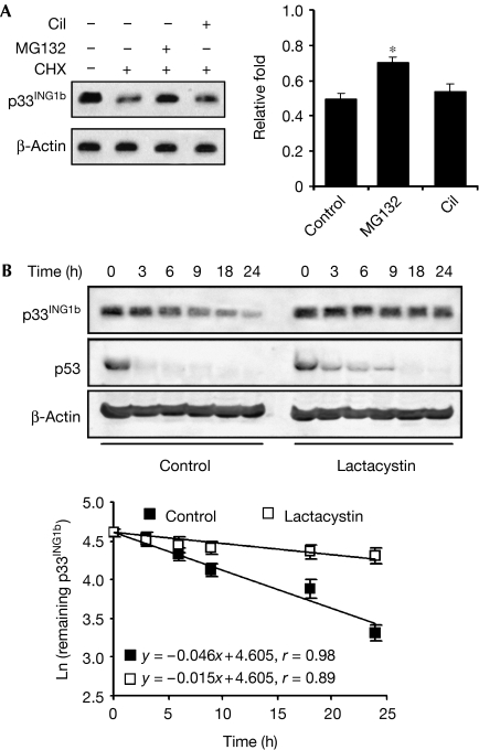
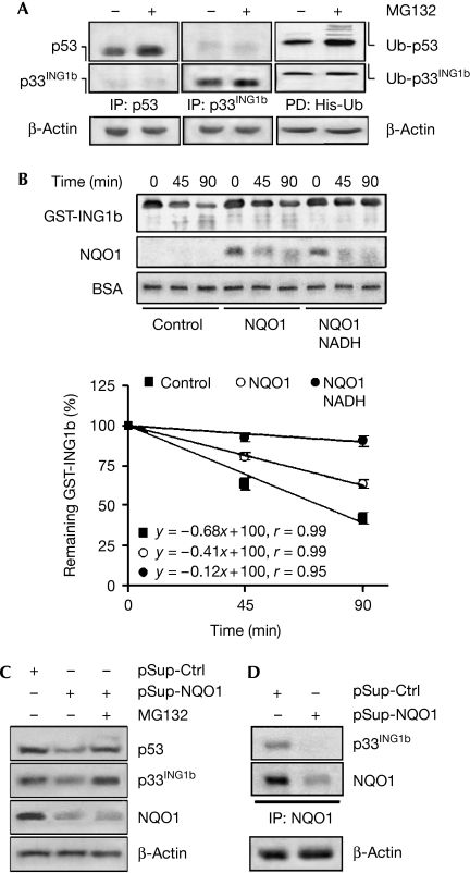
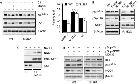
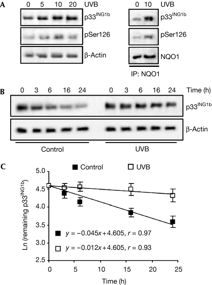
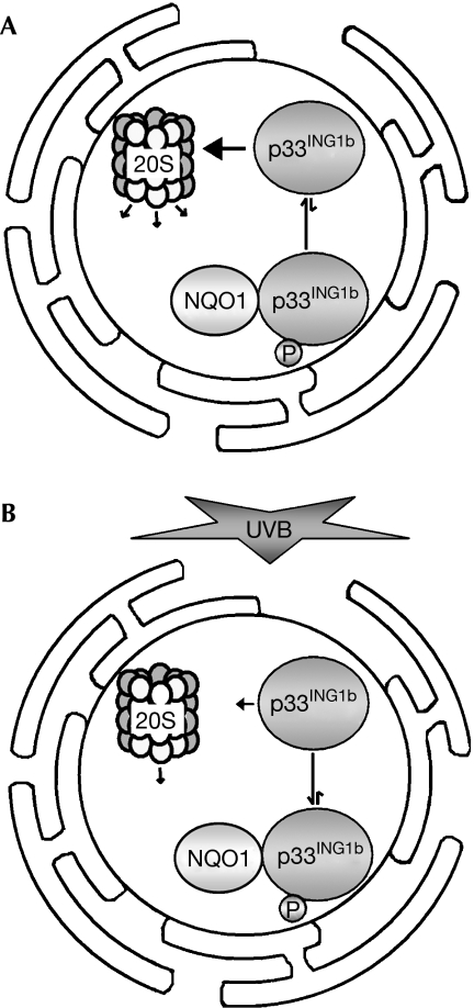
The tumour suppressor p33(ING1b) ((ING1b) for inhibitor of growth family, member 1b) is important in cellular stress responses, including cell-cycle arrest, apoptosis, chromatin remodelling and DNA repair; however, its degradation pathway is still unknown. Recently, we showed that genotoxic stress induces p33(ING1b) phosphorylation at Ser 126, and abolishment of Ser 126 phosphorylation markedly shortened its half-life. Therefore, we suggest that Ser 126 phosphorylation modulates the interaction of p33(ING1b) with its degradation machinery, stabilizing this protein. Combining the use of inhibitors of the main degradation pathways in the nucleus (proteasome and calpains), partial isolation of the proteasome complex, and in vitro interaction and degradation assays, we set out to determine the degradation mechanism of p33(ING1b). We found that p33(ING1b) is degraded in the 20S proteasome and that NAD(P)H quinone oxidoreductase 1 (NQO1), an oxidoreductase previously shown to modulate the degradation of p53 in the 20S proteasome, inhibits the degradation of p33(ING1b). Furthermore, ultraviolet irradiation induces p33(ING1b) phosphorylation at Ser 126, which, in turn, facilitates its interaction with NQO1.


Conflict of interest statement

The authors declare that they have no conflict of interest.

Figures

Figure 1
Figure 1
p33ING1b is degraded in the proteasome. (A) p33ING1b protein levels were determined by western blot in nuclear extracts of cells treated for 16 h with CHX or CHX plus MG132 or CiI. The density of the bands was determined and plotted as the relative fold compared with the untreated control. The bars represent the average±s.d. from three independent experiments; *P<0.01. (B) Effect of lactacystin on the half-life of p33ING1b. p33ING1b levels were determined in whole-cell extracts of cells treated with CHX (Control) or CHX plus lactacystin for different time points by western blot analysis. p53 levels were determined as a positive control of proteasomal degradation. The degradation rate of p33ING1b was determined by densitometric analysis of bands as described in the Methods. Values are means±s.d. CHX, cycloheximide; CiI, calpain inhibitor I; ING1b, inhibitor of growth family, member 1b.
Figure 2
Figure 2
p33ING1b is degraded in the 20S proteasome complex. (A) Nuclear extracts from cells treated for 16 h in the presence (+) or absence (−) of MG132 were immunoprecipitated (IP) with p53 and p33ING1 antibodies. Nuclear extracts expressing polyhistidine-tagged ubiquitin (His-Ub) were pulled down (PD) with Ni-NTA agarose beads and probed against p33ING1b. p53 was used as a positive control of proteasomal degradation. β-Actin was detected as an input control in aliquots of nuclear extracts before the assay. (B) NQO1 protects p33ING1b against degradation in the 20S proteasome in vitro. Isolated GST-ING1b was incubated with purified 20S proteasome at 37°C for different time points in the absence or presence of isolated NQO1 with or without 1 mM NADH. Samples were blotted and probed with antibodies against p33ING1b and NQO1. BSA (added to stabilize the enzymes) was detected as a negative control of degradation in the 20S proteasome. The degradation rate of p33ING1b was determined by densitometric analysis of the bands. Values are means±s.d. (C,D) NQO1 modulates the degradation of p33ING1b in situ. (C) Nuclear extracts from cells transfected with a plasmid encoding either NQO1-specific shRNA (pSup-NQO1) or control shRNA (pSup-Ctrl) and treated with MG132 and CHX for 16 h were blotted and probed with a p33ING1b antibody. The blots were also probed for NQO1 to ensure successful knockdown. p53 was used as a positive control of proteasomal degradation. (D) Nuclear extracts from cells transfected with either pSup-NQO1 or pSup-Ctrl were immunoprecipitated with an NQO1 antibody, blotted and probed for p33ING1b or NQO1. β-Actin was detected as an input control in aliquots of nuclear extracts before immunoprecipitation. BSA, bovine serum albumin; CHX, cycloheximide; GST, glutathione S-transferase; ING1b, inhibitor of growth family, member 1b; NQO1, NAD(P)H quinone oxidoreductase 1; shRNA, short hairpin RNA.
Figure 3
Figure 3
NQO1 preferentially binds to phosphorylated p33ING1b. (A) Phosphorylation at Ser 126 does not alter the degradation pathway of p33ING1b. Nuclear extracts obtained from cells transfected with pCI-NeoING1b (WT) or pCI-NeoS126A (S126A), and treated for 16 h with CHX alone (control) or combined with MG132 or CiI, were blotted and probed for p33ING1b or p53. The intensity of p33ING1b signal in each band was quantified and plotted as the relative fold compared with that of untreated cells. Values represent average±s.d. from three independent experiments. *P<0.01; **P<0.001. (B) NQO1 preferentially binds to p33ING1b phosphorylated at Ser 126. Aliquots of 50 μg of nuclear extracts prepared from cells expressing WT, S126A or S126E mutant p33ING1b were blotted. p33ING1b and NQO1 expression and phosphorylation at Ser 126 were determined by western blot using specific antibodies (left panel). Aliquots of 250 μg nuclear extracts were immunoprecipitated (IP) with an NQO1 antibody and the immunocomplexes were analysed by western blotting for p33ING1b, Ser 126 phosphorylation or NQO1 (right panel). (C) GST or GST-ING1b was incubated with isolated NQO1 in the absence or presence of NADH. The complexes isolated by GST pull-down were blotted, and NQO1 and GST-ING1b levels were determined by western blot and that of GST by Ponceau S staining. (D) Nuclear extracts from cells co-transfected with a plasmid encoding NQO1-specific shRNA (pSup-NQO1) or control shRNA (pSup-Ctrl) and pCI-NeoING1b (WT) or pCI-NeoS126A, and treated with MG132 and CHX for 16 h were blotted and probed for p33ING1b and NQO1. p53 was used as a positive control of proteasomal degradation. CiI, calpain inhibitor I; CHX, cycloheximide; GST, glutathione S-transferase; ING1b, inhibitor of growth family, member 1b; NQO1, NAD(P)H quinone oxidoreductase 1; shRNA, short hairpin RNA; WT, wild type.
Figure 4
Figure 4
UVB irradiation induces binding of p33ING1b to NQO1 through phosphorylation at Ser 126. (A) p33ING1b expression and phosphorylation at Ser 126 were determined in nuclear extracts of cells irradiated with different dosages of UVB (mJ/cm2) and treated with CHX by western blot using specific antibodies (left panel). Aliquots of two nuclear extracts (250 μg each) were immunoprecipitated (IP) with an NQO1 antibody and the levels of p33ING1b or NQO1 or phosphorylation at Ser 126 in the immunocomplexes were assessed (right panel). (B) Effect of UVB irradiation on the half life of p33ING1b. MMRU cells were subjected UVB irradiation (10 mJ/cm2) and treated with CHX. p33ING1b protein levels were determined in nuclear extracts from these cells by western blot. (C) The degradation rate of p33ING1b was determined by densitometric analysis of the bands. Values are means±s.d. CHX, cycloheximide; ING1b, inhibitor of growth family, member 1b; NQO1, NAD(P)H quinone oxidoreductase 1.
Figure 5
Figure 5
Proposed model for the effect of UVB irradiation on the mechanism of p33ING1b degradation. NQO1 preferentially binds to p33ING1b phosphorylated at Ser 126. (A) Under the conditions of non-ultraviolet stress, p33ING1b shows a low degree of phosphorylation at Ser 126 and is degraded readily in the 20S proteasome complex in the nucleus. (B) On UVB irradiation, p33ING1b is efficiently phosphorylated at Ser 126, inducing its binding to NQO1, which, in turn, stabilizes p33ING1b. ING1b, inhibitor of growth family, member 1b; NQO1, NAD(P)H quinone oxidoreductase 1.

Similar articles

Cited by

References

    1. Asher G, Shaul Y (2005) p53 proteasomal degradation: poly-ubiquitination is not the whole story. Cell Cycle 4: 1015–1018 - PubMed
    1. Asher G, Tsvetkov P, Kahana C, Shaul Y (2005a) A mechanism of ubiquitin-independent proteasomal degradation of the tumor suppressors p53 and p73. Genes Dev 19: 316–321 - PMC - PubMed
    1. Asher G, Bercovich Z, Tsvetkov P, Shaul Y, Kahana C (2005b) 20S proteasomal degradation of ornithine decarboxylase is regulated by NQO1. Mol Cell 17: 645–655 - PubMed
    1. Campos EI, Chin MY, Kuo WH, Li G (2004) Biological functions of the ING family tumor suppressors. Cell Mol Life Sci 61: 2597–2613 - PMC - PubMed
    1. Chehab NH, Malikzay A, Stavridi ES, Halazonetis TD (1999) Phosphorylation of Ser 20 mediates stabilization of human p53 in response to DNA damage. Proc Natl Acad Sci USA 96: 13777–13782 - PMC - PubMed

Publication types

MeSH terms