. 2008 Feb 29;4(2):e1000006.
doi: 10.1371/journal.ppat.1000006.

Interleukin-6 is crucial for recall of influenza-specific memory CD4 T cells

Affiliations

Interleukin-6 is crucial for recall of influenza-specific memory CD4 T cells

Maria Paula Longhi et al. PLoS Pathog. .

Abstract

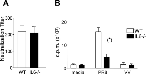
Currently, our understanding of mechanisms underlying cell-mediated immunity and particularly of mechanisms that promote robust T cell memory to respiratory viruses is incomplete. Interleukin (IL)-6 has recently re-emerged as an important regulator of T cell proliferation and survival. Since IL-6 is abundant following infection with influenza virus, we analyzed virus-specific T cell activity in both wild type and IL-6 deficient mice. Studies outlined herein highlight a novel role for IL-6 in the development of T cell memory to influenza virus. Specifically, we find that CD4+ but not CD8+ T cell memory is critically dependent upon IL-6. This effect of IL-6 includes its ability to suppress CD4+CD25+ regulatory T cells (Treg). We demonstrate that influenza-induced IL-6 limits the activity of virus-specific Tregs, thereby facilitating the activity of virus-specific memory CD4+ T cells. These experiments reveal a critical role for IL-6 in ensuring, within the timeframe of an acute infection with a cytopathic virus, that antigen-specific Tregs have no opportunity to down-modulate the immune response, thereby favoring pathogen clearance and survival of the host.


Conflict of interest statement

The authors have declared that no competing interests exist.

Figures

Figure 1
Figure 1. IL-6 production post-infection with influenza virus.
WT mice were infected i.n. with 20 HAU H17 influenza virus. Three and eight days after infection, serum levels of IL-6, IL-10, MCP-1/CCL2, IFN-γ, TNF-α, and IL-12 were measured in uninfected (A) and infected (B) mice by Cytometric Bead Array.
Figure 2
Figure 2. Nucleoprotein (NP)-Tetramer-positive CD8+ T cells in the spleens of influenza virus-infected mice.
Representative dot plots showing staining of spleen cells recovered from a WT and IL-6−/− mouse 10 days post i.n. infection with 20 HAU H17 influenza virus. The cells were stained with CD8-specific mAbs and either the NP-Tetramer (Db-ASNENMDAM, NP-Tet) or an irrelevant tetramer comprising Db and a peptide derived from Lymphocytic choriomeningitis virus (Db-KAVYNFATC, GP-Tet). The plots are representative of 3 mice per group.
Figure 3
Figure 3. Influenza specific CD8+ T cell activity in WT and IL-6−/− mice.
Peptide-specific CTL assays were carried out at 2 weeks (A) and 8 weeks (B) post i.n. infection with H17 influenza virus. B16 target cells were pulsed with the NP68 peptide prior to use in a standard 5-h 51Cr release assay. Total number of CD8+T cells recovered from lungs of WT and IL-6−/− mice following primary (C) or secondary (D) i.n. challenge with H17 influenza virus. Mice were analyzed individually, and values shown are the mean±SD (n = 3 mice/group). The results are representative of three independent experiments. Statistical significance was evaluated using the Student's t test.
Figure 4
Figure 4. Influenza specific CD4+ T cell activity in WT and IL-6−/− mice.
Proliferation assays were carried out using CD4+ T cells purified from spleens of WT and IL-6−/− mice 2 weeks (A) and 8 weeks (B) post i.n. infection with 20 HAU H17 influenza virus. Effectors were incubated with irradiated WT splenocytes alone or WT splenocytes infected with inactivated H17 influenza or vaccinia virus. [3H]-Thymidine was added on day 5 and proliferation measured by thymidine incorporation after 18 hrs. Mice were analyzed individually and values shown are the mean±SEM (n = 3 mice/group). Spleen cells were isolated 8 weeks after primary H17 infection and influenza-specific CD4+ T cells were analyzed for intracellular TNF-α by flow cytometry (C). Each symbol represents an individual mouse. Kinetic analysis of total CD4+ T cells infiltrated in lungs from WT and IL6−/− mice following primary (D) and secondary (E) i.n. challenge with 20 HAU H17 influenza virus. Mice were analyzed individually and values shown are the mean±SD (n = 3 mice/group). Statistical significance was evaluated using the Students t test.
Figure 5
Figure 5. Neutralizing activity in the serum of the H17 influenza virus memory mice.
(A) Serum from WT and IL-6−/− mice was harvested at 6–8 weeks post i.n. infection with 20 HAU of H17. MDCK cells and serum samples were cultured in the presence of 10 HAU of H17 and the viability of the cells determined 3 days later using the alamar blue assay. The presence of neutralizing antibodies in serum was determined by comparing cell viability of MDCKs cultured with serum from naïve mice, with viability of MDCKs cultured with serum from previously infected mice (see Materials and Methods for calculation). Results are presented as the mean neutralization index score±SEM at each dilution (n = 6–7 mice per group). (B) T cell cross-reactivity of PR8 and H17 was measured in a CD4+ T cell proliferation assay. Mice were infected i.n. with 20 HAU H17 virus and 8 weeks later CD4+ T cells were purified and stimulated in vitro with PR8-infected APCs. Proliferation was measured by [3H]-thymidine incorporation at day 5. Each bar represents the mean value±SEM (n = 3 wells/group). Statistical significance was evaluated using the Students t test.
Figure 6
Figure 6. Challenge of H17 influenza virus immune mice with the heterologous virus, PR8.
WT and IL-6−/− mice infected 6 weeks previously with H17 influenza virus were rechallenged i.n. with 20 HAU of the PR8 virus. Four days post secondary infection the lungs were harvested and the number of CD4+ T cells analyzed by flow cytometry (A). Proliferation and cytokine production was measured in splenocytes of H17-immune mice re-challenged 4 days previously with PR8 virus (B). The cells were labelled with CFSE and incubated for 6 days with H17 influenza-infected APCs as described in materials and methods. Specific proliferation was measured CFSE dilution and IFNγ production on gated CD4+ cells. The dot plots show a representative of 5 mice per group.
Figure 7
Figure 7. Influenza-specific suppressor activity in WT and IL-6−/− mice.
Mice were infected i.n. with 20 HAU of influenza virus (H17). Eight weeks after infection, splenocytes from WT and IL6−/− mice were harvested. (A) Purified CD4+ T cells were tested for proliferation against inactivated H17 before (ND) or after (D) depletion of CD25+ T cells. Influenza-specific proliferation in both populations was measured by [3H]-thymidine incorporation at day 5. Each symbol represents an individual mouse and the lines join responses in undepleted (ND) and CD25-depleted (D) CD4+ T cell populations. A stimulation index greater than 2 was considered a positive response. (B) CD4+CD25+ cells from WT and IL-6−/− mice, infected 2 or 8 weeks previously, were isolated from splenocytes and their suppressive capacity was evaluated by incubation at a ratio of 1∶1 with CD4+ T cells from a WT mouse infected 2 weeks previously with H17 influenza virus. The cells were stimulated with either APCs exposed to 1 µg/ml anti-CD3 mAbs (B) or influenza infected APCs (C and D). Proliferation was measured by [3H]-thymidine incorporation at day 5. CD4+CD25+ cells from 4 individual WT and IL-6−/−mice were analyzed and each bar represents the mean value±SEM of each group. Statistical significance was evaluated using the Students t test.

References

    1. Kishimoto T. Interleukin-6: from basic science to medicine–40 years in immunology. Annu Rev Immunol. 2005;23:1–21. - PubMed
    1. Salomon R, Hoffmann E, Webster RG. Inhibition of the cytokine response does not protect against lethal H5N1 influenza infection. Proc Natl Acad Sci U S A. 2007;104:12479–12481. - PMC - PubMed
    1. Szretter KJ, Gangappa S, Lu X, Smith C, Shieh WJ, et al. Role of host cytokine responses in the pathogenesis of avian H5N1 influenza viruses in mice. J Virol. 2007;81:2736–2744. - PMC - PubMed
    1. Jones SA. Directing transition from innate to acquired immunity: defining a role for IL-6. J Immunol. 2005;175:3463–3468. - PubMed
    1. Ito H, Takazoe M, Fukuda Y, Hibi T, Kusugami K, et al. A pilot randomized trial of a human anti-interleukin-6 receptor monoclonal antibody in active Crohn's disease. Gastroenterology. 2004;126:989–996. discussion 947. - PubMed

Publication types

MeSH terms