. 2008 Jul;116(1):25-35.
doi: 10.1007/s00401-008-0372-4. Epub 2008 Apr 4.

Clinical, neuropathological and genotypic variability in SNCA A53T familial Parkinson's disease. Variability in familial Parkinson's disease

Affiliations

Clinical, neuropathological and genotypic variability in SNCA A53T familial Parkinson's disease. Variability in familial Parkinson's disease

K Markopoulou et al. Acta Neuropathol. 2008 Jul.

Abstract

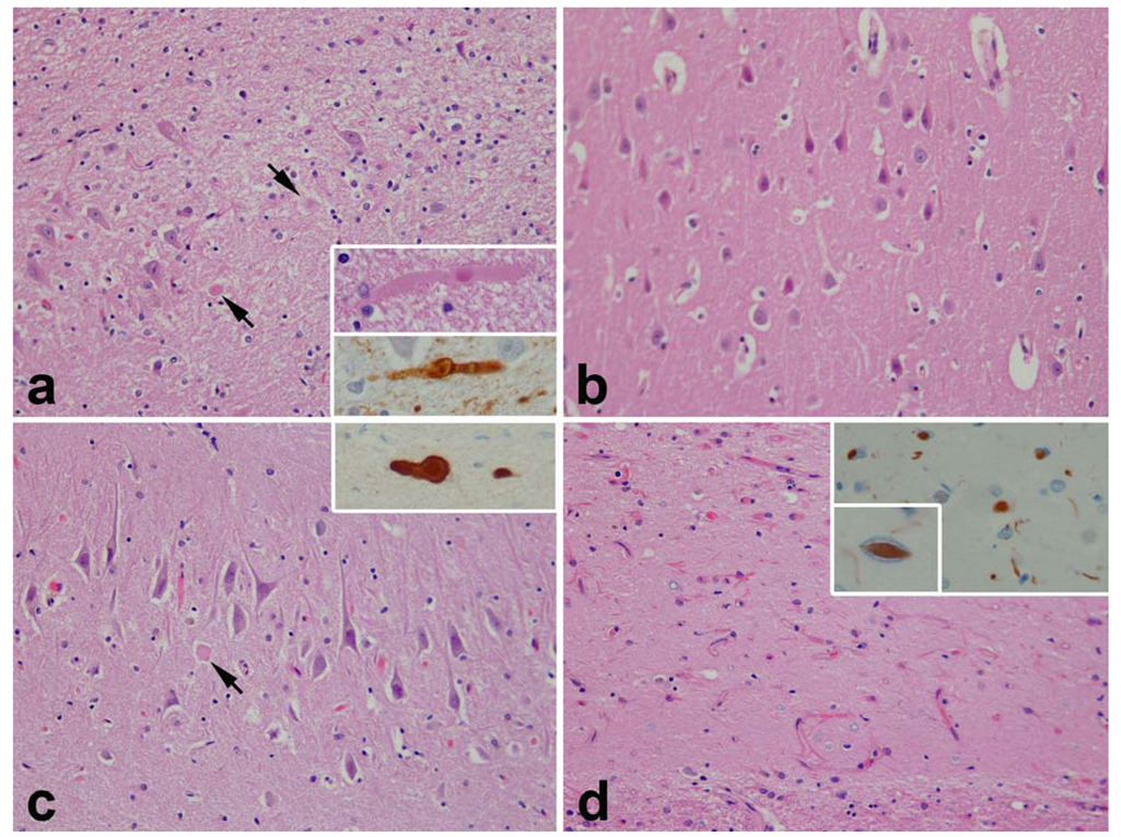
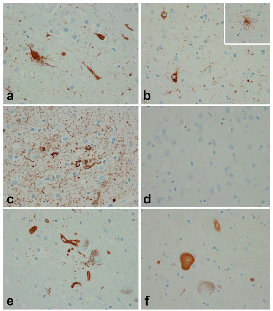
Individuals with familial Parkinson's disease (PD) due to a monogenic defect can show considerable clinical and neuropathological variability. To identify factors underlying this variability, histopathological analysis was performed in two clinically different A53T alpha-synuclein heterozygotes from Family H, a multigenerational alpha-synuclein A53T kindred. To determine whether additional genetic factors could contribute to phenotypic variability, Family H and another multigenerational A53T kindred were analyzed for parkin polymorphisms. We identified a previously described variant in parkin exon 4 associated with increased PD risk (S167N). The two A53T heterozygotes had markedly different neuropathology and different parkin genotypes: A N167 homozygote had early onset rapidly progressive disease, early dementia, myoclonus and sleep disorder, while a S167 homozygote had late onset, slowly progressive disease and late dementia. Both had brainstem, cortical, and intraneuritic Lewy bodies (LB). The N167 individual had widespread cortical neurofibrillary degeneration, while the S167 individual had only medial temporal lobe neurofibrillary degeneration. The N167 individual had severe neuronal loss in CA2 associated with Lewy neurites (LN), while the S167 individual had severe neuronal loss in CA1 associated with TDP-43 immunoreactive neuronal inclusions. These findings implicate TDP-43 in the pathology of familial PD and suggest that parkin may act as a modifier of the A53T alpha-synuclein phenotype of familial PD. Furthermore, they suggest a mechanism by which a rare genetic variant that is associated with a minor increase of PD risk in the heterozygous state may, in the homozygous state, exacerbate a disease phenotype associated with a highly penetrant dominant allele.


Figures

Fig. 1
Fig. 1
Family H and HEL-1 pedigrees. Filled squares/circles represent affected individuals with the α-synuclein A53T mutation. (a) Family H pedigree. The arrows point to IV-5, who is a α-synuclein A53T heterozygote and has S167 (wild-type) Parkin and to individual V-16, who is a α-synuclein A53T heterozygote and has N167 Parkin. (b) The HEL-1 pedigree.
Fig. 2
Fig. 2
Molecular analysis of PRKN alleles in Family H. The sequence traces of parkin exon 4 and the alleles present at parkin and α-synuclein loci in three members of in a nuclear family from Family H. Symbols are as in Figure 1. The filled lower quadrant in V-18 indicates α-synuclein A53T heterozygosity. The males all are heterozygous for the G209A (A53T) mutation in exon 4 of α-synuclein. Individual IV-10 is a heterozygote at PRKN (G/A, S167N), and developed symptoms at age 56. Individual V-16 is a PRKN homozygote (A/A, N167) and developed symptoms at age 29. Individual V-18 is a PRKN heterozygote (G/A, S167N) and remained asymptomatic at age 25.
Fig. 3
Fig. 3
Macroscopic findings of individual IV-5. The external convexity shows focal atrophy of the superior temporal gyrus (STG, arrow). Coronal sections show medial temporal atrophy (MTA, arrow) and atrophy of the hippocampus consistent with hippocampal sclerosis (HpScl, arrow). Transverse sections of midbrain and pons show loss of neuromelanin pigmentation in the SN (arrow) and LC (arrow).
Fig. 4
Fig. 4
Hippocampal pathology in V-16 (a and b) and in IV-5 (c and d). Both individuals had focal neuronal loss and gliosis. In V-16 it was marked in CA2 (a) and associated with dystrophic axons (arrows; and at higher magnification in insets stained with H&E and α-synuclein), while CA1 was unremarkable (b). In IV-5 neuronal loss was minimal in CA2 (c) although there were a few dystrophic axons (arrow, and at higher magnification in inset stained with α-synuclein), but it was very severe in CA1 (d) and accompanied by TDP-43 immunoreactive neurites and neuronal cytoplasmic inclusions (inset) and lentiform intranuclear inclusions (small inset). TDP-43 images are from inferior temporal gyrus. (All figures ×400 original magnification)
Fig. 5
Fig. 5
Tau pathology in V-16 (a, c and e) and in IV-5 (b, d and f). Despite the young age of V-16 (35 years) there were NFTs and pretangles in the entorrhinal cortex (a) and striking tau pathology in neurons and neurites in the inferior temporal cortex (c). In IV-5 the tau pathology was sparse and limited to medial temporal lobe structures (b) and consistent with argyrophilic grain disease. Note the presence of pretangles and grains in the amygdala (b) as well as tau-positive ramified astrocytes typical of argyrophilic grain disease (inset in b). There was no tau pathology in the inferior temporal cortex (d). In both V-16 and IV-5 there was focal tau immunoreactivity in LBs and intraneuritic LBs (e and f). Intraneuritic LBs in SN show tau immunoreactivity at the periphery with pale or no staining at the center of the LB. (All figures ×400 original magnification)
Fig. 6
Fig. 6
α-Synuclein pathology in V-16 (a, c, e and g) and in IV-5 (b, d, f and h). The SN in both individuals shows marked neuronal loss. In V-16 axonal spheroids are evident (arrows). Classical LBs (insets in a and b) were uncommon in brainstem nuclei (inset in a -- locus ceruleus; inset in b -- oculomotor nucleus). Immunohistochemistry for α-synuclein shows prominent axonal spheroids in the SN in both individuals (c and d). The basal ganglia had prominent spheroids in V-16 (e). Glial cytoplasmic inclusions were prominent in IV-5 (f), but detected in both individuals (glial inclusions at higher magnification in V-16 -- inset in e -- and in IV-5 -- inset in f). Both individuals had cortical α-synuclein pathology most prominent in lower cortical layers, with a mixture of LN, dystrophic axons and cytoplasmic inclusions. Cortical pathology was worse in V-16 (g) than in IV-5 (h). (All figures ×400 original magnification)

References

    1. Amador-Ortiz C, Ahmed Z, Zehr C, Dickson DW. Hippocampal sclerosis dementia differs from hippocampal sclerosis in frontal lobe degeneration. Acta Neuropathol. 2007;113:245–252. - PMC - PubMed
    1. Amador-Ortiz C, Lin W-L, Ahmed Z, Personett D, Davies P, Duara R, Graff-Redford NR, Hutton ML, Dickson DW. TDP-43 immunoreactivity in hippocampal sclerosis and Alzheimer's disease. Ann Neurol. 2007;61:435–445. - PMC - PubMed
    1. Arai T, Hasegawa M, Akiyama H, Ikeda K, Nonaka T, Mori H, Mann D, Tsuchiya K, Yoshida M, Hashizume Y, Oda T. TDP-43 is a component of ubiquitin-positive tau-negative inclusions in frontotemporal lobar degeneration and amyotrophic lateral sclerosis. Biochem Biophys Res Commun. 2006;351:602–611. - PubMed
    1. Ayala YM, Pantano S, D'Ambroglio A, Buratti E, Brindisi A, Marchetti C, Romano M, Baralle FE. Human, Drosophila and C.elegans TDP-43: Nucleic acid binding properties and splicing regulatory function. J Mol Biol. 2005;348:575–588. - PubMed
    1. Baptista MJ, Cookson MR, Miller DW. Parkin and alpha-synuclein: opponent actions in the pathogenesis of Parkinson's disease. Neuroscientist. 2004;10:63–72. - PubMed

Publication types