Obstructive urolithiasis, unilateral hydronephrosis, and probable nephrolithiasis in a 12-year-old Clydesdale gelding
- PMID: 18390103
- PMCID: PMC2249726
Obstructive urolithiasis, unilateral hydronephrosis, and probable nephrolithiasis in a 12-year-old Clydesdale gelding
Abstract
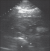
A 12-year-old Clydesdale gelding was presented for colic and dysuria. Obstructive urolithiasis and chronic renal disease were diagnosed via transurethral endoscopy and percutaneous ultrasonography. Nephroliths, hydronephrosis, and peri-ureteral fibrosis were present. Surgical intervention was declined and the gelding was managed medically with antibiotics and dietary modification.
Urolithiase obstructive, hydronéphrose unilatérale et lithiase rénale probable chez un Clydesdale hongre âgé de 12 ans. Un Clydesdale hongre âgé de 12 ans a été présenté pour colique et dysurie. Une urolithiase obstructive et une maladie rénale chronique ont été diagnostiquées par endoscopie transurétrale et échographie percutanée. La présence de néphrolithes, d’hydronéphrose et de fibrose péri-urétrale a été remarquée. La possibilité d’une intervention chirurgicale n’a pas été retenue et l’animal a été traité par antibiothérapie et modification de la diète.
(Traduit par Docteur André Blouin)
Figures


Similar articles
-
Chronic renal failure associated with nephrolithiasis, ureterolithiasis, and renal dysplasia in a 2-year-old quarter horse gelding.Vet Radiol Ultrasound. 1999 Jul-Aug;40(4):361-4. doi: 10.1111/j.1740-8261.1999.tb02126.x. Vet Radiol Ultrasound. 1999. PMID: 10463829
-
Diagnostic and management considerations for nephrolithiasis in the gravid patient.Clin Nephrol. 2016 Feb;85(2):70-6. doi: 10.5414/CN108770. Clin Nephrol. 2016. PMID: 26709525 Review.
-
Urolithiasis and nephrolithiasis.JAAPA. 2017 Sep;30(9):49-50. doi: 10.1097/01.JAA.0000522145.52305.aa. JAAPA. 2017. PMID: 28858017 No abstract available.
-
Kidney and Ureteral Stones.Emerg Med Clin North Am. 2019 Nov;37(4):637-648. doi: 10.1016/j.emc.2019.07.004. Epub 2019 Aug 19. Emerg Med Clin North Am. 2019. PMID: 31563199 Review.
-
Ureteropyeloscopic removal of a nephrolith from a 19 year old Hanoverian gelding.Vet Surg. 2022 Jul;51 Suppl 1:O53-O59. doi: 10.1111/vsu.13815. Epub 2022 May 10. Vet Surg. 2022. PMID: 35535970
References
-
- Laverty S, Pascoe JR, Ling GV, Lavoie JP, Ruby AL. Urolithiasis in 68 horses. Vet Surg. 1992;21:56–62. - PubMed
-
- Schott HC. Obstructive disease of the urinary tract. In: Reed SM, Bayley WM, Sellon DC, editors. Equine Internal Medicine. 2. St. Louis: WB Saunders; 2004. pp. 1259–1269.
-
- Ehmen SJ, Divers TJ, Gillette D, Reef VB. Obstructive nephrolithiasis and ureterolithiasis associated with chronic renal failure in horses: Eight cases (1981–1987) J Am Vet Med Assoc. 1990;97:249–253. - PubMed
-
- Newton SA, Cheeseman MT, Edwards GB. Bilateral renal and ureteral calculi in a 10-year old gelding. Vet Rec. 1999;144:383–385. - PubMed
Publication types
MeSH terms
Substances
LinkOut - more resources
Full Text Sources
Medical
