. 2008 Jun;190(11):3824-34.
doi: 10.1128/JB.00138-08. Epub 2008 Apr 4.

Cyclic AMP in mycobacteria: characterization and functional role of the Rv1647 ortholog in Mycobacterium smegmatis

Affiliations

Cyclic AMP in mycobacteria: characterization and functional role of the Rv1647 ortholog in Mycobacterium smegmatis

Bob Kennedy M Dass et al. J Bacteriol. 2008 Jun.

Abstract

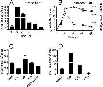
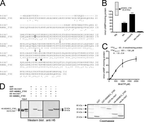
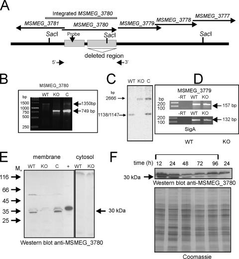
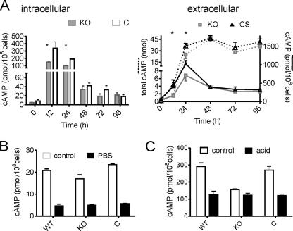
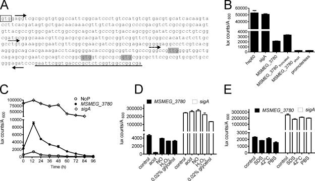
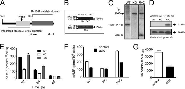
Mycobacterial genomes are endowed with many eukaryote-like nucleotide cyclase genes encoding proteins that can synthesize 3',5'-cyclic AMP (cAMP). However, the roles of cAMP and the need for such redundancy in terms of adenylyl cyclase genes remain unknown. We measured cAMP levels in Mycobacterium smegmatis during growth and under various stress conditions and report the first biochemical and functional characterization of the MSMEG_3780 adenylyl cyclase, whose orthologs in Mycobacterium tuberculosis (Rv1647) and Mycobacterium leprae (ML1399) have been recently characterized in vitro. MSMEG_3780 was important for producing cAMP levels in the logarithmic phase of growth, since the DeltaMSMEG_3780 strain showed lower intracellular cAMP levels at this stage of growth. cAMP levels decreased in wild-type M. smegmatis under conditions of acid stress but not in the DeltaMSMEG_3780 strain. This was correlated with a reduction in MSMEG_3780 promoter activity, indicating that the effect of the reduction in cAMP levels on acid stress was caused by a decrease in the transcription of MSMEG_3780. Complementation of the DeltaMSMEG_3780 strain with the genomic integration of MSMEG_3780 or the Rv1647 gene could restore cAMP levels during logarithmic growth. The Rv1647 promoter was also acid sensitive, emphasizing the biochemical and functional similarities in these two adenylyl cyclases. This study therefore represents the first detailed biochemical and functional analysis of an adenylyl cyclase that is important for maintaining cAMP levels in mycobacteria and underscores the subtle roles that these genes may play in the physiology of the organism.


Figures

FIG. 1.
FIG. 1.
cAMP levels in M. smegmatis during growth and stress conditions. (A) Intracellular cAMP levels during growth of wild-type M. smegmatis. (B) Total amount of extracellular cAMP (solid line) present in a culture (total volume, 10 ml). Extracellular cAMP (dotted line) of the same cultures is expressed per 108 cells. C. M. smegmatis cells were cultured for 30 h, resuspended in the medium indicated, and cultured for 5 h, following which the intracellular cAMP level was measured. (D) M. smegmatis cells were cultured for 30 h, and cells were resuspended in fresh medium and cultured for another 1.5 h at 37°C or at 42°C. Alternatively, cells were resuspended in medium containing 0.05% SDS or in PBS and cultured for 1.5 h. The intracellular cAMP level was measured at the end of 1.5 h. All data shown represent the means ± standard errors of the means (SEM) of duplicate determinations, with each assay being performed three times.
FIG. 2.
FIG. 2.
Expression, purification, and characterization of MSEMG_3786 adenylyl cyclase and interaction with Rv1647. (A) Sequence similarity of the Rv1647 and the MSMEG_3780 proteins. The boxed residue represents the annotated start of MSMEG_3780 according to the Sanger Centre annotation. The predicted amino acid sequences were aligned using ClustalW. Dashed arrows represent substrate-specifying residues; gray arrows represent metal-binding residues; black arrows represent transition-state stabilizing residues. (B) Purified MSMEG_3780 (∼500 ng) was assayed in the presence of either 10 mM Mg or 10 mM Mn as a free metal along with 1 mM ATP. CHAPS (0.5%) or NaCl (500 mM) was added in some assays, along with 10 mM Mn and 1 mM MnATP as a substrate, and assays were carried out at pH 9. The inset shows purified MSMEG_3780 used for adenylyl cyclase assays. Values represent the means ± SEM of duplicate determinations, with each assay being performed three times. (C) MSMEG_3780 activity was assayed in the presence of various concentrations of MnATP in the presence of 10 mM free Mn. The level of cAMP produced was measured by radioimmunoassay. All data shown represent the means ± SEM of triplicate determinations, with each assay being performed twice. (D) A GST pull-down assay was performed as described in Materials and Methods. Hexahistidine-tagged proteins that interacted with the GST fusion protein were detected by Western blot analysis using an anti-His6 antibody. An equal aliquot of proteins that were taken for interaction was subjected to SDS-polyacrylamide gel electrophoresis, and the gel was stained to ensure that approximately equal amounts of protein were used for the experiment. The result shown is representative of experiments performed twice.
FIG. 3.
FIG. 3.
Generation and characterization of the ΔΜSMEG_3780 strain and expression of MSMEG_3780 during growth. (A) Diagrammatic representation of the genome region of MSMEG_3780 indicating the presence of SacI sites used for analysis and the region encompassing the catalytic domain of MSMEG_3780 that was deleted in the knockout strain. Arrows indicate the 5′ and 3′ primers used for PCR analysis. (B) PCR was performed using primers flanking the MSMEG_3780 gene with template genomic DNA prepared from wild-type (WT), knockout (KO), or complement (C) strains. The wild-type strain should amplify a fragment of ∼1,300 kb, while the knockout strain should amplify a fragment of ∼800 bp in length. The complement strain would show both fragments as a result of the insertion of the promoter-MSMEG_3780 fragment at the att site. (C) Southern blot analysis was performed on genomic DNA prepared from the three strains following digestion with SacI. The probe was prepared from a region containing the promoter of the MSMEG_3780 gene, which was also present in the fragment that was inserted at the att site in the complement strain. (D) Reverse transcription (RT)-PCR analysis of the MSMEG_3779 gene of RNA prepared from the wild-type and knockout strains. RNA was prepared from the two strains and reverse transcribed, and PCR was performed using primers specific to the MSMEG_3779 gene or sigA for normalization. (E) Western blot analysis was performed with cytosolic and membrane fractions prepared from wild-type, ΔΜSMEG_3780, and complemented strains (50 μg protein) using a specific antibody raised to the GST-MSMEG_3780 protein. The + indicates a lane loaded with 20 ng of purified His-tagged MSMEG_3780. Mw, weight-average molecular weight (in thousands). (F) Membrane protein was prepared from the wild-type and ΔΜSMEG_3780 strains and analyzed by Western blotting using MSMEG_3780 antibody. The Coomassie-stained gel shows the normalization of total protein taken for the Western blot analysis.
FIG. 4.
FIG. 4.
cAMP levels in the ΔΜSMEG_3780 strain. (A) Intra- and extracellular cAMP levels during growth of wild-type, ΔΜSMEG_3780 (knockout [KO]), and complemented (C) strains. * represents values that are significantly different (P < 0.05) in the knockout strain compared to those of wild-type (WT) and complemented strains (CS). Data shown are the means ± SEM of duplicate determinations, with the experiment being repeated three times. (B) Intracellular cAMP levels were measured in the indicated strains under conditions similar to those detailed in the legend of Fig. 1C. (C) Intracellular cAMP levels were measured in the indicated strains under conditions similar to those detailed in the legend of Fig. 1D.
FIG. 5.
FIG. 5.
Identification of the MSMEG_3780 promoter and its activity during growth and stress conditions. (A) Sequence of a part of the MSMEG_3780 gene indicating promoters used for analysis. Forward arrows indicate the 5′ end of the MS_3780, MS_3780truncated (dotted arrow), or MS_3780short (dashed arrow) promoter, and the reverse arrow indicates the 3′ ends of the promoter constructs. The clear box indicates the translational start codon as annotated by TIGR, while shaded nucleotides indicate alternative start codons based on the promoter analysis. Underlined bases indicate sequences that encode the beginning of the catalytic domain of MSMEG_3780. (B) Regions containing the putative promoter of MSMEG_3780 were cloned upstream of the luxAB gene, and luciferase activity was measured. The activities of the hsp60 and sigA promoters and a promoterless construct were also monitored. (C) Activities of the promoterless construct (NoP) and MSMEG_3780 and sigA promoters during growth. M. smegmatis cells were transformed with the indicated plasmids, and luciferase activity was monitored as described in Materials and Methods. (D) M. smegmatis cells were cultured for 30 h, and cells were resuspended in medium as indicated and cultured for another 5 h, following which luciferase activity was monitored. (E) M. smegmatis cells were cultured for 30 h, and cells were resuspended in medium for another 1.5 h at 37°C or at 42°C. Alternatively, cells were resuspended in medium containing 0.05% SDS or in PBS and cultured for 1.5 h, followed by the estimation of luciferase activity. All data shown are the means ± SEM of duplicate determinations of experiments performed three times.
FIG. 6.
FIG. 6.
Characterization of the ΔMSMEG_3780 strain complemented with Rv1647. (A) Diagrammatic representation of the DNA inserted into the ΔMSMEG_3780 strain containing the MSMEG_3780 promoter driving the expression of the catalytic domain of the Rv1647 gene. The SacI sites used for analysis are shown, and arrows indicate the positions of primers used for PCR. (B) PCR analysis of genomic DNA prepared from the wild-type (WT), knockout (KO), and complement (RvC) strains using Rv1647-specific primers. Also shown is a PCR of the genomic DNA with primers specific to the 16S gene. (C) Southern blot analysis of the DNA prepared from wild-type, knockout, and complement strains was performed following SacI digestion. The probe used was a region present in the MSMEG_3780 promoter. The fragments hybridizing in wild-type and knockout genomic DNAs are identical to those shown in Fig. 3, while a convenient SacI site that was introduced into the Rv1647 fragment used for integration allowed the detection of a unique fragment in complement genomic DNA. (D) Western blot analysis using membranes prepared from wild-type, knockout, and complement strains at 30 h of growth. Total protein (50 μg) in the blot was normalized using an affinity-purified antibody to Rv1647 and a monoclonal antibody to the gyrase A subunit. (E) Intracellular cAMP levels were measured in the wild-type, knockout, and complement strains. Data shown are the means ± SEM of duplicate determinations of experiments performed twice. (F) Intracellular cAMP levels in wild-type, knockout, and complement strains following acid stress for 5 h. Values shown are the means ± SEM. (G) The Rv1647 promoter was fused to luxAB, and the promoter activity was measured in control cells and cells cultured under acidic conditions for 5 h. Data shown represent the means ± SEM of duplicate determinations of experiments performed twice.

Similar articles

Cited by

References

    1. Ahuja, N., P. Kumar, and R. Bhatnagar. 2004. The adenylate cyclase toxins. Crit. Rev. Microbiol. 30187-196. - PubMed
    1. Antoni, F. A. 2000. Molecular diversity of cyclic AMP signalling. Front. Neuroendocrinol. 21103-132. - PubMed
    1. Bai, G., M. A. Gazdik, D. D. Schaak, and K. A. McDonough. 2007. The Mycobacterium bovis BCG cyclic AMP receptor-like protein is a functional DNA binding protein in vitro and in vivo, but its activity differs from that of its M. tuberculosis ortholog, Rv3676. Infect. Immun. 755509-5517. - PMC - PubMed
    1. Bai, G., L. A. McCue, and K. A. McDonough. 2005. Characterization of Mycobacterium tuberculosis Rv3676 (CRPMt), a cyclic AMP receptor protein-like DNA binding protein. J. Bacteriol. 1877795-7804. - PMC - PubMed
    1. Baker, D. A., and J. M. Kelly. 2004. Structure, function and evolution of microbial adenylyl and guanylyl cyclases. Mol. Microbiol. 521229-1242. - PubMed

Publication types

MeSH terms

LinkOut - more resources