. 2008 Apr 15;180(8):5423-9.
doi: 10.4049/jimmunol.180.8.5423.

Abrogation of anti-retinal autoimmunity in IL-10 transgenic mice due to reduced T cell priming and inhibition of disease effector mechanisms

Affiliations

Abrogation of anti-retinal autoimmunity in IL-10 transgenic mice due to reduced T cell priming and inhibition of disease effector mechanisms

Rajeev K Agarwal et al. J Immunol. .

Abstract

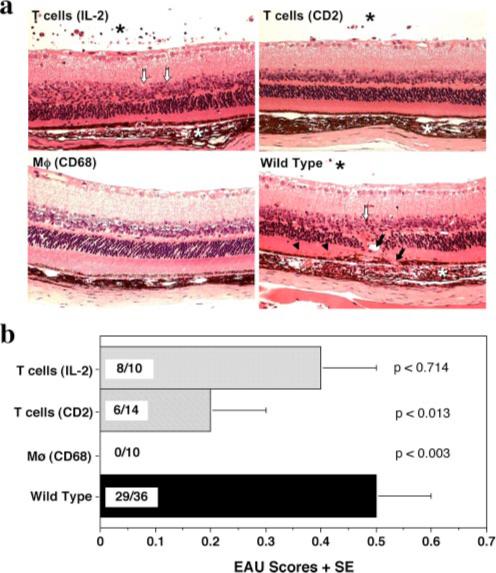
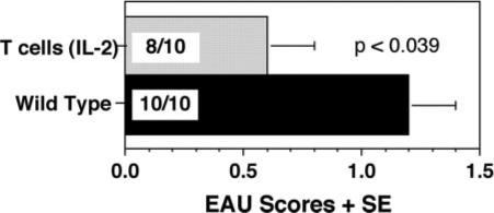
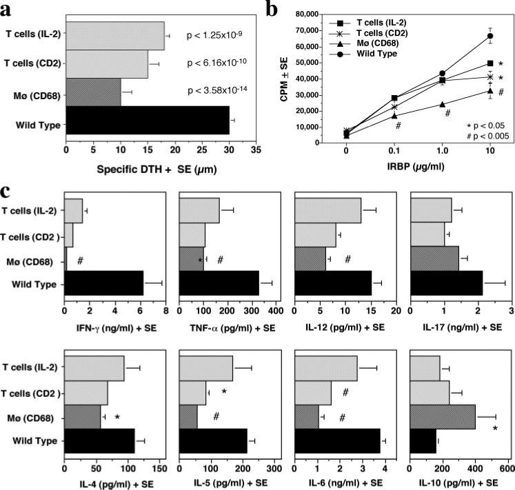
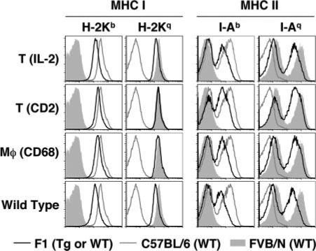
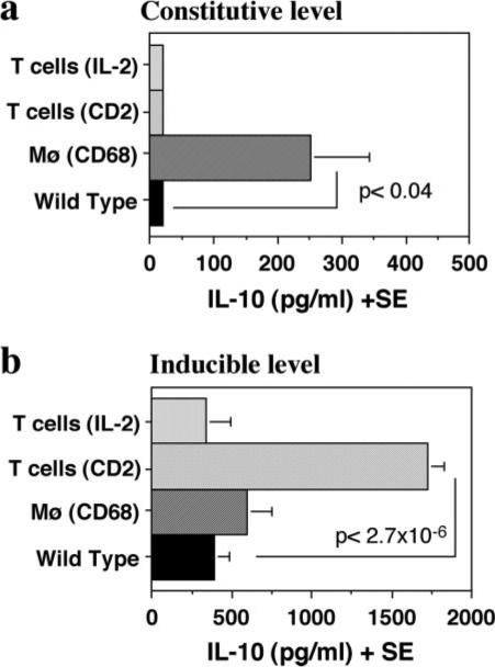
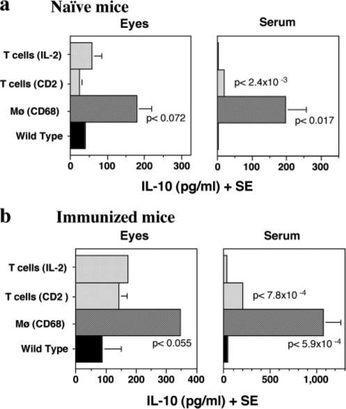
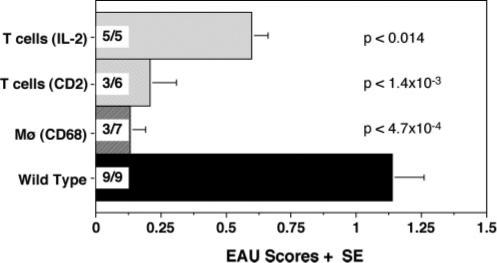
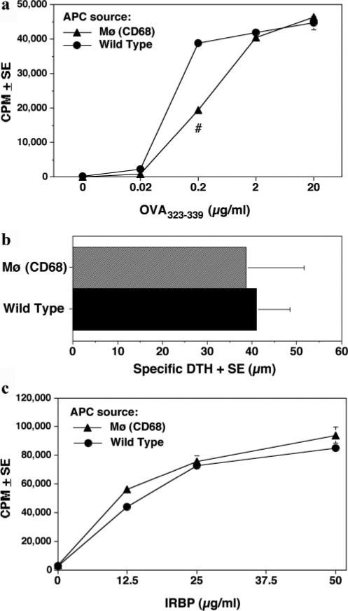
Experimental autoimmune uveitis (EAU) induced by immunization of animals with retinal Ags is a model for human uveitis. The immunosuppressive cytokine IL-10 regulates EAU susceptibility and may be a factor in genetic resistance to EAU. To further elucidate the regulatory role of endogenous IL-10 in the mouse model of EAU, we examined transgenic (Tg) mice expressing IL-10 either in activated T cells (inducible) or in macrophages (constitutive). These IL-10-Tg mice and non-Tg wild-type controls were immunized with a uveitogenic regimen of the retinal Ag interphotoreceptor retinoid-binding protein. Constitutive expression of IL-10 in macrophages abrogated disease and reduced Ag-specific immunological responses. These mice had detectable levels of IL-10 in sera and in ocular extracts. In contrast, expression of IL-10 in activated T cells only partially protected from EAU and marginally reduced Ag-specific responses. All IL-10-Tg lines showed suppression of Ag-specific effector cytokines. APC from Tg mice constitutively expressing IL-10 in macrophages exhibited decreased ability to prime naive T cells, however, Ag presentation to already primed T cells was not compromised. Importantly, IL-10-Tg mice that received interphotoreceptor retinoid-binding protein-specific uveitogenic T cells from wild-type donors were protected from EAU. We suggest that constitutively produced endogenous IL-10 ameliorates the development of EAU by suppressing de novo priming of Ag-specific T cells and inhibiting the recruitment and/or function of inflammatory leukocytes, rather than by inhibiting local Ag presentation within the eye.


Figures

FIGURE 1
FIGURE 1
Abrogation of EAU in IL-10-Tg mice. IL-10-Tg and non-Tg mice on the (C57BL/6 × FVB/N) F1 background were immunized with 150 μg of IRBP and injected with 0.5 μg of PTX. Eyes were harvested 3 wk later. a, EAU histopathology in IL-10-Tg and non-Tg littermates. Note normal appearance of retina in CD68/IL-10-Tg mouse vs inflammatory cells in choroid and vitreous (*), photoreceptor outer segment shortening and nuclear layer disorganization (open arrows), choroidal neovascularization (arrows) and subretinal hemorrhages (arrowheads) in non-Tg, IL-2/IL-10- or CD2/IL-10-Tg mouse. b, EAU scores. The average + SE is shown as the EAU score within a group. Incidence is shown within each bar as EAU positive/total mice in the group. These data are a composite of at least two experiments. p values compared with WT are indicated in the graph.
FIGURE 2
FIGURE 2
Genetic background and/or gene dose effect of IL-10 in IL-2/IL-10-Tg mice. Homozygous IL-2/IL-10-Tg and WT mice on the C57BL/6 background were immunized with IRBP (150 μg) and injected with PTX (0.5 μg). Eyes were harvested 21 days later. Note significant protection from disease in IL-2/IL-10-Tg mice compared with WT controls. EAU scores are average of two experiments.
FIGURE 3
FIGURE 3
Ag-specific immunological responses in IL-10-Tg mice. a, DTH response. Mice immunized with IRBP and PTX were ear challenged on day 19 with 10 μg IRBP in PBS, and the ear swelling was measured 48 h later. The data are a composite of two experiments. IL-2/IL-10, n = 10; CD2/IL-10, n = 9; CD68/IL-10, n = 10; WT, n = 30. p values compared with WT are indicated in the graph. b, LN cell proliferation. Draining LNs collected on day 21 after immunization were pooled within each group. Cells were stimulated with graded concentrations of IRBP (0−10 μg/ml) in triplicates and were pulsed with [3H]thymidine for the last 18 h of the 60 h culture. Proliferation is represented as mean count per minutes (cpm) ± SE. The data are from a representative experiment of two. c, Agspecific cytokine profiles. Supernatants were collected from 48 h cultures of splenocytes stimulated with 10 μg/ml IRBP and were assayed for the indicated cytokines. Shown is a representative experiment of two. Statistically significant differences compared with WT are indicated as *, significant (p < 0.05) or #, highly significant (p < 0.005).
FIGURE 4
FIGURE 4
MHC class I and II staining of splenocytes from IL-10 Tg and non-Tg mice. Cells were gated on the lymphocyte population. MHC class I (H-2Kb and H-2Kq) or MHC class II (I-Ab and I-Aq) levels are shown as histograms (black line) overlaid to WT FVB/N (filled histogram) or C57BL/6 (gray line) parental strains. Shown is a representative experiment of two, each using two randomly selected individuals per genotype.
FIGURE 5
FIGURE 5
Spontaneous vs induced levels of IL-10. Splenocytes from naive IL-10-Tg and non-Tg mice were cultured with or without soluble anti-CD3 Ab and supernatants were collected at 48 h to measure IL-10 levels. The data are average of three separate experiments. Statistical significance is calculated using independent t test. a, Spontaneous levels of IL-10. Supernatants from unstimulated cultures were used. b, Induced levels of IL-10. Supernatants from anti-CD3-stimulated culture (1.0 μg/ml) were used. Statistically significant differences compared with WT controls are indicated.
FIGURE 6
FIGURE 6
IL-10 levels in ocular extracts and sera of naive and immunized IL-10-Tg mice. a, IL-10 levels in naive mice. b, IL-10 levels in IRBP-immunized mice on day 21 post immunization. Data are a composite of at least two independent experiments. p values compared with WT controls are indicated in the graphs.
FIGURE 7
FIGURE 7
Adoptive transfer of uveitogenic effector T cells into IL-10-Tg mice. Donor WT (C57BL/6 × FVB/N) F1 mice were immunized with 150 μg IRBP and 0.2 μg PTX. Two weeks later, cells from spleens and draining lymph nodes were harvested and stimulated in vitro for 72 h with 30 μg/ml IRBP in the presence of irradiated WT splenocytes. Activated T cells were injected i.p. into IL-10-Tg or WT mice. Eyes were collected histopathological evaluation of EAU 14 days after transfer. Incidence is shown within each bar as EAU positive/total mice in the group. Data are a composite of two experiments. Statistical significances are shown in the graph.
FIGURE 8
FIGURE 8
Ag presenting function of IL-10 Tg APC to naive and primed T cells. a, Naive T cells from OT-II mice were stimulated in the presence of APC from IL-10-Tg or WT F1 mice with various concentrations of OVA peptide. Cells were incubated for 60 h and were pulsed with [3H]thymidine for the last 18 h. Proliferation is represented as mean cpm ± SE. #, p < ( 0.005. b, Local adoptive transfer of WT T cells and IRBP-pulsed APC (PEC) from CD68/IL-10-Tg or WT mice 48 h posttransfer (n = 5 per group). c, In vitro proliferation of T cells from IRBP-immunized WT mice to various concentrations of IRBP (0−50 μg/ml) in the presence of irradiated APC from spleens of either naive CD68/IL-10-Tg or WT mice. Proliferation assay was done as in a. All data are representative of three independent experiments. No statistical significance in b or c.

References

    1. Gritz DC, Wong IG. Incidence and prevalence of uveitis in Northern California; the Northern California Epidemiology of Uveitis Study. Ophthalmology. 2004;111:491–500. - PubMed
    1. Agarwal RK, Caspi RR. Rodent models of experimental autoimmune uveitis. Methods Mol. Med. 2004;102:395–419. - PubMed
    1. Gery I, Nussenblatt RB, Chan CC, Caspi RR. The Molecular Pathology of Autoimmune Diseases. Taylor and Francis; New York, NY: 2002. Autoimmune diseases of the eye. pp. 978–998.
    1. Caspi RR. Mechanisms underlying autoimmune uveitis. Drug Discov. Today: Dis. Mech. 2006;3:199–206.
    1. Tang J, Zhu W, Silver PB, Su SB, Chan CC, Caspi RR. Autoimmune uveitis elicited with antigen-pulsed dendritic cells has a distinct clinical signature and is driven by unique effector mechanisms: initial encounter with autoantigen defines disease phenotype. J. Immunol. 2007;178:5578–5587. - PubMed

Publication types

MeSH terms