. 2008 Apr 15;180(8):5653-61.
doi: 10.4049/jimmunol.180.8.5653.

Mechanism of IL-1beta-induced increase in intestinal epithelial tight junction permeability

Affiliations

Mechanism of IL-1beta-induced increase in intestinal epithelial tight junction permeability

Rana Al-Sadi et al. J Immunol. .

Abstract

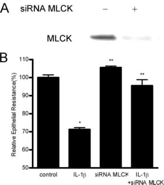
The IL-1beta-induced increase in intestinal epithelial tight junction (TJ) permeability has been postulated to be an important mechanism contributing to intestinal inflammation of Crohn's disease and other inflammatory conditions of the gut. The intracellular and molecular mechanisms that mediate the IL-1beta-induced increase in intestinal TJ permeability remain unclear. The purpose of this study was to elucidate the mechanisms that mediate the IL-1beta-induced increase in intestinal TJ permeability. Specifically, the role of myosin L chain kinase (MLCK) was investigated. IL-1beta caused a progressive increase in MLCK protein expression. The time course of IL-1beta-induced increase in MLCK level correlated linearly with increase in Caco-2 TJ permeability. Inhibition of the IL-1beta-induced increase in MLCK protein expression prevented the increase in Caco-2 TJ permeability. Inhibition of the IL-1beta-induced increase in MLCK activity also prevented the increase in Caco-2 TJ permeability. Additionally, knock-down of MLCK protein expression by small interference RNA prevented the IL-1beta-induced increase in Caco-2 TJ permeability. The IL-1beta-induced increase in MLCK protein expression was preceded by an increase in MLCK mRNA expression. The IL-1beta-induced increase in MLCK mRNA transcription and subsequent increase in MLCK protein expression and Caco-2 TJ permeability was mediated by activation of NF-kappaB. In conclusion, our data indicate that the IL-1beta increase in Caco-2 TJ permeability was mediated by an increase in MLCK expression and activity. Our findings also indicate that the IL-1beta-induced increase in MLCK protein expression and Caco-2 TJ permeability was mediated by an NF-kappaB-dependent increase in MLCK gene transcription.


Figures

FIGURE 1
FIGURE 1
Time course effect of IL-1β on Caco-2 myosin L chain kinase (MLCK) protein expression, transepithelial resistance (TER), and paracellular permeability. The Caco-2 MLCK protein expression was determined by Western blot analysis as described in Materials and Methods. The effect of IL-1β (10 ng/ml) on Caco-2 TER and mucosal-to-serosal flux of paracellular marker inulin (2 μM) were measured sequentially over the 48-h experimental period as described in Materials and Methods. A, Time course effect of IL-1β on Caco-2 MLCK protein expression (β-actin was used as an internal control for protein loading). B, T ime course effect of IL-1β on Caco-2 TER (means ± SE, n = 4). C, Time course effect of IL-1β on mucosal-to-serosal inulin flux (means ± SE, n = 4). *, p < 0.05 vs control. D, Graph of IL-1β effect on MLCK protein expression and Caco-2 TER (r = 0.99). E, Graph of IL-1β effect on MLCK protein expression and inulin flux (r = 0.97).
FIGURE 2
FIGURE 2
The effect of protein synthesis inhibitor, cycloheximide (CHX) on IL-1β-induced increase in MLCK protein expression and drop in Caco-2 TER. A, Cycloheximide (5 μM) pretreatment prevented the IL-1β-induced up-regulation in MLCK protein expression as assessed by Western blot analysis. B, Cycloheximide prevented the IL-1β-induced drop in Caco-2 TER (means ± SE, n = 4). *, p < 0.001 vs control; **, p < 0.001 vs IL-1β treatment.
FIGURE 3
FIGURE 3
Effect of IL-1β on Caco-2 MLCK activity. A, Time course effect of IL-1β on MLCK activity of filter-grown Caco-2 monolayers was determined by ELISA-based in vitro kinase activity measurements as described in Materials and Methods. After appropriate time period, MLCK activity was assessed by measuring the in vitro phosphorylation of MLC (S-19). IL-1β treatment resulted in a time-dependent increase in Caco-2 MLCK activity. *, p < 0.05 vs control. B, Time course effect of IL-1β on phosphorylation of MLC (P-MLC) in filter-grown Caco-2 monolayers over the 48-h experimental period as determined by Western blot analysis. C, Pretreatment with cycloheximide (5 μM) inhibited the IL-1β-induced increase in MLCK kinase activity, cycloheximide alone did not affect MLCK enzyme activity. *, p < 0.001 vs control; **, p < 0.001 vs IL-1β treatment.
FIGURE 4
FIGURE 4
Effect of MLCK inhibitors ML-7 and ML-9 on IL-1β-induced increase in Caco-2 MLCK activity and drop in Caco-2 TER. Filter-grown Caco-2 monolayers were treated with IL-1β (10 ng/ml) for a 48-h experimental period. ML-7 (10 μM) and ML-9 (20 μM) significantly prevented the IL-1β (10 ng/ml) induced increase in MLCK activity (in vitro kinase assay) and drop in Caco-2 TER (n = 4) (B). *, p < 0.001 vs control; **, p < 0.001 vs IL-1β treatment.
FIGURE 5
FIGURE 5
Effect of siRNA induced MLCK knock-down on IL-1β-induced drop on Caco-2 TER. Caco-2 monolayers were transfected with MLCK siRNA for a 96-h time period as described in Materials and Methods. A, MLCK siRNA transfection resulted in a near complete depletion in MLCK protein expression as determined by Western blot analysis. B, MLCK siRNA transfection prevented the IL-1β-induced drop in Caco-2 TER (n = 4). *, p < 0.001 vs control; **, p < 0.001 vs IL-1β treatment.
FIGURE 6
FIGURE 6
IL-1β effect on Caco-2 MLCK mRNA expression. A, Time course effect of IL-1β on Caco-2 MLCK mRNA expression was determined by real-time PCR as described in the Materials and Methods. MLCK mRNA level was expressed relative to the control level which was assigned a value of 1. The average copy number of MLCK mRNA in controls was 4.63 × 10¹¹. *, p < 0.001 vs control. B, Actinomycin-D (100 ng/ml) effect on IL-1β-induced increase in MLCK mRNA expression (6 h of IL-1β treatment). Actinomycin-D significantly inhibited the IL-1β increase in MLCK mRNA expression. *, p < 0.05 vs control; **, p < 0.05 vs IL-1β treatment. C, Actinomycin-D significantly prevented the IL-1β-induced up-regulation of MLCK protein expression as determined by Western blot analysis. D, Actinomycin-D also prevented the IL-1β-induced drop in Caco-2 TER (n = 4). *, p < 0.001 vs control; **, p < 0.001 vs IL-1β treatment.
FIGURE 7
FIGURE 7
IL-1β effect on NF-κB activation. A, IL-1β effect on NF-κB activation as assessed by NF-κB p65 nuclear translocation. NF-κB p65 cytoplasmic-to-nuclear translocation was determined by immunofluorescent staining with anti-NF-κB p65 Ab. Filter-grown Caco-2 monolayers were treated with 10 ng/ml IL-1β for 30 min. B, Effect of NF-κB inhibitor, PDTC (100 μM), on IL-1β-induced increase in MLCK mRNA expression. Caco-2 cells were pretreated with PDTC for 1 h before IL-1β treatment (6-h experimental period). MLCK mRNA level was determined by realtime PCR (n = 6). *, p < 0.05 vs control; **, p < 0.05 vs IL-1β treatment. C, Effect of PDTC (100 μM) on IL-1β up-regulation of MLCK protein expression. Filter-grown Caco-2 cells were treated with PDTC 1 h before IL-1β treatment for 48 h. MLCK protein expression was assessed by Western blot analysis. D, Effect of PDTC (100 μM) on IL-1β-induced drop in Caco-2 TER. *, p < 0.001 vs control; **, p < 0.001 vs IL-1β treatment.
FIGURE 8
FIGURE 8
Effect of siRNA induced knock-down of NF-κB p65 on IL-1β-induced increase in MLCK expression and drop in Caco-2 TER and. Filter-grown Caco-2 monolayers were transfected with NF-κB p65 siRNA for a 96-h time period as described in Materials and Methods. A, NF-κB p65 siRNA transfection resulted in a near complete knock-down of NF-κB p65 expression. B, siRNA NF-κB p65 transfection prevented the IL-1β-induced increase in MLCK protein expression. C, NF-κB p65 siRNA transfection prevented the IL-1β-induced drop in Caco-2 TER. *, p < 0.001 vs control; **, p < 0.001 vs IL-1β treatment.
FIGURE 9
FIGURE 9
Proposed scheme of the IL-1β-induced increase in intestinal epithelial tight junction (TJ) permeability.

References

    1. Hollander D. Crohn's disease: a permeability disorder of the tight junction? Gut. 1988;29:1621–1624. - PMC - PubMed
    1. Ma TY. Intestinal epithelial barrier dysfunction in Crohn's disease. Proc. Soc. Exp. Biol. Med. 1997;214:318–327. - PubMed
    1. Berkes J, Viswanathan VK, Savkovic SD, Hecht G. Intestinal epithelial responses to enteric pathogens: effects on the tight junction barrier, ion transport, and inflammation. Gut. 2003;52:439–451. - PMC - PubMed
    1. Hollander D. Intestinal permeability, leaky gut, and intestinal disorders. Curr. Gastroenterol. Rep. 1999;1:410–416. - PubMed
    1. Anderson JM, Van Itallie CM. Tight junctions and the molecular basis for regulation of paracellular permeability. Am. J. Physiol. 1995;269:G467–G475. - PubMed

Publication types

MeSH terms