. 2008 Apr 15;180(8):5689-98.
doi: 10.4049/jimmunol.180.8.5689.

Rapid changes in microRNA-146a expression negatively regulate the IL-1beta-induced inflammatory response in human lung alveolar epithelial cells

Affiliations

Rapid changes in microRNA-146a expression negatively regulate the IL-1beta-induced inflammatory response in human lung alveolar epithelial cells

Mark M Perry et al. J Immunol. .

Abstract

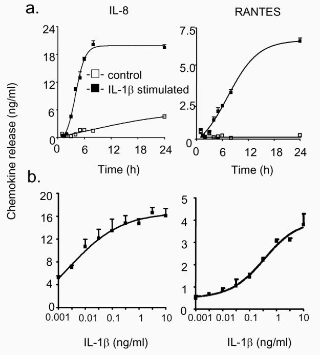
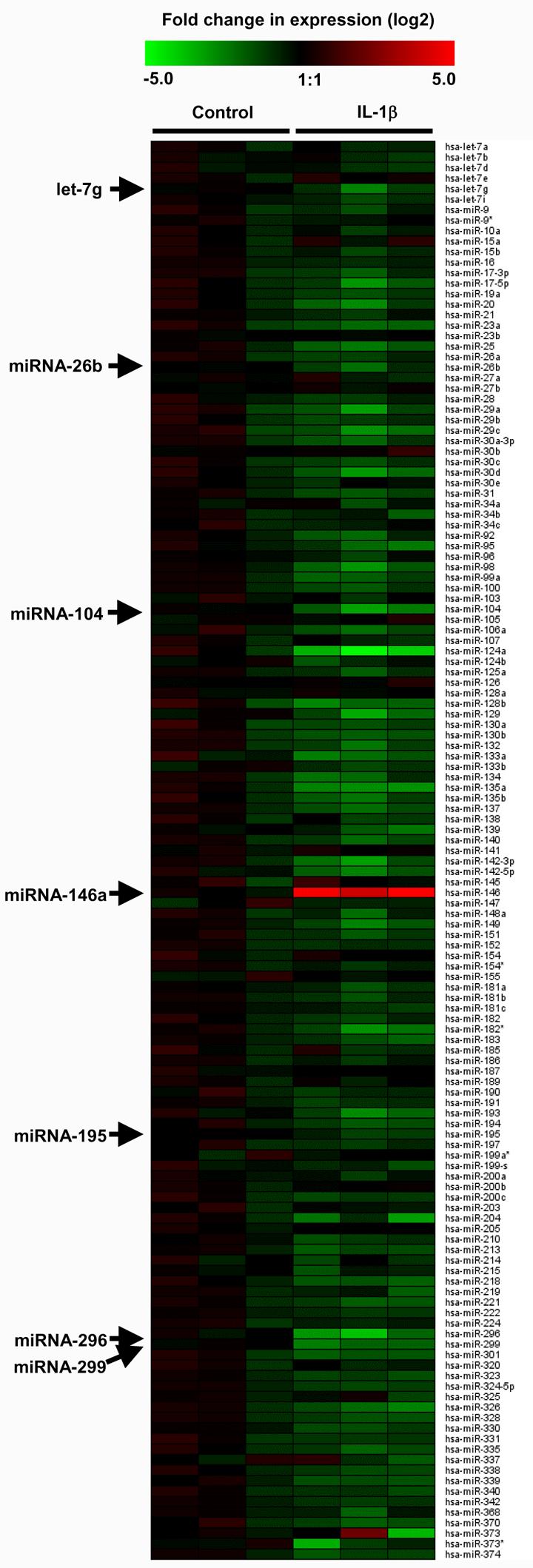
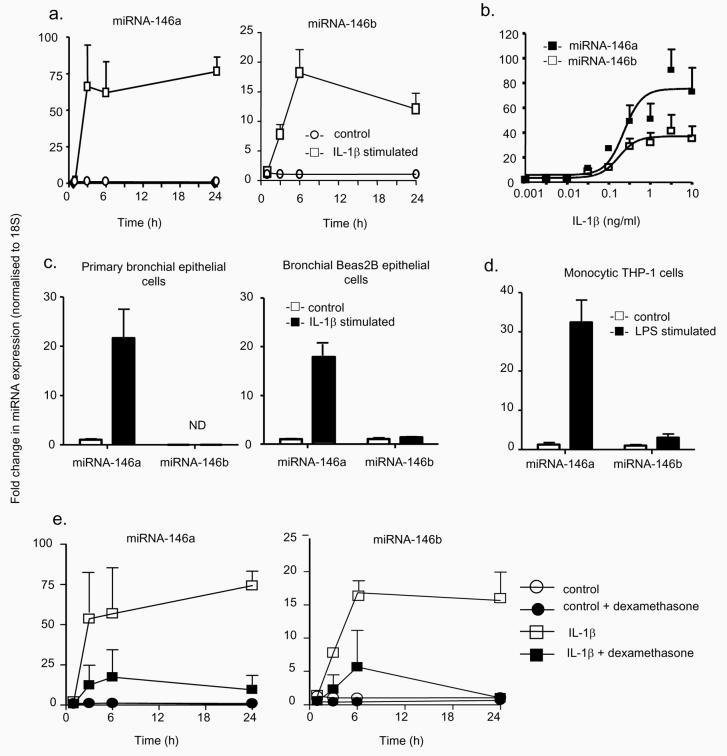
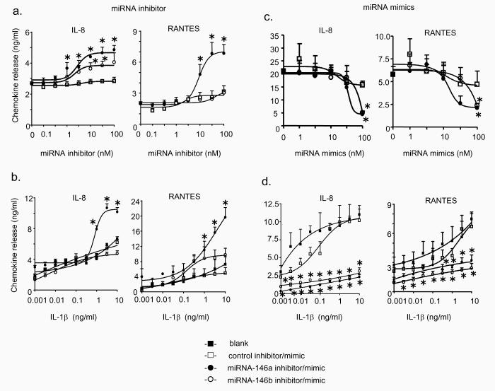
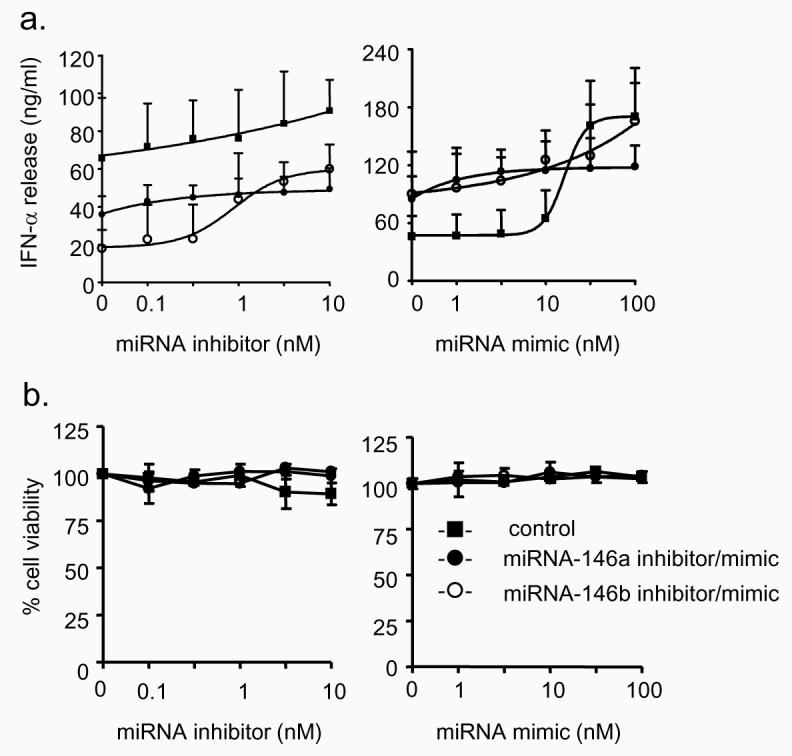
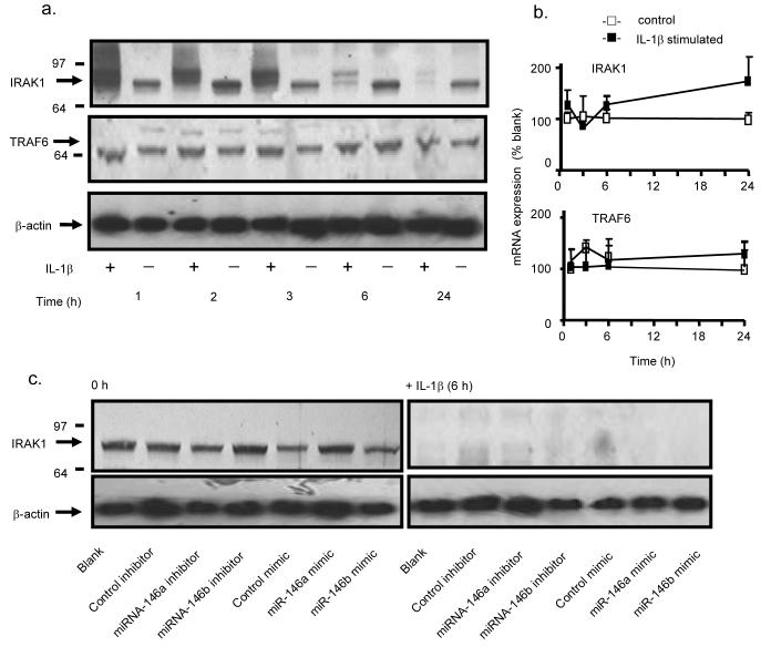
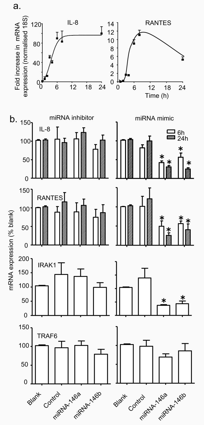
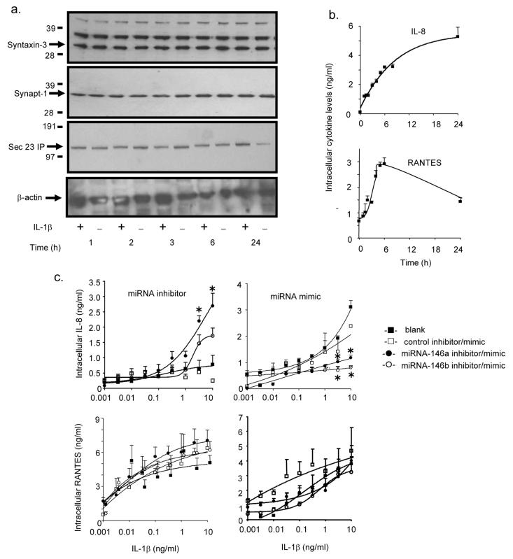
Posttranscriptional regulation of gene expression by microRNAs (miRNAs) has been implicated in the regulation of chronic physiological and pathological responses. In this report, we demonstrate that changes in the expression of miRNAs can also regulate acute inflammatory responses in human lung alveolar epithelial cells. Thus, stimulation with IL-1beta results in a rapid time- and concentration-dependent increase in miRNA-146a and, to a lesser extent, miRNA-146b expression, although these increases were only observed at high IL-1beta concentration. Examination of miRNA function by overexpression and inhibition showed that increased miRNA-146a expression negatively regulated the release of the proinflammatory chemokines IL-8 and RANTES. Subsequent examination of the mechanism demonstrated that the action of miRNA-146a was mediated at the translational level and not through the down-regulation of proteins involved in the IL-1beta signaling pathway or chemokine transcription or secretion. Overall, these studies indicate that rapid increase in miRNA-146a expression provides a novel mechanism for the negative regulation of severe inflammation during the innate immune response.


Figures

Figure 1
Figure 1
Time- and concentration-dependency of IL-1β-induced IL-8 and RANTES release. A549 cells were exposed to either buffer or 1 ng/ml IL-1β for the indicated time (a) or to the indicated IL-1β concentration for 24 h (b) prior to measurement of IL-8 and RANTES release. The results are expressed as the mean ± SEM of 3 individual samples and are representative of 3 independent experiments.
Figure 2
Figure 2
IL-1β-induced changes in miRNA expression. A549 cells were exposed to IL-1β (1 ng/ml) for 3 h and the profile of expression of 156 miRNAs was measured using TaqMan RT-PCR. The log2 transformed values of the fold-change in expression compared to time-matched saline controls are represented as a heat map where red and green indicate an increase and decrease in miRNA expression, respectively.
Figure 3
Figure 3
Characterisation of the mechanism of miRNA-146a and miRNA-146b expression. The time- and concentration-dependent induction of miRNA-146a and miRNA-146b in A549 cells was determined following exposure to 1 ng/ml IL-1β for the indicated time (a) or to the indicated IL-1β concentration for 6 h (b) and the increases in miR-146a and miR-146b expression were determined by RT-PCR. To confirm the observations in A549 cells, the levels of miRNA-146a and miR-146b were measured at 6 h in IL-1β-stimulated primary human bronchial epithelial cells and transformed human bronchial Beas2B epithelial cells (c) or in LPS-stimulated monocytic THP-1 cells (d). Alternatively, control- and IL-1β-(1ng/ml) stimulated A549 cells were pre-treated with dexamethasone (1 μM) for 60 min, and the expression of miRNA-146a and miRNA-146b was determined at the indicated time points (e). These results are expressed as the mean ± SEM of 3 independent experiments. ND = not detected.
Figure 4
Figure 4
Effect of inhibitors and mimics of miRNA-146a and miRNA-146b upon IL-1β–induced IL-8 and RANTES release. A549 cells were exposed to transfection reagent alone (blank), control inhibitor or mimic, miR-146a inhibitor or mimic and miR-146b inhibitor or mimic for 4 h and then exposed to either 1 ng/ml IL-1β (a,c) or the indicated IL-1β concentration (b,d) and the concentration of IL-8 and RANTES in the supernatant was determined at 6 h and 24 h, respectively. The results are expressed as the mean ± SEM of 3 individual samples and are representative of at least 3 independent experiments. * p < 0.05 versus time-matched (a,c) or concentration-matched (b,d) transfected controls.
Figure 5
Figure 5
Effect of miRNA-146a and miRNA-146b inhibitors and mimics upon IFN-α release and cell viability. A549 cells exposed to control miRNA inhibitor/mimic, miR-146a inhibitor/mimic or miR-146b inhibitor/mimic for 4 h and then exposed to 1 ng/ml IL-1β for 24h. Supernatant samples were then removed for measurement of IFN-α (a) and the cell viability of the remaining cells was determined (b). The results are expressed as the mean ± SEM of 3 individual samples and the graphs are representative of 3 independent experiments.
Figure 6
Figure 6
Role of miRNA-146a and miRNA-146b in the IL-1β signalling pathway. A549 cells were exposed IL-1β (1 ng/ml) and the expression of IRAK1 and TRAF6 protein (a) and mRNA (b) were detected at the indicated time points. In separate studies, A549 cells were transfected with an inhibitor or mimic of miRNA-146a, miRNA-146b or the relevant control miRNA and the effect upon IRAK1 protein expression was determined at 0 h and at 6 h following IL-1β stimulation (c). The results in panel (a) and (c) are representative of a least 3 independent experiments whilst panel (b) gives the mean ± SEM of 3 independent experiments.
Figure 7
Figure 7
Role of miRNA-146a and miRNA-146b during IL-1β-induced IL-8 and RANTES transcription. A549 cells were stimulated with IL-1β and the expression of IL-8 and RANTES mRNA was determined at the indicated time (a). Alternatively, cells were transfected for 4 h with transfection reagent alone (blank), control inhibitor or mimic (control), miR-146a inhibitor or mimic (miRNA-146a) and miR-146b inhibitor or mimic (miRNA-146b) for 4 h, exposed to 1 ng/ml IL-1β and the expression of IL-8, RANTES, IRAK1 and TRAF6 mRNA was determined at 6 h and/or 24 h. The results are expressed as the mean ± SEM of at least 3 independent experiments. * p < 0.05 versus blanks.
Figure 8
Figure 8
Role of miRNA-146a and miRNA-146b during IL-1β-induced IL-8 and RANTES secretion. A549 cells were stimulated with IL-1β (1 ng/ml) and at the indicated times, the intracellular protein expression of syntaxin-3, synaptotagmin-1 or sec23-IP was determined by Western blotting (a) and IL-8 and RANTES by ELISA (b). Alternatively, cells were transfected for 4 h with transfection reagent alone (blank), control inhibitor or mimic, miR-146a inhibitor or mimic and miR-146b inhibitor or mimic for 4 h and then exposed to the indicated IL-1β concentration and the intracellular IL-8 and RANTES protein expression was determined at 6 h. The results are the mean ± SEM of 3 individual samples and are representative of at least 3 independent experiments. * p < 0.05 versus blanks.

References

    1. Braddock M, Quinn A. Targeting IL-1 in inflammatory disease: new opportunities for therapeutic intervention. Nat.Rev.Drug Discov. 2004;3:330–339. - PubMed
    1. Li X, Qin J. Modulation of Toll-interleukin 1 receptor mediated signaling. J.Mol Med. 2005;83:258–266. - PubMed
    1. Liew FY, Xu D, Brint EK, O'Neill LA. Negative regulation of toll-like receptor-mediated immune responses. Nat Rev.Immunol. 2005;5:446–458. - PubMed
    1. Ambros V. The functions of animal microRNAs. Nature. 2004;431:350–355. - PubMed
    1. He L, Hannon GJ. MicroRNAs: small RNAs with a big role in gene regulation. Nat.Rev.Genet. 2004;5:522–531. - PubMed

Publication types