. 2008 Jul;84(1):292-301.
doi: 10.1189/jlb.1207832. Epub 2008 Apr 7.

Priming of neutrophil oxidative burst in diabetes requires preassembly of the NADPH oxidase

Affiliations

Priming of neutrophil oxidative burst in diabetes requires preassembly of the NADPH oxidase

Kazuhiro Omori et al. J Leukoc Biol. 2008 Jul.

Abstract

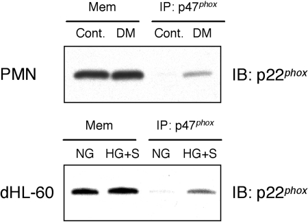
Hyperglycemia associated with diabetes mellitus results in the priming of neutrophils leading to oxidative stress that is, in part, responsible for diabetic complications. p47phox, a NADPH oxidase cytosolic subunit, is a key protein in the assembly of the NADPH oxidase leading to superoxide generation. Little is known about the priming mechanism of oxidative pathways in neutrophils of people with diabetes. In this study, the kinetics of p47phox activation was investigated by comparing neutrophils from diabetic and healthy subjects, and the mechanism of hyperglycemia-induced changes was studied by using neutrophil-like HL-60 cells as a model. In resting neutrophils from diabetic subjects, p47phox prematurely translocates to the cell membrane and preassembles with p22phox, a NADPH oxidase membrane subunit. This premature p47phox translocation and preassembly with p22phox were also observed in HL-60 cells cultured with high glucose (HG; 25 mM) and with the specific ligand for the receptor for advanced glycation end products (RAGE), S100B. Phosphorylation of ERK1/2, but not p38 MAPK, was the primary signaling pathway, as evidenced by PD98059 suppressing the translocation of p47phox in HL-60 cells incubated with HG and S100B. HL-60 cells cultured in HG and S100B exhibited a 1.8-fold increase in fMLP-induced superoxide generation compared with those cultured in normal glucose (5.5 mM). These data suggest that HG and increased AGE prime neutrophils and increase oxidative stress inducing the translocation of p47phox to the cell membrane and preassembly with p22phox by stimulating a RAGE-ERK1/2 pathway.


Figures

Figure 1
Figure 1
Neutrophil function and p47phox translocation in diabetic subjects. (A) Superoxide generation in neutrophils from diabetic [diabetes mellitus (DM)] subjects is enhanced upon fMLP stimulation. Isolated neutrophils from diabetic and control subjects were stimulated with fMLP (1 μM) or PBS for 5 min. Superoxide release was measured as the SOD-inhibitable reduction of cytochrome c and expressed as the percent of control [Cont.; fMLP stimulation=100% (0.84±0.11 nmole O2/106 cells/min); *, P<0.05; n=13 subjects in each group]. (B) Membrane-associated proteins and whole cell lysates were extracted from unstimulated neutrophils from diabetic and healthy subjects and were analyzed by Western blotting with anti-p47phox, p67phox, Rac2, and p22phox antibodies as described in Materials and Methods. Western blots reveal that there is no difference in the total expression of p47phox in unstimulated neutrophils from diabetic and healthy subjects. p47phox is translocated to the cell membrane-rich fraction in neutrophils from diabetic subjects (P=0.041). Expression of p67phox, Rac2, and p22phox is not significantly different between healthy and diabetic subjects. The same membrane was stripped and reprobed with antiactin antibody. The levels of actin were evaluated as a loading control. The relative density of p47phox, p67phox, Rac2, and p22phox bands was quantified by densitometry, and the data were normalized to actin. Results are expressed as mean ± sem of 16 subjects in each group. IB, Immunoblot.
Figure 2
Figure 2
fMLP-induced superoxide generation in neutrophil-like HL-60 cells, which were cultured in NG or HG, with or without the RAGE-specific ligand S100B for 24 h. Cells were stimulated with fMLP (100 nM) or PBS for 5 min, and superoxide generation was quantified as the SOD-inhibitable reduction of ferricytochrome C as described in Materials and Methods. Neutrophil-like HL-60 cells cultured in NG with S100B or in HG with S100B exhibited greater fMLP-induced superoxide generation than the cells cultured in NG alone. Results are expressed as mean ± sem of at least six different experiments (*, P<0.05, compared with the cells cultured in NG).
Figure 3
Figure 3
p47phox translocation to the cell membrane in neutrophil-like HL-60 cells. Membrane-associated proteins and whole cell lysates were extracted from unstimulated, neutrophil-like HL-60 cells cultured in NG or HG, with or without S100B for 24 h, and were analyzed by Western blotting with the anti-p47phox, p67phox, Rac2, or p22phox antibodies as described in Materials and Methods. (A and C) There is no difference of the expression of total p47phox, p67phox, Rac2, and p22phox in unstimulated, neutrophil-like HL-60 cells regardless of the culture condition. (B and C) p47phox translocated to the cell membrane-rich fraction in neutrophil-like HL-60 cells cultured in HG with S100B (HG+S, P=0.008, vs. NG, P=0.016, vs. NG with S100B (S; NG+S), P=0.009, vs. HG, P=0.016, vs. NG+MN) and NG with S100B (P=0.042 vs. NG), and p67phox, Rac2, and p22phox expression did not change. The same membrane was stripped and reprobed with antiactin antibody. The levels of actin were evaluated as a loading control. p47phox, p67phox, Rac2, and p22phox were quantified by densitometry, and the data were normalized to actin. Results are expressed as mean ± sem of at least three different experiments. *, P < 0.05.
Figure 4
Figure 4
Confocal microscopic evaluation of p47phox translocation to the cell membrane in neutrophil-like HL-60 cells. Unstimulated, neutrophil-like HL-60 cells cultured in NG or HG, with or without S100B for 24 h, were fixed and attached to glass slides. The cells were incubated with anti-p47phox antibody and visualized using confocal microscopy. (A) NG, (B) NG with S100B; NG + S, (C) HG, (D) HG with S100B; HG + S, (E) NG + MN, (F) HG with S100B and PD98059 (PD); HG + S + PD. The results from three independent experiments are represented by the image. (Original scale bars: 5 μm.)
Figure 5
Figure 5
p47phox preassembled with p22phox in the cell membrane (Mem) fraction in unstimulated neutrophils from diabetic subjects or AGE-stimulated, neutrophil-like HL-60 cells. Unstimulated neutrophils from diabetic or healthy subjects and neutrophil-like HL-60 cells cultured in NG or HG with S100B for 24 h were fractionated to membrane and cytosolic fractions. The membrane-rich fraction samples were immunoprecipitated (IP) with p47phox mAb, and immunoprecipitated samples were analyzed by Western blotting using anti-p22phox antibody as described in Materials and Methods. Western blotting data reveal that there is no difference in the expression of p22phox in the membrane-rich fraction of unstimulated neutrophils from diabetic and healthy subjects. p47phox is preassembled with p22phox in the cell membrane-rich fraction in unstimulated neutrophils from diabetic subjects as well as unstimulated, neutrophil-like HL-60 cells cultured with AGE. Results are representative of at least three independent experiments. PMN, Polymorphonuclear neutrophil; dHL-60, differentiated neutrophil-like HL-60 cells.
Figure 6
Figure 6
AGE induces the continuous phosphorylation of ERK1/2, but not p38 MAPK. Neutrophil-like HL-60 cells cultured in NG or HG were stimulated with S100B (5 μg/ml) for the indicated periods of time. Whole cell lysates were analyzed by Western blotting using antibodies to phospho-ERK1/2 and phospho-p38 MAPK as described in Materials and Methods. Stimulation with S100B for 5 min resulted in the rapid phosphorylation of ERK1/2 and p38 MAPK. The phosphorylation of ERK1/2 continued for 240 min, particularly ERK1. However, the phosphorylation of p38 MAPK was undetectable after 15 min. There was no detectable difference between NG and HG. The same membrane was stripped and reprobed with antiactin antibody. The levels of actin were evaluated as a loading control. Results are representative of three independent experiments.
Figure 7
Figure 7
PD98059 suppresses p47phox translocation. Membrane-associated proteins were extracted from unstimulated, neutrophil-like HL-60 cells cultured in NG or HG, with or without PD98059 (25 μM) or SB203580 (10 μM) for 24 h, and were analyzed by Western blotting with anti-p47phox and p22phox antibodies as described in Materials and Methods. PD98059 inhibited p47phox translocation to the cell membrane-rich fraction of HL-60 cells cultured in NG with S100B (NG+S) and completely inhibited p47phox translocation to the cell membrane-rich fraction of HL-60 cells cultured in HG with S100B (HG+S; Fig. 7A). SB203580 did not suppress p47phox translocation to the cell membrane (Fig. 7B). PD98059 and SB203580 did not alter p22phox expression (Fig. 7). The same membrane was stripped and reprobed with antiactin antibody, where actin was used as the loading control. Results are representative of three independent experiments.

References

    1. Zimmet P., Alberti K. G., Shaw J. Global and societal implications of the diabetes epidemic. Nature. 2001;414:782–787. - PubMed
    1. Ritz E., Orth S. R. Nephropathy in patients with type 2 diabetes mellitus. N Engl J Med. 1999;341:1127–1133. - PubMed
    1. Yamagishi S., Nakamura K., Matsui T., Takenaka K., Jinnouchi Y., Imaizumi T. Cardiovascular disease in diabetes. Mini Rev Med Chem. 2006;6:313–318. - PubMed
    1. Renard C., Van Obberghen E. Role of diabetes in atherosclerotic pathogenesis What have we learned from animal models? Diabetes Metab. 2006;32:15–29. - PubMed
    1. Pop-Busui R., Sima A., Stevens M. Diabetic neuropathy and oxidative stress. Diabetes Metab Res Rev. 2006;22:257–273. - PubMed

Publication types

MeSH terms

Substances