. 2008 Jun;76(6):2379-91.
doi: 10.1128/IAI.01143-07. Epub 2008 Apr 7.

Inheritance of immune polarization patterns is linked to resistance versus susceptibility to Cryptococcus neoformans in a mouse model

Affiliations

Inheritance of immune polarization patterns is linked to resistance versus susceptibility to Cryptococcus neoformans in a mouse model

Gwo-hsiao Chen et al. Infect Immun. 2008 Jun.

Abstract

Genetic background variation between inbred strains accounts for different levels of susceptibility to Cryptococcus neoformans in the mouse infection model. To elucidate the inheritance of immunophenotypic traits and their associations with clearance outcomes during cryptococcal infection, we compared C57BL/6, BALB/c, and their first-generation hybrid, CB6F1 (F1), mice. Mice from each group were infected with C. neoformans (10(4) CFU) and analyzed at weekly intervals over a 6-week period. BALB/c mice progressively cleared the cryptococcal infection in the lungs and showed a Th1-skewed immune response: a Th1-shifted cytokine profile, modest lung pathology, and no significant elevation in the systemic immunoglobulin E (IgE) level. In contrast, C57BL/6 mice developed a chronic infection with a Th2-skewed immune response: a Th2-shifted cytokine profile, pulmonary eosinophilia, severe lung pathology, elevated serum IgE, fungemia, and cryptococcal dissemination in the central nervous system. F1 mice demonstrated intermediate resistance to C. neoformans, with a stronger resemblance to the immunophenotype of the resistant (BALB/c) mice. F1 mice also demonstrated enhanced pulmonary recruitment of lymphocytes, especially CD8(+) T cells, in comparison to both parental strains, suggesting positive heterosis. We conclude that the inheritance of traits responsible for early cytokine induction in the infected lungs and dendritic-cell maturation/activation status in draining nodes is responsible for the intermediate immune response polarization and clearance outcome observed initially in the lungs of F1 mice. The enhanced pulmonary lymphocyte recruitment could be responsible for a gradual shutdown of the undesirable Th2 arm of the immune response and subsequently improved anticryptococcal resistance in F1 mice.


Figures

FIG. 1.
FIG. 1.
Pulmonary clearance of C. neoformans infection in BALB/c, F1, and C57BL/6 mice. Mice were inoculated intratracheally with 104 CFU of C. neoformans 52D, and the pulmonary C. neoformans burden was evaluated at weeks 1 to 6 postinfection. Values represent means ± SEM (n = 6 to 20 mice/group/time point). *, significant difference from results for F1 mice; **, significant difference from results for BALB/c mice. Data were pooled from three parallel experiments.
FIG. 2.
FIG. 2.
Systemic dissemination of C. neoformans from the lung in BALB/c, F1, and C57BL/6 mice. Mice were inoculated intratracheally with 104 CFU of C. neoformans 52D, and the spleen (A) and brain (B) C. neoformans burden were evaluated at week 3 postinfection. Values represent means ± SEM (n = 6 mice/group/time point). *, significant difference from results for F1 mice; **, significant difference from results for BALB/c mice. Data were pooled from three parallel experiments.
FIG. 3.
FIG. 3.
Leukocyte recruitment in C. neoformans-infected BALB/c, F1, and C57BL/6 mice. Lung leukocytes were isolated from individual mice and enumerated at weeks 0, 1, 2, 4, and 6 postinfection. Values represent means ± SEM (n = 6 to 20 mice/group/time point). *, significant difference from results for F1 mice; **, significant difference from results for BALB/c mice. Data were pooled from five parallel experiments.
FIG. 4.
FIG. 4.
Lymphocyte subset recruitment following pulmonary C. neoformans 52D infection. Samples of leukocyte suspensions from infected mice were stained with fluorochrome-labeled antibodies specific for CD4+, CD8+, and B220+ lymphocytes and analyzed by flow cytometry as described in Materials and Methods. Results are illustrated as percentages or absolute numbers of each lymphocyte subset in the total lung leukocyte sample. Values represent means ± SEM (n = 4 to 6 mice/group/time point). *, significant difference from results for F1 mice; **, significant difference from results for BALB/c mice. Data were pooled from two parallel experiments.
FIG. 5.
FIG. 5.
T2 hallmarks: pulmonary eosinophilia and elevated levels of serum IgE. (A) Pulmonary eosinophils following C. neoformans 52D infection. Aliquots of leukocyte suspension were cytospun onto slides and cell subsets enumerated at weeks 0, 1, 2, 3, and 5 postinfection. Results are illustrated as numbers of eosinophils in the total lung leukocyte isolate. Values represent means ± SEM (n = 6 to 20 mice/group/time point). (B) Total serum IgE levels were evaluated using serum samples from BALB/c, F1, and C57BL/6 mice. Samples were evaluated for the concentration of total IgE using the ELISA. Values represent means ± SEM (n = 6 to 10 mice/group/time point). *, significant difference from results for F1 mice; **, significant difference from results for BALB/c mice. Data were pooled from three parallel experiments.
FIG. 6.
FIG. 6.
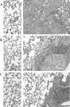
Photomicrographs of lung histology from C57BL/6 mice, F1 mice (B), and BALB/c mice (C). Left, uninfected lung sections; right, C. neoformans-infected lung sections at week 6 postinfection. Note the multinucleated giant macrophages composing a large portion of the mixed leukocyte infiltrate consolidating the infected lungs in C57BL/6 mice, less-diffuse inflammatory response and a mixture of giant cells and tightly packed mononuclear cell infiltrate in F1 mice, and tightly packed mononuclear cell infiltrate and largely normal lung morphology in BALB/c mice. Pictures show hematoxylin-and-eosin-stained lung sections at an objective power of ×20.
FIG. 7.
FIG. 7.
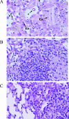
High-power photomicrographs of lung pathologies from C57BL/6 mice (A), F1 mice (B), or BALB/c mice (C) at week 6 postinfection. Note the multinucleated giant macrophages containing both intracellular YM1/YM2 crystals (YM) and cryptococcal organisms (*), consistent with the presence of AAMs, as well as small red granulocytes consistent with eosinophils (Eo) for C57BL/6 mice; the tight mononuclear cell infiltrate and the absence of AAMs and eosinophils in BALB/c mice, consistent with Th1 granulomatous inflammation; and intermediate lung pathology shown in the lungs of F1 mice. Pictures show hematoxylin-and-eosin-stained lung sections at an objective power of ×40.
FIG. 8.
FIG. 8.
Cytokine production in BAL fluid from C. neoformans-infected BALB/c, F1, and C57BL/6 mice. (A) BAL samples were collected at 7 days postinfection and analyzed for cytokines by ELISA. (B) The IFN-γ/IL-4 ratio was shown to illustrate a general trend in immune response polarization. Data were pooled from two parallel experiments (n = 4 to 6). Values represent means ± SEM in pg/ml supernatant. *, significant difference from results for F1 mice; **, significant difference from results for BALB/c mice.
FIG. 9.
FIG. 9.
Cytokine production in pulmonary leukocyte culture supernatants from C. neoformans-infected BALB/c, F1, and C57BL/6 mice at weeks 0, 2, and 3 postinfection. Isolated pulmonary leukocytes (5 × 10⁶/ml) were cultured for 24 h postharvest, and cytokines were evaluated by ELISA. Values represent means ± SEM in pg/ml supernatant (n = 6 and above for each of the analyzed time points). *, significant difference from results for F1 mice; **, significant difference from results for BALB/c mice.
FIG. 10.
FIG. 10.
Phenotypical analysis of DC in lung draining lymph nodes isolated from BALB/c, F1, and C57BL/6 mice. (A) Gating strategy for DC in the lung draining lymph nodes. CD11c+ MHCII+ populations were gated to analyze the proportion of cells with high costimulatory molecule expression. Note the increased density of DC (CD11c+ MHCII+ population) in the nodes in response to C. neoformans infection. (B) Overlaid histograms of BALB/c (light gray line), F1 (black line), C57BL/6 (solid black area), and uninfected F1 mice (solid gray area). Percentages of DC with high expression of CD40, DC80, and CD86 molecules are listed below each respective histogram. Values represent means ± SEM (n = 6 mice/group). **, significant difference from results for BALB/c mice. Data were pooled from two parallel experiments.

References

    1. Aguirre, K., J. Crowe, A. Haas, and J. Smith. 2004. Resistance to Cryptococcus neoformans infection in the absence of CD4+ T cells. Med. Mycol. 4215-25. - PubMed
    1. Arora, S., Y. Hernandez, J. R. Erb-Downward, R. A. McDonald, G. B. Toews, and G. B. Huffnagle. 2005. Role of IFN-gamma in regulating T2 immunity and the development of alternatively activated macrophages during allergic bronchopulmonary mycosis. J. Immunol. 1746346-6356. - PubMed
    1. Casadevall, A., and J. R. Perfect. 1998. Cryptococcus neoformans, 1st ed., vol. 1. ASM Press, Washington, DC.
    1. Casadevall, A., and L. A. Pirofski. 2003. The damage-response framework of microbial pathogenesis. Nat. Rev. Microbiol. 117-24. - PMC - PubMed
    1. Chen, G. H., R. A. McDonald, J. C. Wells, G. B. Huffnagle, N. W. Lukacs, and G. B. Toews. 2005. The gamma interferon receptor is required for the protective pulmonary inflammatory response to Cryptococcus neoformans. Infect. Immun. 731788-1796. - PMC - PubMed

Publication types

MeSH terms

LinkOut - more resources