

Role of Porphyromonas gingivalis SerB in gingival epithelial cell cytoskeletal remodeling and cytokine production
- PMID: 18391005
- PMCID: PMC2423092
- DOI: 10.1128/IAI.00156-08
Role of Porphyromonas gingivalis SerB in gingival epithelial cell cytoskeletal remodeling and cytokine production
Abstract
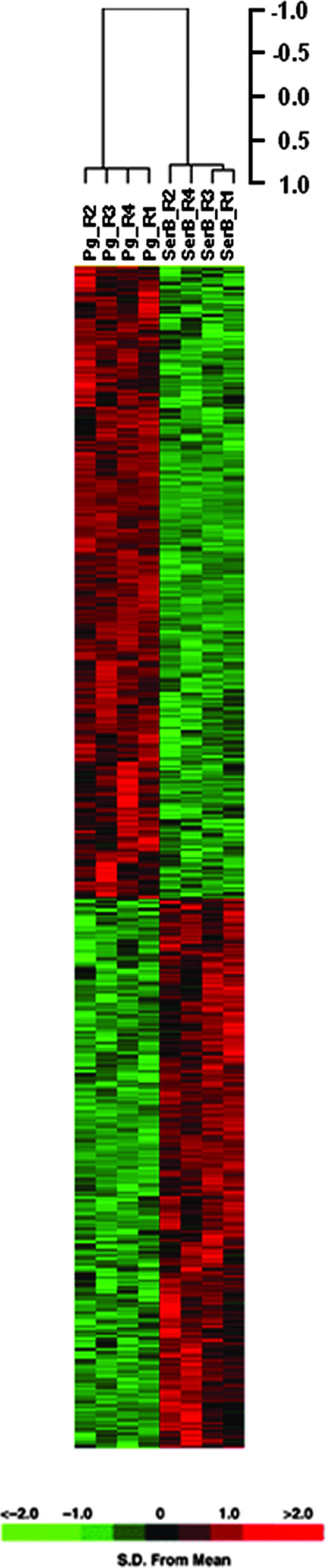
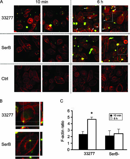
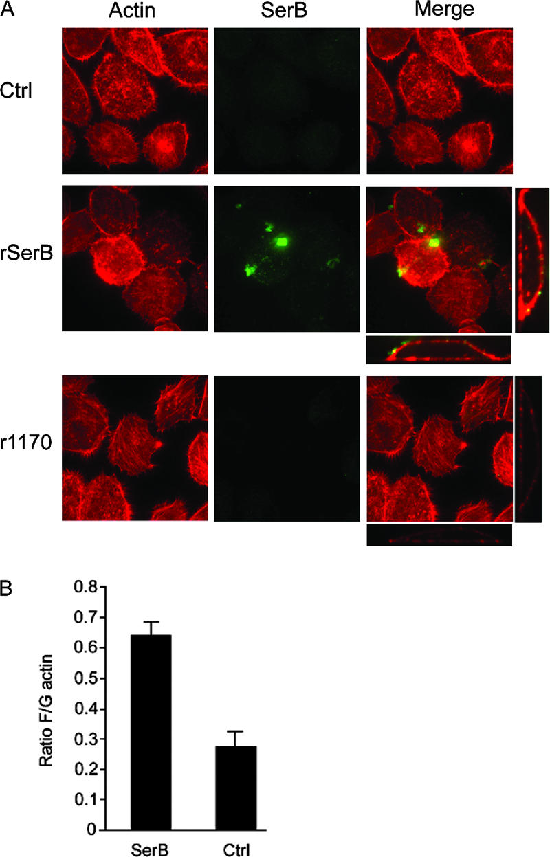
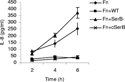
The SerB protein of Porphyromonas gingivalis is a HAD family serine phosphatase that plays a critical role in entry and survival of the organism in gingival epithelial cells. SerB is secreted by P. gingivalis upon contact with epithelial cells. Here it is shown by microarray analysis that SerB impacts the transcriptional profile of gingival epithelial cells, with pathways involving the actin cytoskeleton and cytokine production among those significantly overpopulated with differentially regulated genes. Consistent with the transcriptional profile, a SerB mutant of P. gingivalis exhibited defective remodeling of actin in epithelial cells. Interaction between gingival epithelial cells and isolated SerB protein resulted in actin rearrangement and an increase in the F/G actin ratio. SerB protein was also required for P. gingivalis to antagonize interleukin-8 accumulation following stimulation of epithelial cells with Fusobacterium nucleatum. SerB is thus capable of modulating host cell signal transduction that impacts the actin cytoskeleton and cytokine production.
Figures

Similar articles
-
Porphyromonas gingivalis SerB-mediated dephosphorylation of host cell cofilin modulates invasion efficiency.Cell Microbiol. 2012 Apr;14(4):577-88. doi: 10.1111/j.1462-5822.2011.01743.x. Epub 2012 Feb 2. Cell Microbiol. 2012. PMID: 22212282 Free PMC article.
-
Role of Porphyromonas gingivalis phosphoserine phosphatase enzyme SerB in inflammation, immune response, and induction of alveolar bone resorption in rats.Infect Immun. 2010 Nov;78(11):4560-9. doi: 10.1128/IAI.00703-10. Epub 2010 Aug 30. Infect Immun. 2010. PMID: 20805334 Free PMC article.
-
The serine phosphatase SerB of Porphyromonas gingivalis suppresses IL-8 production by dephosphorylation of NF-κB RelA/p65.PLoS Pathog. 2013;9(4):e1003326. doi: 10.1371/journal.ppat.1003326. Epub 2013 Apr 18. PLoS Pathog. 2013. PMID: 23637609 Free PMC article.
-
Differential regulation of cytokine genes in gingival epithelial cells challenged by Fusobacterium nucleatum and Porphyromonas gingivalis.Microb Pathog. 2004 Dec;37(6):303-12. doi: 10.1016/j.micpath.2004.10.003. Epub 2004 Dec 8. Microb Pathog. 2004. PMID: 15619426
-
Interleukin-8 and intercellular adhesion molecule 1 regulation in oral epithelial cells by selected periodontal bacteria: multiple effects of Porphyromonas gingivalis via antagonistic mechanisms.Infect Immun. 2001 Mar;69(3):1364-72. doi: 10.1128/IAI.69.3.1364-1372.2001. Infect Immun. 2001. PMID: 11179300 Free PMC article.
Cited by
-
Treponema denticola-Induced RASA4 Upregulation Mediates Cytoskeletal Dysfunction and MMP-2 Activity in Periodontal Fibroblasts.Front Cell Infect Microbiol. 2021 May 19;11:671968. doi: 10.3389/fcimb.2021.671968. eCollection 2021. Front Cell Infect Microbiol. 2021. PMID: 34094999 Free PMC article.
-
New Trends in the Impact of Periodontal Treatment on Early Cardiovascular Diseases Outcomes: Insights and Future Perspectives.Rev Cardiovasc Med. 2023 Oct 8;24(10):287. doi: 10.31083/j.rcm2410287. eCollection 2023 Oct. Rev Cardiovasc Med. 2023. PMID: 39077574 Free PMC article. Review.
-
Identification of PGN_1123 as the Gene Encoding Lipid A Deacylase, an Enzyme Required for Toll-Like Receptor 4 Evasion, in Porphyromonas gingivalis.J Bacteriol. 2019 May 8;201(11):e00683-18. doi: 10.1128/JB.00683-18. Print 2019 Jun 1. J Bacteriol. 2019. PMID: 30782639 Free PMC article.
-
Genetic transformation of an obligate anaerobe, P. gingivalis for FMN-green fluorescent protein expression in studying host-microbe interaction.PLoS One. 2011 Apr 15;6(4):e18499. doi: 10.1371/journal.pone.0018499. PLoS One. 2011. PMID: 21525983 Free PMC article.
-
The M. tuberculosis HAD phosphatase (Rv3042c) interacts with host proteins and is inhibited by Clofazimine.Cell Mol Life Sci. 2016 Sep;73(17):3401-17. doi: 10.1007/s00018-016-2177-2. Epub 2016 Mar 17. Cell Mol Life Sci. 2016. PMID: 26984196 Free PMC article.
References
-
- Arsura, M., G. R. Panta, J. D. Bilyeu, L. G. Cavin, M. A. Sovak, A. A. Oliver, V. Factor, R. Heuchel, F. Mercurio, S. S. Thorgeirsson, and G. E. Sonenshein. 2003. Transient activation of NF-κB through a TAK1/IKK kinase pathway by TGF-beta1 inhibits AP-1/SMAD signaling and apoptosis: implications in liver tumor formation. Oncogene 22412-425. - PubMed
-
- Belton, C. M., P. C. Goodwin, S. Fatherazi, M. M. Schubert, R. J. Lamont, and K. T. Izutsu. 2004. Calcium oscillations in gingival epithelial cells infected with Porphyromonas gingivalis. Microbes Infect. 6440-447. - PubMed
-
- Belton, C. M., K. T. Izutsu, P. C. Goodwin, Y. Park, and R. J. Lamont. 1999. Fluorescence image analysis of the association between Porphyromonas gingivalis and gingival epithelial cells. Cell Microbiol. 1215-223. - PubMed
-
- Bernard, O. 2007. Lim kinases, regulators of actin dynamics. Int. J. Biochem. Cell Biol. 391071-1076. - PubMed
Publication types
MeSH terms
Substances
Associated data
- Actions
Grants and funding
LinkOut - more resources
Full Text Sources
Molecular Biology Databases
