. 2008 Jun;76(6):2642-50.
doi: 10.1128/IAI.01639-07. Epub 2008 Apr 7.

In LipL32, the major leptospiral lipoprotein, the C terminus is the primary immunogenic domain and mediates interaction with collagen IV and plasma fibronectin

Affiliations

In LipL32, the major leptospiral lipoprotein, the C terminus is the primary immunogenic domain and mediates interaction with collagen IV and plasma fibronectin

Pricila Hauk et al. Infect Immun. 2008 Jun.

Abstract

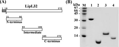
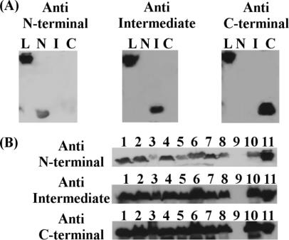
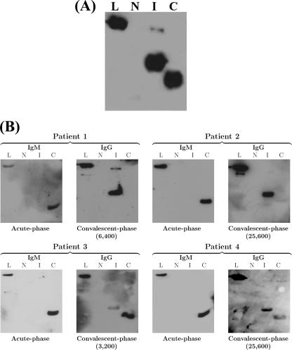
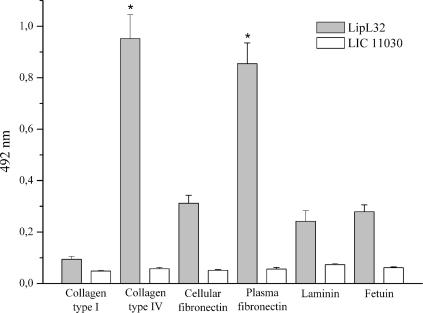
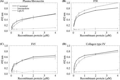
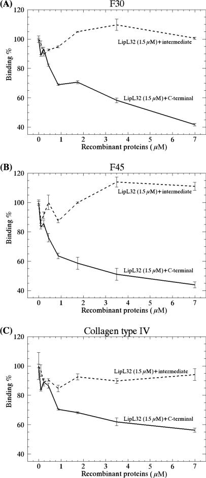
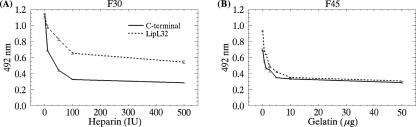
LipL32 is the major leptospiral outer membrane lipoprotein expressed during infection and is the immunodominant antigen recognized during the humoral immune response to leptospirosis in humans. In this study, we investigated novel aspects of LipL32. In order to define the immunodominant domains(s) of the molecule, subfragments corresponding to the N-terminal, intermediate, and C-terminal portions of the LipL32 gene were cloned and the proteins were expressed and purified by metal affinity chromatography. Our immunoblot results indicate that the C-terminal and intermediate domains of LipL32 are recognized by sera of patients with laboratory-confirmed leptospirosis. An immunoglobulin M response was detected exclusively against the LipL32 C-terminal fragment in both the acute and convalescent phases of illness. We also evaluated the capacity of LipL32 to interact with extracellular matrix (ECM) components. Dose-dependent, specific binding of LipL32 to collagen type IV and plasma fibronectin was observed, and the binding capacity could be attributed to the C-terminal portion of this molecule. Both heparin and gelatin could inhibit LipL32 binding to fibronectin in a concentration-dependent manner, indicating that the 30-kDa heparin-binding and 45-kDa gelatin-binding domains of fibronectin are involved in this interaction. Taken together, our results provide evidence that the LipL32 C terminus is recognized early in the course of infection and is the domain responsible for mediating interaction with ECM proteins.


Figures

FIG. 1.
FIG. 1.
(A) Schematic representation of LipL32 protein. Shown are the signal peptide (amino acids 1 to 19), N-terminal domain (amino acids 21 to 92), intermediate domain (amino acids 93 to 184), and C-terminal domain (amino acids 185 to 272). The asterisk indicates the cysteine to be lipidated. (B) Purification of recombinant proteins. SDS-PAGE (15%) of purified recombinant proteins obtained by metal affinity chromatography. Lane 1, LipL32; lane 2, N-terminal fragment; lane 3, intermediate fragment; lane 4, C-terminal fragment; lane M, molecular mass marker.
FIG. 2.
FIG. 2.
Specificity of antisera against LipL32 fragments. Sera from mice immunized with recombinant LipL32 subfragments recognize the corresponding fragments (N, N terminal; I, intermediate; C, C terminal), as well as the intact protein (L, LipL32) (A), and native LipL32 from whole-cell extracts of L. interrogans serovars Icterohaemorrhagiae (column 1), Copenhageni (column 2), Bratislava (column 3), Hardjo (column 4), Autumnalis (column 5), Pomona (column 6), Pyrogenes (column 7), and Canicola (column 8) and L. kirchneri serovar Grippotyphosa (column 10) (B). Sera also recognized the recombinant protein (column 11) but failed to react with the whole-cell extract of L. biflexa serovar Patoc (column 9).
FIG. 3.
FIG. 3.
Recombinant LipL32 protein subfragments and reactivity with sera from mice (A) and leptospirosis patients (B). Immunoblot analyses of N-terminal (N), intermediate (I), C-terminal (C), and intact LipL32 (L) recombinant proteins (2 μg/lane). Membranes were probed with serum from mice immunized with recombinant LipL32 (A) or with sera obtained from leptospirosis patients 1 to 4 (Table 1) during the acute and convalescent phases of illness (B). Blots were developed with goat anti-mouse IgG-peroxidase conjugate (A) or goat anti-human IgM-peroxidase conjugate (acute phase) and goat anti-human IgG-peroxidase conjugate (convalescent phase) (B). MAT titers for each patient are indicated.
FIG. 4.
FIG. 4.
Binding of recombinant LipL32 to ECM macromolecules. Wells were coated with 1 μg of laminin, collagen type I, collagen type IV, cellular fibronectin, plasma fibronectin, and the control protein fetuin. Recombinant protein attachment was assessed by an ELISA-based method. One microgram of recombinant protein was added per well. Optical densities were determined at 492 nm. Data represent the mean ± the standard deviation of three independent experiments, each performed in triplicate. For statistical analyses, the attachment of LipL32 to the ECM components was compared to the attachment of the protein to fetuin by the two-tailed t test (*, P < 0.0001).
FIG. 5.
FIG. 5.
Binding of LipL32, its C-terminal domain, and its intermediate domain to plasma fibronectin (intact molecule and F30 and F45 proteolytic fragments) and to collagen IV as a function of protein concentration. Panels: A, binding to plasma fibronectin; B, binding to F30; C, binding to F45; D, binding to collagen IV. Recombinant protein concentrations ranged from 0 to 4 μM. Each point represents the mean absorbance value at 492 nm ± the standard error of three independent experiments.
FIG. 6.
FIG. 6.
Inhibition of LipL32 binding to ECM by the C-terminal fragment. Wells were coated with F30 (A), F45 (B), or collagen IV (C), and increasing concentrations of the C-terminal or intermediate domain (0 to 7 μM) were added in the presence of a fixed LipL32 concentration (1.5 μM). Optical densities were determined at 492 nm. Data represent the mean ± the standard error of three independent experiments, each performed in triplicate.
FIG. 7.
FIG. 7.
Inhibition of LipL32 binding to F30 and F45 by heparin and gelatin. Binding of LipL32 and its C-terminal domain (2 μM) to F30 (A) and F45 (B) was assayed in the presence of increasing amounts of heparin (0 to 500 IU) or gelatin (0 to 50 μg). Optical densities were determined at 492 nm. Data represent the mean ± the standard error of three independent experiments.

Similar articles

Cited by

References

    1. Barbosa, A. S., P. A. E. Abreu, F. O. Neves, M. V. Atzingen, M. M. Watanabe, M. L. Vieira, Z. M. Morais, S. A. Vasconcellos, and A. L. T. O. Nascimento. 2006. A newly identified leptospiral adhesin mediates attachment to laminin. Infect. Immun. 746356-6364. - PMC - PubMed
    1. Bhandari, P., and J. Gowrishankar. 1997. An Escherichia coli host strain useful for efficient overproduction of cloned gene products with NaCl as the inducer. J. Bacteriol. 1794403-4406. - PMC - PubMed
    1. Bharti, A. R., J. E. Nally, J. N. Ricaldi, M. A. Matthias, M. M. Diaz, M. A. Lovett, P. N. Levett, R. H. Gilman, M. R. Willig, E. Gotuzzo, and J. M. Vinetz on behalf of the Peru-United States Leptospirosis Consortium. 2003. Leptospirosis: a zoonotic disease of global importance. Lancet Infect. Dis. 3757-771. - PubMed
    1. Bomfim, M. R. Q., A. I. Ko, and M. C. Koury. 2005. Evaluation of the recombinant LipL32 in enzyme-linked immunosorbent assay for the serodiagnosis of bovine leptospirosis. Vet. Microbiol. 10989-94. - PubMed
    1. Boonyod, D., Y. Poovorawan, P. Bhattarakosol, and C. Chirathaworn. 2005. LipL32, an outer membrane protein of Leptospira, as an antigen in a dipstick assay for diagnosis of leptospirosis. Asian Pac. J. Allergy Immunol. 23133-141. - PubMed

Publication types

MeSH terms

LinkOut - more resources